. 2016 Nov 18;11(11):3043-3051.
doi: 10.1021/acschembio.6b00588. Epub 2016 Sep 15.

Thermal Unthreading of the Lasso Peptides Astexin-2 and Astexin-3

Affiliations

Thermal Unthreading of the Lasso Peptides Astexin-2 and Astexin-3

Caitlin D Allen et al. ACS Chem Biol. .

Abstract

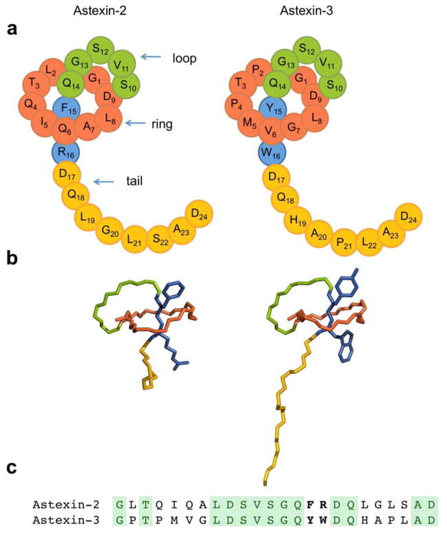
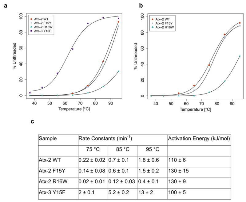
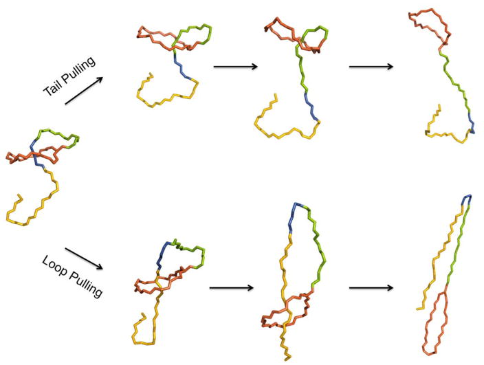
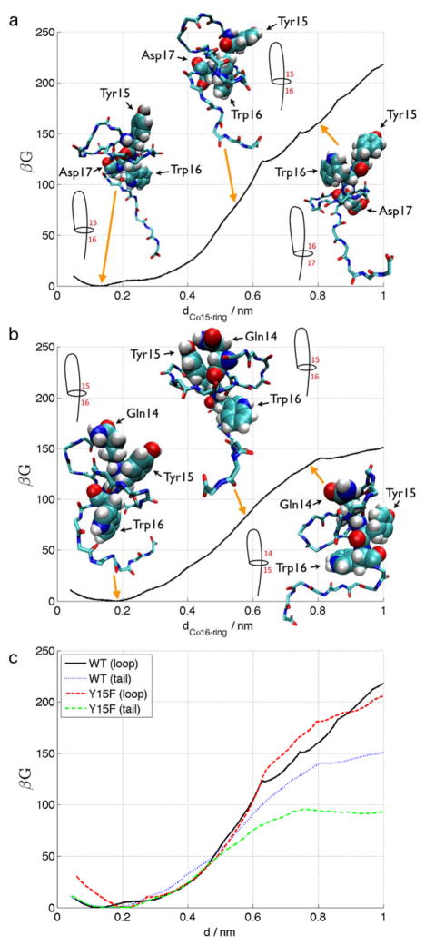
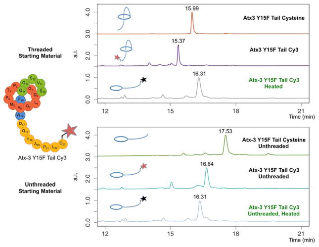
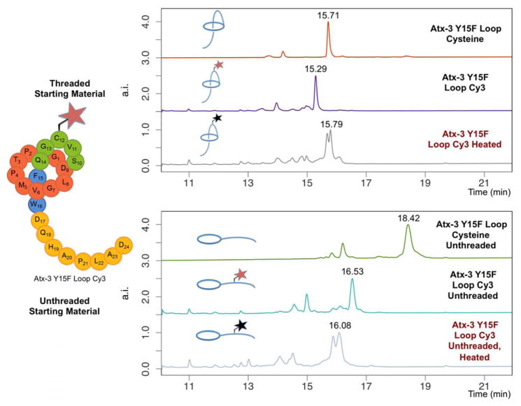
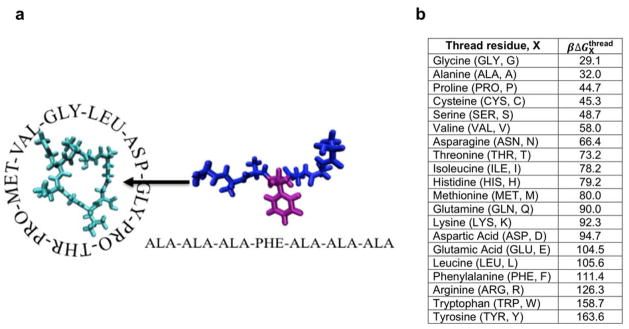
Lasso peptides are a class of knot-like polypeptides in which the C-terminal tail of the peptide threads through a ring formed by an isopeptide bond between the N-terminal amine group and a side chain carboxylic acid. The small size (∼20 amino acids) and simple topology of lasso peptides make them a good model system for studying the unthreading of entangled polypeptides, both with experiments and atomistic simulation. Here, we present an in-depth study of the thermal unthreading behavior of two lasso peptides astexin-2 and astexin-3. Quantitative kinetics and energetics of the unthreading process were determined for variants of these peptides using a series of chromatography and mass spectrometry experiments and biased molecular dynamics (MD) simulations. In addition, we show that the Tyr15Phe variant of astexin-3 unthreads via an unprecedented "tail pulling" mechanism. MD simulations on a model ring-thread system coupled with machine learning approaches also led to the discovery of physicochemical descriptors most important for peptide unthreading.


Figures

Figure 1
Figure 1. Sequence and structure of astexin-2 and astexin-3
(a) Cartoon representation of the peptides. Red represents the ring of the peptide, green is the loop, blue are steric lock residues, and orange is the C-terminal tail. (b) NMR structures of astexin-2 ΔC4 (left), from PDB file 2N6U and full-length astexin-3, from PDB file 2N6V (right). Sidechains of steric lock residues are shown and the color scheme is as in part A. (c) Sequence comparison of astexins-2 and -3. Identities are highlighted in green, steric lock residues are in bold type.
Figure 2
Figure 2. Thermal denaturation curves, rate constants, and activation energies for thermolabile astexin-2 ΔC3 and -3 variants
Thermal denaturation curves for peptides heated for (a) one hour or (b) two hours, at the indicated temperatures and then analyzed by HPLC. The % unthreaded was determined by integrating the chromatograms. (c) Rate constants and activation energies
Figure 3
Figure 3. Schematic of two directions for lasso peptide unthreading
Top: unthreading via pulling on the C-terminal tail. Bottom: unthreading via loop pulling. The color scheme is as in Figure 1; position of steric lock residues are marked in blue.
Figure 4
Figure 4. Potential of mean force (PMF) computed by umbrella sampling molecular dynamics simulations for the unthreading of astexin-3 WT and Y15F mutants
(a) Unthreading of astexin-3 WT by loop pulling in which the Trp16 steric lock residue passes through the ring. Complete passage of Trp16 occurs at dCα15-ring ≈ 0.8 nm. We report the PMF (Gibbs free energy) G rendered dimensionless by the reciprocal temperature β = 1/kBT where kB is Boltzmann’s constant and T = 298 K. The arbitrary zero of free energy of each PMF curve was specified to coincide with its global minimum, and uncertainties estimated by 100 rounds of bootstrap resampling are smaller than 1 kBT. The PMF curves are superposed with representative snapshots of the peptide along the unthreading trajectory rendered using VMD and schematic line diagrams to illustrate which tail residues are spanning the ring in each image. (b) Unthreading of astexin-3 WT by tail pulling in which the Tyr15 steric lock passes through the ring. Complete passage of Tyr15 occurs at dCα16-ring ≈ 0.75 nm. (c) Composite plot of the PMF curves for loop and tail pulling of the astexin-3 wild-type (WT) and Y15F variant. d corresponds to dCα15-ring in the case of loop pulling, and to dCα16-ring in the case of tail pulling. For both variants of the peptide, the free energy barrier for tail pulling (slipping residue 15) is significantly lower than that for loop pulling (slipping residue 16): ΔGTyr15WT130kBTvs.ΔGTrp16WT160kBT for the WT, and ΔGPhe15Y15F95kBTvs.ΔGTrp16Y15F180kBT for Y15F.
Figure 5
Figure 5. Astexin-3 Y15F constructs for studying unthreading directionality
The cysteine sidechains are highlighted in bright yellow and the steric lock sidechains are in blue as in Figure 1. The bulky sulfo-Cy3 moiety attached as a secondary steric lock is in light pink.
Figure 6
Figure 6. Probing unthreading of astexin-3 Y15F using tail-labeled peptide
Top panel: from top to bottom, astexin-3 Y15F tail cysteine is labeled first with Cy3 and then heated, resulting in an unthreaded species. Bottom panel: from top to bottom, astexin-3 Y15F tail cysteine is first unthreaded by heating then labeled with Cy3 followed by heating again to hydrolyze the maleimide. The final product is identical to what is observed in the top panel, indicating that the Cy3 group on the tail does not prevent unthreading. Red stars represent non-hydrolyzed Cy3 groups, black stars are maleimide hydrolyzed Cy3.
Figure 7
Figure 7. Probing unthreading of astexin-3 Y15F using loop-labeled peptide
Top panel: from top to bottom, astexin-3 Y15F loop cysteine was labeled with Cy3 and subsequently heated at 95 °C for 1 hr. Bottom panel: from top to bottom, astexin-3 Y15F loop cysteine was fully unthreaded by heating, followed by conjugation with Cy3 and further heating to hydrolyze the maleimide. In contrast to the results in Figure 6, these two treatments result in different species, indicating that conjugation of Cy3 in the loop region prevents full unthreading of the peptide. Red stars represent non-hydrolyzed Cy3 groups, black stars are maleimide-hydrolyzed Cy3.
Figure 8
Figure 8. Ring-thread system comprising the nine-residue Gly-Pro-Thr-Pro-Met-Val-Gly-Leu-Asp astexin-3 “ring” excised from the native peptide and a seven-residue Ala-Ala-Ala-X-Ala-Ala-Ala “thread” in which X was exchanged for each of the 20 natural amino acids
(a) This image illustrates the X = Phe case. The thread was computationally docked and threaded through the ring using artificial umbrella biasing potentials, and the umbrella sampling data analyzed using the WHAM approach to estimate the free energy barrier for passage of each amino acid residue through the ring free of the confounding effects of its local environment within the astexin-3 peptide chain. (b) Calculated free energy barriers for passage of the X residue within the AAAXAAA thread through the excised Gly-Pro-Thr-Pro-Met-Val-Gly-Leu-Asp astexin-3 ring by umbrella sampling molecular dynamics simulations. Values are reported in units of kBT = β−1 where kB is Boltzmann’s constant and T = 298 K. Uncertainties in the free energy barriers were quantified by performing five independent runs of the AAAFAAA system from which we estimated an uncertainty in the measured barrier of ±8.3 kBT.

References

    1. Jamroz M, Niemyska W, Rawdon EJ, Stasiak A, Millett KC, Sulkowski P, Sulkowska JI. KnotProt: a database of proteins with knots and slipknots. Nucleic Acids Res. 2015;43:D306–D314. - PMC - PubMed
    1. Lim NCH, Jackson SE. Molecular knots in biology and chemistry. J Phys: Conds Matter. 2015;27 - PubMed
    1. Lim K, Zhang H, Tempczyk A, Krajewski W, Bonander N, Toedt J, Howard A, Eisenstein E, Herzberg O. Structure of the YibK methyltransferase from Haemophilus influenzae (HI0766): A cofactor bound at a site formed by a knot. Proteins: Struct Funct, Genet. 2003;51:56–67. - PubMed
    1. Wagner JR, Brunzelle JS, Forest KT, Vierstra RD. A light-sensing knot revealed by the structure of the chromophore-binding domain of phytochrome. Nature. 2005;438:325–331. - PubMed
    1. Sayre TC, Lee TM, King NP, Yeates TO. Protein stabilization in a highly knotted protein polymer. Protein Eng Des Sel. 2011;24:627–630. - PMC - PubMed

Publication types

LinkOut - more resources