

Three-dimensional cone-beam CT sialography in non tumour salivary pathologies: procedure and results
- PMID: 27588734
- PMCID: PMC5595048
- DOI: 10.1259/dmfr.20150431
Three-dimensional cone-beam CT sialography in non tumour salivary pathologies: procedure and results
Abstract
Objectives: Non-tumour salivary diseases are common. Imaging studies are essential for their diagnosis and before undergoing an endoscopic or surgical treatment. In this study, we aimed at presenting our procedure and results obtained with three-dimensional CBCT (3D-CBCT) sialography for non-tumour salivary gland diseases.
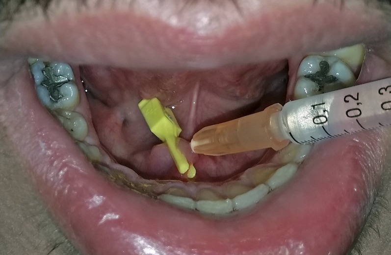
Methods: Patients with parotid or submandibular salivary symptoms were examined by 3D-CBCT sialography. They received an intraductal injection of 0.5 mL of water-soluble contrast medium maintained in the gland, followed by examination in a NewTom wide-field CBCT device. Images were processed with multiplanar and 3D reconstructions.
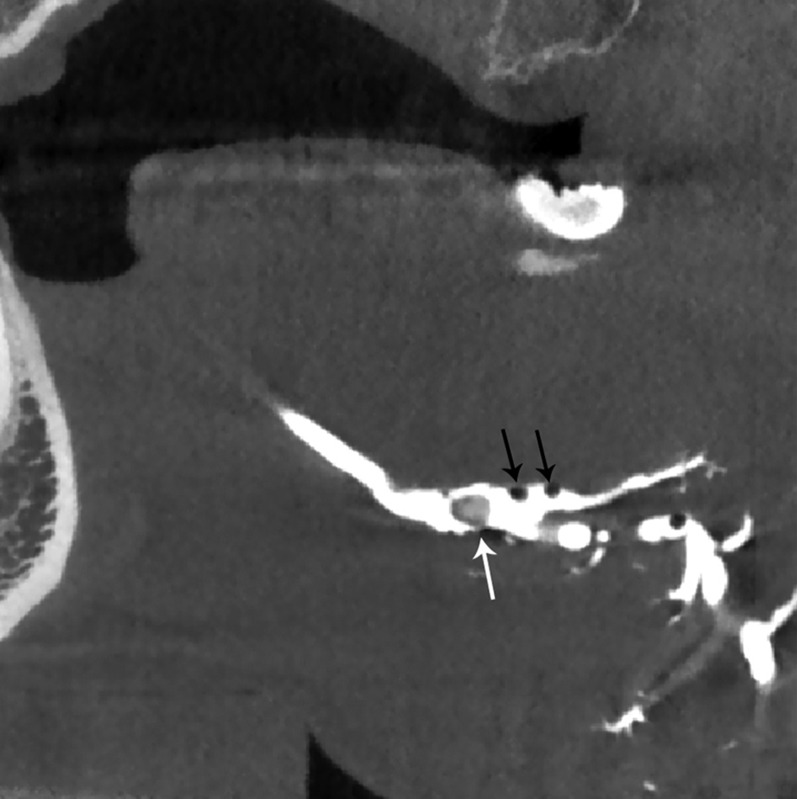
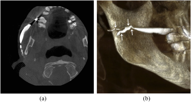
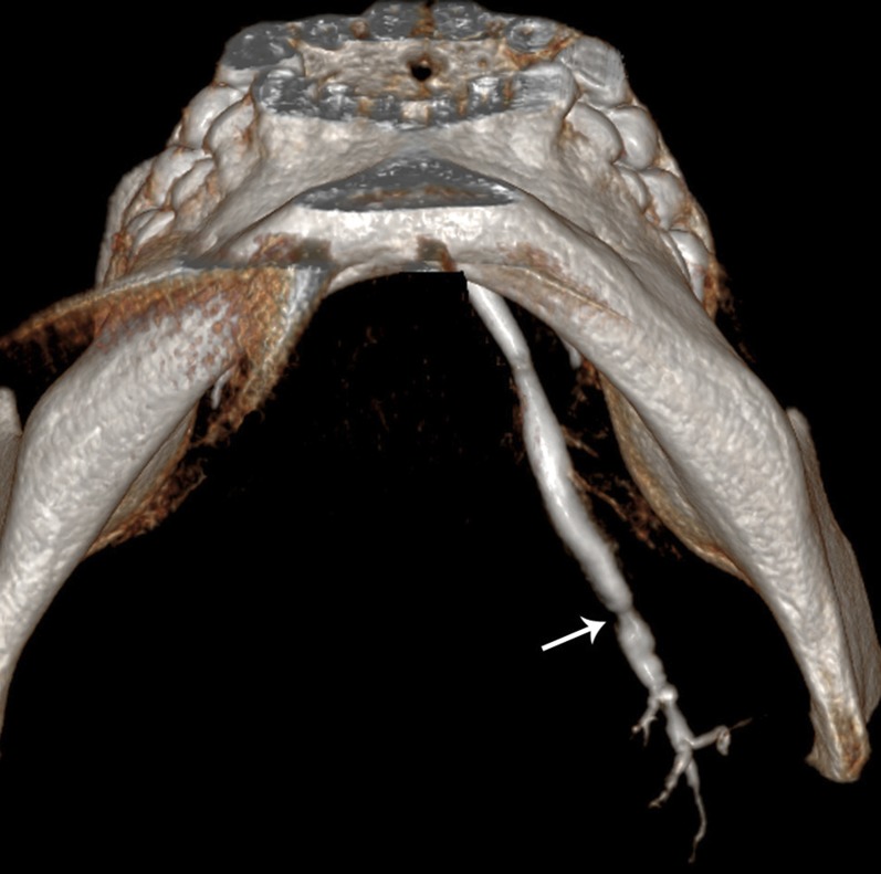
Results: A ductal exploration could be performed until the fourth divisions. The main lesions found were stones, stenosis, dilatations and "dead tree" appearance of the ductal system. No side effects of the catheterization or the iodine contrast were reported, nor tissue damages related to the contrast keeping technique.
Conclusions: 3D-CBCT sialography seems to represent a reliable non-invasive diagnostic tool for ductal salivary diseases. More studies are needed to assess the value of 3D-CBCT sialography compared with conventional imaging.
Keywords: CBCT; diagnostic imaging; salivary gland disease; sialography.
Figures

References
-
- Iro H, Zenk J, Escudier MP, Nahlieli O, Capaccio P, Katz P, et al. . Outcome of minimally invasive management of salivary calculi in 4,691 patients. Laryngoscope 2009; 119: 263–8. doi: https://doi.org/10.1002/lary.20008 - DOI - PubMed
-
- Katz P. New techniques for the treatment of salivary lithiasis: sialoendoscopy and extracorporal lithotripsy: 1773 cases. [In French.] Ann Otolaryngol Chir Cervicofac 2004; 121: 123–32. - PubMed
-
- Marchal F. Salivary gland endoscopy: new limits? [In French.] Rev Stomatol Chir Maxillofac 2005; 106: 244–9. - PubMed
-
- Avrahami E, Englender M, Chen E, Shabtay D, Katz R, Harell M. CT of submandibular gland sialolithiasis. Neuroradiology 1996; 38: 287–90. doi: https://doi.org/10.1007/BF00596550 - DOI - PubMed
-
- Katz P. Imagerie normale des glandes salivaires. In: EMC Radiodiagnostic—Coeur poumons. Paris: Elsevier Masson SAS; 2006.
MeSH terms
Substances
LinkOut - more resources
Full Text Sources
Other Literature Sources
Medical
