. 2017 Feb 1;595(3):825-838.
doi: 10.1113/JP273051. Epub 2016 Oct 2.

Development of an excitatory kisspeptin projection to the oxytocin system in late pregnancy

Affiliations

Development of an excitatory kisspeptin projection to the oxytocin system in late pregnancy

Alexander J Seymour et al. J Physiol. .

Abstract

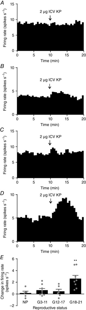
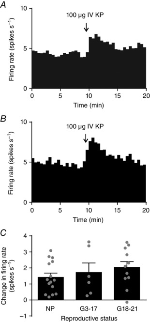
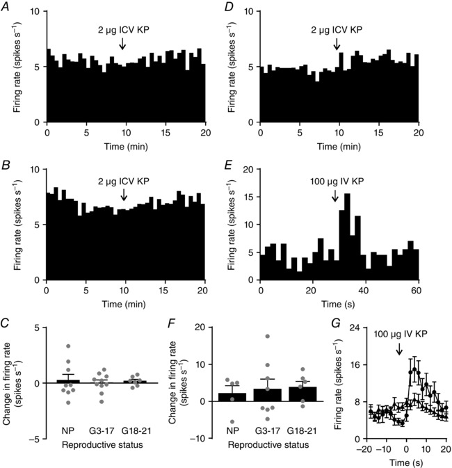
Key points: Oxytocin release from the posterior pituitary gland stimulates uterine contraction during birth but the central mechanisms that activate oxytocin neurones for birth are not well characterized. We found that that kisspeptin fibre density around oxytocin neurones increases in late-pregnant rats. These kisspeptin fibres originated from hypothalamic periventricular nucleus neurones that upregulated kisspeptin expression in late pregnancy. Oxytocin neurones were excited by central kisspeptin administration in late-pregnant rats but not in non-pregnant rats or early- to mid-pregnant rats. Our results reveal the emergence of a new excitatory kisspeptin projection to the oxytocin system in late pregnancy that might contribute to oxytocin neurone activation for birth.

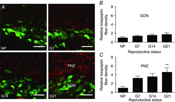
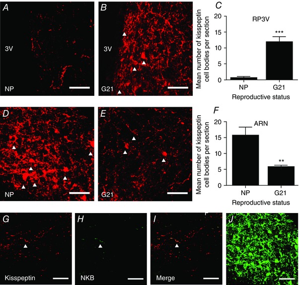
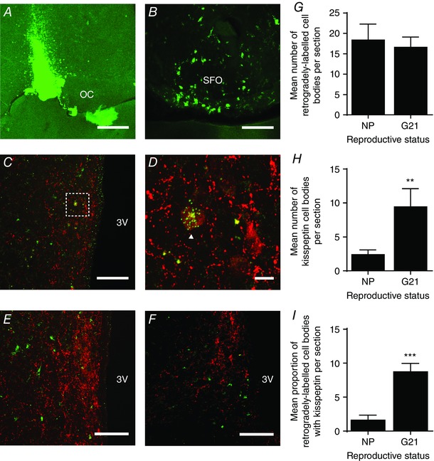
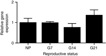
Abstract: The hormone oxytocin promotes uterine contraction during parturition. Oxytocin is synthesized by magnocellular neurones in the hypothalamic supraoptic and paraventricular nuclei and is released into the circulation from the posterior pituitary gland in response to action potential firing. Systemic kisspeptin administration increases oxytocin neurone activity to elevate plasma oxytocin levels. Here, immunohistochemistry revealed that rats on the expected day of parturition (day 21 of gestation) had a higher density of kisspeptin-positive fibres in the perinuclear zone surrounding the supraoptic nucleus (which provides dense glutamatergic and GABAergic innervation to the supraoptic nucleus) than was evident in non-pregnant rats. Retrograde tracing showed the kisspeptin projections to the perinuclear zone originated from the hypothalamic periventricular nucleus. Quantitative RT-PCR showed that kisspeptin receptor mRNA, Kiss1R mRNA, was expressed in the perinuclear zone-supraoptic nucleus and that the relative Kiss1R mRNA expression does not change over the course of pregnancy. Finally, intracerebroventricular administration of kisspeptin increased the firing rate of oxytocin neurones in anaesthetized late-pregnant rats (days 18-21 of gestation) but not in non-pregnant rats, or in early- or mid-pregnant rats. Taken together, these results suggest that kisspeptin expression is upregulated in the periventricular nucleus projection to the perinuclear zone of the supraoptic nucleus towards the end of pregnancy. Hence, this input might activate oxytocin neurones during parturition.

Keywords: kisspeptin; oxytocin; pregnancy; supraoptic nucleus; vasopressin.


Figures

Figure 1
Figure 1. Kisspeptin fibre density in the perinuclear zone of non‐pregnant and pregnant rats
A, representative dual‐label confocal images showing kisspeptin immunoreactive fibres (red) and oxytocin immunoreactive neurones (green) in the supraoptic nucleus (SON) and perinuclear zone (PNZ) of a non‐pregnant rat (NP), an early‐pregnant rat (G7), a mid‐pregnant rat (G14) and a late‐pregnant rat (G21). Scale bars = 50 μm. B, relative kisspeptin fibre density within the supraoptic nucleus. One‐way ANOVA showed no significant effect of reproductive status (F 3,43 = 1.46, P = 0.24). C, relative kisspeptin fibre density within the perinuclear zone. One‐way ANOVA showed a significant effect of reproductive status (F 3,43 = 3.95, P = 0.01); ** P ≤ 0.01 compared to non‐pregnant rats, Bonferroni's post hoc test. [Colour figure can be viewed at wileyonlinelibrary.com]
Figure 2
Figure 2. Kisspeptin and neurokinin B expression in non‐pregnant and pregnant rats
A and B, representative confocal images showing kisspeptin expression in the rostral periventricular area of the third ventricle (RP3V) from a non‐pregnant rat (NP; A) and a late‐pregnant rat (G21; B). White arrowheads indicate labelled cell bodies. C, number of kisspeptin‐positive cell bodies in the RP3V of non‐pregnant rats and late‐pregnant rats; *** P < 0.001, Student's t test. D and E, representative confocal images showing kisspeptin expression in the arcuate nucleus (ARN) of a non‐pregnant rat (D) and a late‐pregnant rat (E). F, number of kisspeptin‐positive cell bodies in the arcuate nucleus of non‐pregnant rats and late‐pregnant rats; ** P < 0.01, Student's t test. G–I, representative confocal images showing limited co‐expression of neurokinin B (NKB) with kisspeptin in supraoptic nucleus/perinuclear zone sections from a late‐pregnant rat. White arrowhead indicates a co‐labelled fibre. J, NKB labelling of cell bodies and fibres in the arcuate nucleus of a non‐pregnant rat. Scale bars in all images = 50 μm. [Colour figure can be viewed at wileyonlinelibrary.com]
Figure 3
Figure 3. Retrograde labelling of kisspeptin neurones in the periventricular nucleus from the supraoptic nucleus/perinuclear zone
A, confocal image showing a positive injection site in the perinuclear zone–supraoptic nucleus, immediately lateral to the optic chiasm (OC). Scale bar = 500 μm. B, confocal image showing retrograde labelling of cell bodies in the subfornical organ (SFO), an area known to project to the supraoptic nucleus. Scale bar = 50 μm. C, confocal image showing kisspeptin and tracer expression in the periventricular nucleus, lateral to the third ventricle (3V). Scale bar = 100 μm. D, high power image of the area bounded by the box in C, showing a kisspeptin‐positive neurone showing punctate tracer labelling (white arrowhead). Scale bar = 10 μm. E and F, representative confocal images showing no retrograde labelling of kisspeptin neurones in the anteroventral periventricular nucleus (E) and arcuate nucleus (F). Scale bars = 100 μm. G, the mean number of retrogradely labelled neurones per section in the periventricular nucleus of non‐pregnant rats (NP) and late‐pregnant rats (G21). H, the mean number of kisspeptin‐positive neurones per section in the periventricular nucleus of non‐pregnant rats and late‐pregnant rats. I, the percentage of retrogradely labelled neurones that were kisspeptin‐positive in the periventricular nucleus of non‐pregnant rats and late‐pregnant rats. *** P < 0.0001 versus non‐pregnant rats, Student's t test. [Colour figure can be viewed at wileyonlinelibrary.com]
Figure 4
Figure 4. Kiss1R mRNA expression in the supraoptic nucleus/perinuclear zone
Kiss1R mRNA expression in the supraoptic nucleus/perinuclear zone non‐pregnant rats and of early‐ (G7) mid‐ (G14) and late‐ (G21) pregnant rats, relative to non‐pregnant rats. There was no significant effect of reproductive status (P = 0.25, one‐way ANOVA).
Figure 5
Figure 5. i.c.v. kisspeptin excites oxytocin neurones only in late‐pregnant rats
A–D, representative ratemeter records (in 30 s bins) showing the effect of intracerebroventricular (i.c.v.) kisspeptin (KP; 2 μg) on the firing rate of oxytocin neurones from a non‐pregnant rat (A; NP), early‐pregnant rat (B; G3–11), mid‐pregnant rat (C; G12–17) and late‐pregnant rat (D; G18–21). E, change in firing rate 2 min before and after i.c.v. kisspeptin administration. One‐way ANOVA showed a significant effect of reproductive status (F 5,37 = 8.60, P < 0.001); ** P < 0.01 compared to non‐pregnant rats, Bonferroni's post hoc test).
Figure 6
Figure 6. i.v. kisspeptin excites oxytocin neurones
A and B, representative ratemeter records (in 30 s bins) showing intravenous (i.v.) kisspeptin (KP; 100 μg) excitation of oxytocin neurones from a non‐pregnant rat (A) and a late‐pregnant rat (B). C, change in firing rate 2 min before and after i.v. kisspeptin administration in early‐ or mid‐pregnant rats (G3–17) and late‐pregnant rats (G18–21). One‐way ANOVA showed no significant effect of reproductive status (F 2,34 = 1.47, P = 0.24).
Figure 7
Figure 7. Effects of kisspeptin on vasopressin neurones
A and B, representative ratemeter records (in 30 s bins) showing no effect of intracerebroventricular (i.c.v.) kisspeptin (KP; 2 μg) on vasopressin neurone activity in a non‐pregnant rat (A) and a late‐pregnant rat (B). C, Change in firing rate 2 min before and after i.c.v. kisspeptin administration in early‐ or mid‐pregnant rats (G3–17) and late‐pregnant rats (G18–21). One‐way ANOVA showed no significant effect of reproductive status (F 2,30 = 0.42, P = 0.66). D, representative ratemeter record (in 30 s bins) from a late‐pregnant rat, showing no sustained effect of intravenous (i.v.) kisspeptin (100 μg) on vasopressin neurone firing rate. E, ratemeter record from the same rat in D in 2 s bins showing a transient increase in vasopressin neurone firing rate after i.v. kisspeptin. F, change in firing rate of vasopressin neurones 2 min before and after i.v. kisspeptin in early‐ or mid‐pregnant rats (G3–17) and late‐pregnant rats (G18–21). One‐way ANOVA showed no significant effect of reproductive status (F 2,26 = 1.76, P = 0.19). G, mean firing rate of the vasopressin neurones shown in F for 20 s before and after i.v. kisspeptin (in 2 s bins); circles show vasopressin neurones that responded to kisspeptin with a short burst of firing, and triangles show vasopressin neurones that were unaffected by i.v. kisspeptin.

Comment in

References

    1. Antonijevic IA, Douglas AJ, Dye S, Bicknell RJ, Leng G & Russell JA (1995). Oxytocin antagonists delay the initiation of parturition and prolong its active phase in rats. J Endocrinol 145, 97–103. - PubMed
    1. Armstrong WE & Stern JE (1997). Electrophysiological and morphological characteristics of neurons in perinuclear zone of supraoptic nucleus. J Neurophysiol 78, 2427–2437. - PubMed
    1. Augustine RA, Bouwer GT, Seymour AJ, Grattan DR & Brown CH (2016). Reproductive regulation of gene expression in the hypothalamic supraoptic and paraventricular nuclei. J Neuroendocrinol 28, DOI: 10.1111/jne.12350. - PubMed
    1. Bianco SD & Kaiser UB (2013). Molecular biology of the kisspeptin receptor: signaling, function, and mutations. Adv Exp Med Biol 784, 133–158. - PubMed
    1. Boudaba C, Di S & Tasker JG (2003). Presynaptic noradrenergic regulation of glutamate inputs to hypothalamic magnocellular neurones. J Neuroendocrinol 15, 803–810. - PubMed

Publication types

LinkOut - more resources