

Multicenter Assessment of Immunohistochemical Methods for Pathological Alpha-Synuclein in Sigmoid Colon of Autopsied Parkinson's Disease and Control Subjects
- PMID: 27589538
- PMCID: PMC5501392
- DOI: 10.3233/JPD-160888
Multicenter Assessment of Immunohistochemical Methods for Pathological Alpha-Synuclein in Sigmoid Colon of Autopsied Parkinson's Disease and Control Subjects
Abstract
Background: Conflicting results from studies of Lewy-type α-synucleinopathy (LTS) in colonic biopsies of subjects with Parkinson's disease (PD) prompted a two-part multicenter assessment. The first assessment, now published (Acta Neuropathol Commun 4 : 35, 2016), examined archived colonic biopsies and found that none of the tested methods was adequately sensitive or specific.
Objective: As the amount of nervous tissue in typical colonic biopsies may be insufficient, and the clinical diagnosis of PD not completely accurate, the objective of the current study was to use instead full-thickness sections of sigmoid colon from autopsy-proven PD and normal subjects.
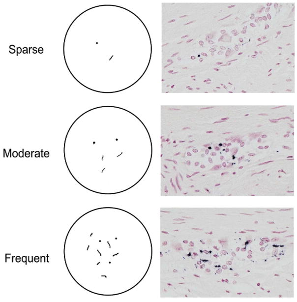
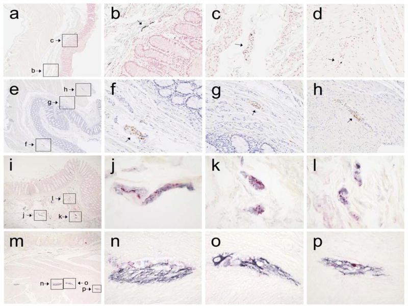
Methods: Seven different immunohistochemical (IHC) methods were used, employing five different primary antibodies and four different combinations of epitope exposure and signal development protocols. Specific staining was defined as being restricted to morphological features consistent with neuronal elements. Stained slides from each subject were independently categorized as being positive or negative for LTS, and their density semi-quantitatively graded, by four raters blinded to diagnosis.
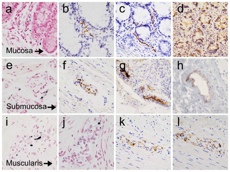
Results: Agreement and mean diagnostic performance varied markedly between raters. With the two most accurate raters, 5 methods achieved diagnostic accuracies of 70% or greater; one method had 100% accuracy and 100% inter-rater agreement. The submucosa had the highest prevalence of pathological LTS staining, followed by the muscularis and mucosa.
Conclusions: The major conclusion of this study is that, when sufficient submucosa and LTS is present, and when specific staining is defined as being consistent with neuronal morphology, adequately-trained raters may reliably distinguish PD colon from control using suitable IHC methods.
Keywords: Lewy body; biopsy; diagnosis; enteric nervous system; gastrointestinal tract; pathology.
Conflict of interest statement
The authors have no conflicts of interest to report.
Figures
References
-
- Qualman SJ, Haupt HM, Yang P, Hamilton SR. Esophageal Lewy bodies associated with ganglion cell loss in achalasia. Similarity to Parkinson’s disease. Gastroenterology. 1984;87:848–56. - PubMed
-
- Kupsky WJ, Grimes MM, Sweeting J, Bertsch R, Cote LJ. Parkinson’s disease and megacolon: concentric hyaline inclusions (Lewy bodies) in enteric ganglion cells. Neurology. 1987;37:1253–5. - PubMed
-
- Wakabayashi K, Takahashi H, Takeda S, Ohama E, Ikuta F. Parkinson’s disease: the presence of Lewy bodies in Auerbach’s and Meissner’s plexuses. Acta Neuropathologica. 1988;76:217–219. - PubMed
-
- Braak H, de Vos RA, Bohl J, Del Tredici K. Gastric alpha-synuclein immunoreactive inclusions in Meissner’s and Auerbach’s plexuses in cases staged for Parkinson’s disease-related brain pathology. Neurosci Lett. 2006;396:67–72. - PubMed
-
- Lebouvier T, Chaumette T, Damier P, Coron E, Touchefeu Y, Vrignaud S, et al. Pathological lesions in colonic biopsies during Parkinson’s disease. Gut. 2008;57:1741–3. - PubMed
Publication types
MeSH terms
Substances
Grants and funding
LinkOut - more resources
Full Text Sources
Other Literature Sources
Medical
Research Materials
