Review
. 2016 Aug 31;8(9):81.
doi: 10.3390/cancers8090081.

Imaging in Colorectal Cancer: Progress and Challenges for the Clinicians

Affiliations
Review

Imaging in Colorectal Cancer: Progress and Challenges for the Clinicians

Eric Van Cutsem et al. Cancers (Basel). .

Abstract

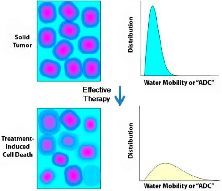
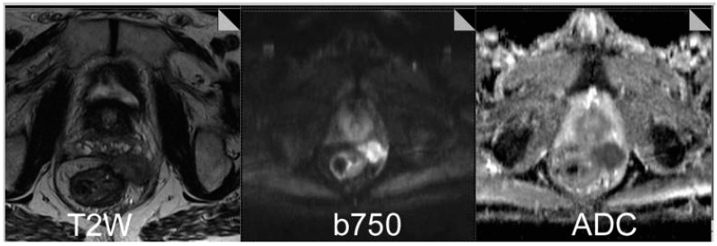
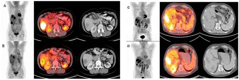
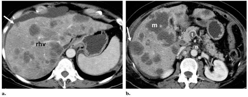
The use of imaging in colorectal cancer (CRC) has significantly evolved over the last twenty years, establishing important roles in surveillance, diagnosis, staging, treatment selection and follow up. The range of modalities has broadened with the development of novel tracer and contrast agents, and the fusion of technologies such as positron emission tomography (PET) and computed tomography (CT). Traditionally, the most widely used modality for assessing treatment response in metastasised colon and rectal tumours is CT, combined with use of the RECIST guidelines. However, a growing body of evidence suggests that tumour size does not always adequately correlate with clinical outcomes. Magnetic resonance imaging (MRI) is a more versatile technique and dynamic contrast-enhanced (DCE)-MRI and diffusion-weighted (DW)-MRI may be used to evaluate biological and functional effects of treatment. Integrated fluorodeoxyglucose (FDG)-PET/CT combines metabolic and anatomical imaging to improve sensitivity and specificity of tumour detection, and a number of studies have demonstrated improved diagnostic accuracy of this modality in a variety of tumour types, including CRC. These developments have enabled the progression of treatment strategies in rectal cancer and improved the detection of hepatic metastatic disease, yet are not without their limitations. These include technical, economical and logistical challenges, along with a lack of robust evidence for standardisation and formal guidance. In order to successfully apply these novel imaging techniques and utilise their benefit to provide truly personalised cancer care, advances need to be clinically realised in a routine and robust manner.

Keywords: angiogenesis; imaging; metastatic colorectal cancer.


Conflict of interest statement

R.G.J. has received honoraria for advisory boards from Roche, Sanofi, Eli Lilley, Amgen and Merck Serono, honoraria for lectures from Roche, Sanofi, and Merck Serono. Financial support to attend international meetings and received funding and free supply of bevacizumab from Roche in a phase II clinical trial. P.F., R.G.H.B.T., H.V. and T.S. have no conflicts of interest.

Figures

Figure 1
Figure 1
Restricted diffusion within rectal cancer with extension into the perirectal space. T2-weighted image demonstrate a well-circumscribed lesion in the perirectal space. Diffusion-weighted image obtained at a b value of 750 demonstrates a high signal, and corresponding ADC map demonstrates relatively restricted diffusion within the tumour. Figure reproduced with permission from Padhani et al. [24].
Figure 1
Figure 1
Restricted diffusion within rectal cancer with extension into the perirectal space. T2-weighted image demonstrate a well-circumscribed lesion in the perirectal space. Diffusion-weighted image obtained at a b value of 750 demonstrates a high signal, and corresponding ADC map demonstrates relatively restricted diffusion within the tumour. Figure reproduced with permission from Padhani et al. [24].
Figure 2
Figure 2
FDG-PET/CT images before (A and C) and 4 weeks after (B and D) 90Y-microsphere radioembolisation in liver-dominant mCRC; (A and B) The illustrated metabolic response was associated with a survival of 12 months after treatment; (C and D) This metabolic non-responder survived 5 months after treatment. Figure reproduced with permission from Sabet et al. [35].
Figure 3
Figure 3
Differentiation of metastases from fat deposition in the liver. Axial portal venous phase contrast-enhanced CT images at the level of the right hepatic vein (rhv) (a) and the pancreatic head (b) show innumerable hypoattenuated lesions throughout the liver. Most of the lesions are round or oval, but the largest (m in b) has a geographic configuration. Because of their low attenuation (<40 HU), the lesions might be mistaken for multifocal fat deposition; however, the mass effect of the lesions, which produces bulging of the liver surface (arrow) and compression of the right hepatic vein, as well as the multiplicity of lesions, their predominant round or oval shape, the thrombus (t in b) in the superior mesenteric vein, and numerous heterogeneous lymph nodes (n in b), are suggestive of malignancy. The lesions were identified as hematogenous metastases from pancreatic adenocarcinoma. Figure reproduced with permission from Hamer et al. [55].

References

    1. Globocan F.S. Colorectal Cancer: Estimated Incidence, Mortality and Prevalence Worldwide in 2012. [(accessed on 30 August 2015)]. Available online: http://globocan.iarc.fr/Pages/fact_sheets_cancer.aspx.
    1. Ferlay J., Steliarova-Foucher E., Lortet-Tieulent J., Rosso S., Coebergh J.W.W., Comber H., Forman D., Bray B. Cancer incidence and mortality patterns in Europe: Estimates for 40 countries in 2012. Eur. J. Cancer. 2013;49:1374–1403. doi: 10.1016/j.ejca.2012.12.027. - DOI - PubMed
    1. Iyer R., Silverman P.M., DuBrow R.A., Charnsangavej C. Imaging in the diagnosis, staging, and follow-up of colorectal cancer. Am. J. Roentgenol. 2002;179:3–13. doi: 10.2214/ajr.179.1.1790003. - DOI - PubMed
    1. Kelloff G., Schilsky R.L., Alberts D.S., Day R.W., Guyton K.Z., Pearce H.L. Colorectal adenomas: A prototype for the use of surrogate end points in the development of cancer prevention drugs. Clin. Cancer Res. 2004;10:3908–3918. doi: 10.1158/1078-0432.CCR-03-0789. - DOI - PubMed
    1. Gollub M., Schwartz L.H., Akhurst T. Update on colorectal cancer imaging. Radiol. Clin. N. Am. 2007;45:85–118. doi: 10.1016/j.rcl.2006.10.003. - DOI - PubMed

LinkOut - more resources