. 2016 Dec;2(1):90.
doi: 10.1186/s40792-016-0219-5. Epub 2016 Sep 2.

Successful resection of giant esophageal liposarcoma by endoscopic submucosal dissection combined with surgical retrieval: a case report and literature review

Affiliations

Successful resection of giant esophageal liposarcoma by endoscopic submucosal dissection combined with surgical retrieval: a case report and literature review

Gosuke Takiguchi et al. Surg Case Rep. 2016 Dec.

Abstract

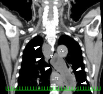
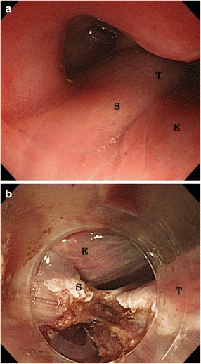
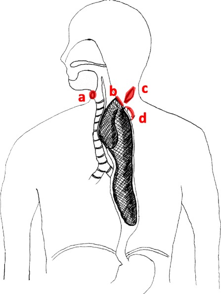
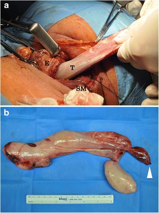
Liposarcoma of the esophagus is very rare. We experienced a huge (27.5 × 11.6 cm) liposarcoma of the esophagus. A 73-year-old man presented with severe dyspnea requiring emergency tracheal intubation. Computed tomography and esophagogastroduodenoscopy showed a large submucosal tumor arising from the esophageal entrance and extending intraluminally to the lower esophagus. We successfully performed endoscopic submucosal dissection (ESD) and esophagotomy to remove the tumor, which preserved swallowing and phonation. The final diagnosis by histopathologic and immunohistologic examination was well-differentiated liposarcoma of the esophagus. Treatment by the combination of ESD and esophagotomy can be performed even for a very large tumor. This method preserves deglutition with a lower risk of recurrent laryngeal nerve paralysis than that with esophagectomy.

Keywords: Combined method; ESD; Esophagotomy; Esophagus; Liposarcoma.


Figures

Fig. 1
Fig. 1
Computed tomography showed giant tumor from the pyriform sinus into the thoracic esophagus intraluminal of the esophagus. The tumor density was mostly low with heterogeneous density in spots (arrow head, T tumor, H heart, Ao aorta)
Fig. 2
Fig. 2
a Esophagogastroduodenoscopy showed that the stalk of the tumor was from the esophageal entrance to oropharynx and the surface was covered with smooth mucous membrane. b The stalk was resected from the base in the endoscopic submucosal dissection procedure (T tumor, S stalk, E esophagus)
Fig. 3
Fig. 3
a Tracheotomy was done for keeping the airway, which was compressed by the tumor. b The stalk of the tumor was easily observed at the cervical esophagus. Endoscopic submucosal dissection was performed, and the tumor was excised from the base. c Skin incision was made at the left neck because the tumor was too large to retrieve from the mouth. d Esophagotomy was performed at cervical region for about 5 cm, and the tumor was retrieved from this incised part
Fig. 4
Fig. 4
Excised specimen. a The skin incision was made at the left neck. Esophagotomy at cervical region for about 5 cm was performed to retrieve the tumor. b The excised specimen consisted of 27 × 11.6 cm branch polypoid mass. Surface of this tumor was smooth and white-colored (arrow head edge of the stalk, T tumor, E esophagus, SM sternocleidomastoid muscle)
Fig. 5
Fig. 5
a, b Histopathological examination showed internal of the tumor consisted of adipose tissue, enriched small vessel, and lymph duct. c, d Immunohistologically, adipose cells were positive for CDK4 (c) and weakly positive for MDM2 (d) in nuclei (HE hematoxylin and eosin stain, CDK4 cyclin-dependent kinase 4, MDM2 murine double minute-2)

References

    1. Fernandez MJ, Davis RP, Nora PF. Gastrointestinal lipoma. Arch Surg. 1983;118:1081–3. doi: 10.1001/archsurg.1983.01390090065015. - DOI - PubMed
    1. Czekajska-Chehab E, Tomaszewska M, Drop A, Dabrowski A, Skomra D, Orłowski T, Kołodziej I, Korobowicz E. Liposarcoma of the esophagus: case report and literature review. Med Sci Monit. 2009;15:123–7. - PubMed
    1. Will U, Lorenz P, Urban H, Meyer F. Curative endoscopic resection of a huge pedunculated esophageal liposarcoma. Endoscopy. 2007;39(Suppl 1):15–6. doi: 10.1055/s-2006-944902. - DOI - PubMed
    1. Yo I, Chung JW, Jeong MH, Lee JJ, An J, Kwon KA, Rim MY, Hahm KB. Huge liposarcoma of esophagus resected by endoscopic submucosal dissection: case report with video. Clin Endosc. 2013;46:297–300. doi: 10.5946/ce.2013.46.3.297. - DOI - PMC - PubMed
    1. Liakakos TD, Troupis TG, Tzathas C, Spirou K, Nikolaou I, Ladas S, Karatzas GM. Primary liposarcoma of esophagus: a case report. World J Gastroenterol. 2006;12:1149–52. doi: 10.3748/wjg.v12.i7.1149. - DOI - PMC - PubMed

LinkOut - more resources