. 2016 Oct 14;291(42):22196-22206.
doi: 10.1074/jbc.M116.749648. Epub 2016 Sep 2.

Role of the Conserved Disulfide Bridge in Class A Carbapenemases

Affiliations

Role of the Conserved Disulfide Bridge in Class A Carbapenemases

Clyde A Smith et al. J Biol Chem. .

Abstract

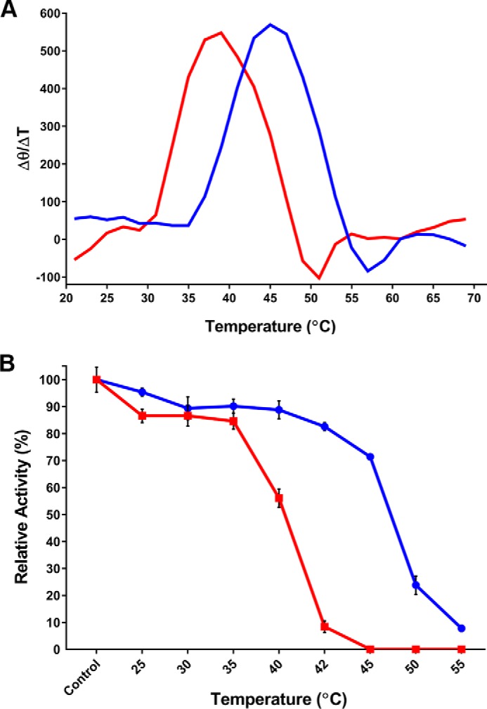
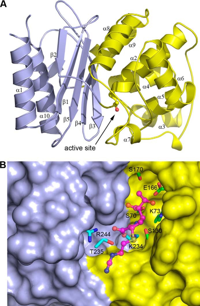
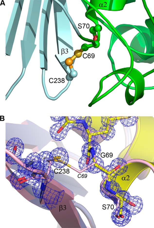
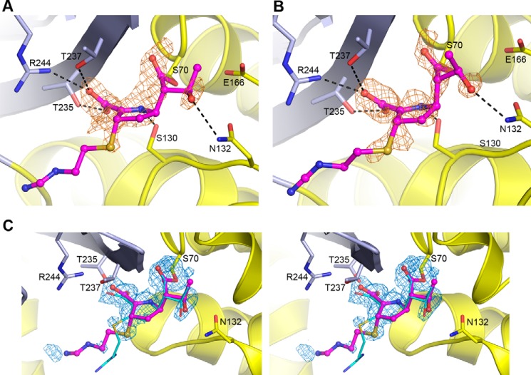
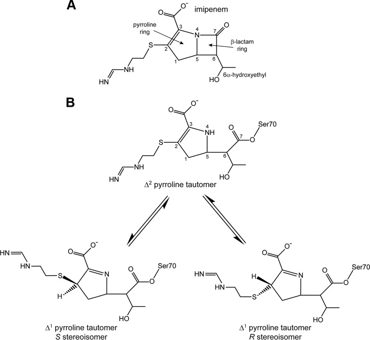
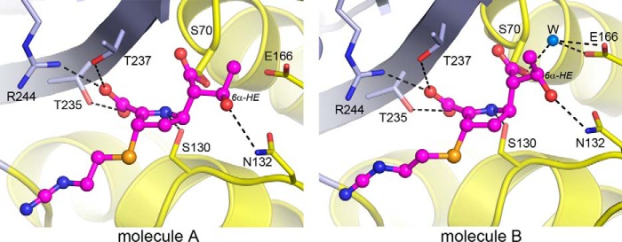
Some members of the class A β-lactamase family are capable of conferring resistance to the last resort antibiotics, carbapenems. A unique structural feature of these clinically important enzymes, collectively referred to as class A carbapenemases, is a disulfide bridge between invariant Cys69 and Cys238 residues. It was proposed that this conserved disulfide bridge is responsible for their carbapenemase activity, but this has not yet been validated. Here we show that disruption of the disulfide bridge in the GES-5 carbapenemase by the C69G substitution results in only minor decreases in the conferred levels of resistance to the carbapenem imipenem and other β-lactams. Kinetic and circular dichroism experiments with C69G-GES-5 demonstrate that this small drop in antibiotic resistance is due to a decline in the enzyme activity caused by a marginal loss of its thermal stability. The atomic resolution crystal structure of C69G-GES-5 shows that two domains of this disulfide bridge-deficient enzyme are held together by an intensive hydrogen-bonding network. As a result, the protein architecture and imipenem binding mode remain unchanged. In contrast, the corresponding hydrogen-bonding networks in NMCA, SFC-1, and SME-1 carbapenemases are less intensive, and as a consequence, disruption of the disulfide bridge in these enzymes destabilizes them, which causes arrest of bacterial growth. Our results demonstrate that the disulfide bridge is essential for stability but does not play a direct role in the carbapenemase activity of the GES family of β-lactamases. This would likely apply to all other class A carbapenemases given the high degree of their structural similarity.

Keywords: antibiotic resistance; carbapenemase; crystal structure; disulfide; enzyme kinetics; enzyme stability; hydrogen bond.


Figures

FIGURE 1.
FIGURE 1.
Thermal stability of GES-5 and the C69G variant. A, the first derivative of the thermal-induced unfolding CD curve of wild type (blue) and C69G (red) GES-5. The change in the CD signal as a function of temperature was monitored at 222 nm. The maxima correspond to the melting temperatures (Tm). B, kinetic thermal stability of wild type (blue circles) and C69G (red squares) GES-5. The activity against ampicillin was measured after incubating the enzymes at various temperatures for 60 min. The activity of control reactions that were run with non-incubated enzymes was defined as 100%.
FIGURE 2.
FIGURE 2.
The structure of the C69G-GES-5 variant enzyme. A, ribbon representation of C69G-GES-5 showing the two structural domains colored light blue (domain 1) and yellow (domain 2) and indicating the secondary structure nomenclature. The location of the active site is indicated, with the side chain of the catalytic serine (Ser70) shown in ball and stick. B, surface representation of C69G-GES-5, with the two structural domains colored light blue (domain 1) and yellow (domain 2). The catalytically important residues in the active site are shown, colored cyan for domain 1 and green for domain 2. The bound imipenem substrate in the C69G-GES-5 complex is shown in magenta ball and stick.
FIGURE 3.
FIGURE 3.
The disulfide bond in GES-5. A, the Cys69–Cys238 disulfide bond linking the N terminus of helix α2 and the C terminus of strand β3 in wild-type GES-5 (12). The two structural domains are light cyan (domain 1) and green (domain 2). B, close-up view of the site of substitution in C69G-GES-5, showing domain 1 on the left in light blue and domain 2 on the right in yellow. The final refined 2FoFc electron density is shown as a blue mesh (contoured at 1.5 σ). The wild-type GES-5 structure is shown as light pink ribbons and sticks superimposed on the C69G-GES-5 structure based upon domain 1 only. The intact C69–Cys238 disulfide in the wild-type structure is shown with the Cys69 labeled in italics.
FIGURE 4.
FIGURE 4.
Imipenem binding in C69G-GES-5. A, initial residual FoFc difference electron density in the active site of C69G-GES-5 soaked in imipenem for molecule A. B, initial residual FoFc difference electron density in the active site of C69G-GES-5 soaked in imipenem for molecule B. In both panels, the density (colored light brown and contoured at 2.5 σ), was calculated following structure solution by molecular replacement but prior to refinement. The location of the final refined imipenem is shown as thin magenta balls and sticks, built as the S stereoisomer of the Δ1 tautomer. The hydrogen bonding interactions with the C7 carbonyl of imipenem in the oxyanion hole are not shown for clarity. The two structural domains are colored light blue (domain 1) and yellow (domain 2). C, stereo view of the omit FoFc difference electron density for the Ser70 side chain and the imipenem in the active site of C69G-GES-5 molecule A. The density (colored blue and contoured at 2.8 σ) was calculated following structure refinement. The final refined imipenem is shown as thin magenta balls and sticks. The imipenem bound to wild-type GES-5 (12) is shown as thin cyan sticks, present in that structure as the Δ2 tautomer.
FIGURE 5.
FIGURE 5.
Chemical structure of imipenem. A, the intact antibiotic with standard atom numbering. B, the hydrolyzed form of imipenem showing the three possible isomers. The S and R stereoisomers of the Δ1 tautomer can interconvert via the Δ2 tautomer.
FIGURE 6.
FIGURE 6.
Conformation differences in the bound imipenem. The acylated imipenem substrate in the two independent C69G-GES-5 molecules present in the asymmetric unit is shown. In molecule B, the rotation of the 6α-hydroxyethyl group (6α-HE) provides sufficient space to allow a water molecule to bind between Glu166 and catalytic Ser70. In molecule A, the carbon atom of the 6α-HE blocks this water-binding site.
FIGURE 7.
FIGURE 7.
The domain interface in GES-5. The molecular surface representation of domain 1 (light cyan) of wild-type GES-5 shows the region of contact (highlighted yellow-green) with the domain 2 (green ribbons). The residues from domain 2 that are involved in interdomain contacts are shown as green balls and sticks.

References

    1. Bush K. (2013) Proliferation and significance of clinically relevant β-lactamases. Ann. N.Y. Acad. Sci. 1277, 84–90 - PubMed
    1. Ambler R. P. (1980) The structure of β-lactamases. Philos. Trans. R. Soc. Lond. B Biol. Sci. 289, 321–331 - PubMed
    1. Jaurin B., and Grundström T. (1981) AmpC cephalosporinase of Escherichia coli K-12 has a different evolutionary origin from that of β-lactamases of the penicillinase type. Proc. Natl. Acad. Sci. U.S.A. 78, 4897–4901 - PMC - PubMed
    1. Ouellette M., Bissonnette L., and Roy P. H. (1987) Precise insertion of antibiotic resistance determinants into Tn21-like transposons: Nucleotide sequence of the OXA-1 β-lactamase gene. Proc. Natl. Acad. Sci. U.S.A. 84, 7378–7382 - PMC - PubMed
    1. Paterson D. L., and Bonomo R. A. (2005) Extended-spectrum β-lactamases: A clinical update. Clin. Microbiol. Rev. 18, 657–686 - PMC - PubMed

LinkOut - more resources