

Transcriptomic and functional pathways analysis of ascorbate-induced cytotoxicity and resistance of Burkitt lymphoma
- PMID: 27590508
- PMCID: PMC5325416
- DOI: 10.18632/oncotarget.11740
Transcriptomic and functional pathways analysis of ascorbate-induced cytotoxicity and resistance of Burkitt lymphoma
Abstract
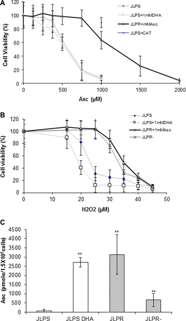
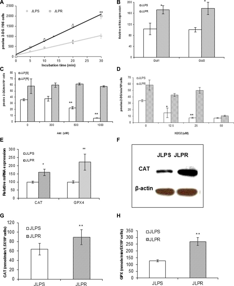
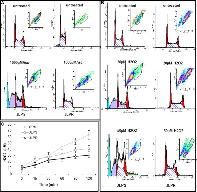
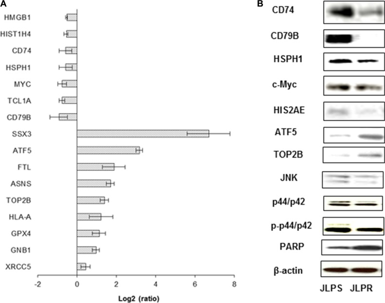
Ascorbate is a pro-oxidant that generates hydrogen peroxide-dependent cytotoxity in cancer cells without adversely affecting normal cells. To determine the mechanistic basis for this phenotype, we selected Burkitt lymphoma cells resistant to ascorbate (JLPR cells) and their ascorbate-sensitive parental cells (JLPS cells). Compared with JLPS cells, the increased glucose uptake in JLPR cells (with upregulated glucose transporters, increased antioxidant enzyme activity, and altered cell cycling) conferred ascorbate-induced cytotoxicity and resistance. Transcriptomic profiles and function pathway analysis identified differentially expressed gene signatures for JLPR cells and JLPS cells, which differential expression levels of five genes (ATF5, CD79B, MHC, Myosin, and SAP18) in ascorbate-resistant cells were related to phosphoinositide 3 kinase, cdc42, DNA methylation and transcriptional repression, polyamine regulation, and integrin-linked kinase signaling pathways. These results suggested that coordinated changes occurred in JLPR cells to enable their survival when exposed to the cytotoxic pro-oxidant stress elicited by pharmacologic ascorbate treatment.
Keywords: Burkitt lymphoma cells; ascorbate; drug resistance; pathway analysis; transcriptomic profiles.
Conflict of interest statement
None
Figures
Similar articles
-
Hepatomas are exquisitely sensitive to pharmacologic ascorbate (P-AscH-).Theranostics. 2019 Oct 18;9(26):8109-8126. doi: 10.7150/thno.35378. eCollection 2019. Theranostics. 2019. PMID: 31754384 Free PMC article.
-
Enhanced antitumor activity of vitamin C via p53 in cancer cells.Free Radic Biol Med. 2012 Oct 15;53(8):1607-15. doi: 10.1016/j.freeradbiomed.2012.07.079. Epub 2012 Aug 4. Free Radic Biol Med. 2012. PMID: 22892142
-
Development of rituximab-resistant lymphoma clones with altered cell signaling and cross-resistance to chemotherapy.Cancer Res. 2007 Feb 1;67(3):1270-81. doi: 10.1158/0008-5472.CAN-06-2184. Cancer Res. 2007. PMID: 17283164
-
Influence of vitamin C and vitamin E on redox signaling: Implications for exercise adaptations.Free Radic Biol Med. 2015 Jul;84:65-76. doi: 10.1016/j.freeradbiomed.2015.03.018. Epub 2015 Apr 2. Free Radic Biol Med. 2015. PMID: 25841784 Review.
-
Mechanisms of anti-cancer effects of ascorbate: Cytotoxic activity and epigenetic modulation.Blood Cells Mol Dis. 2018 Mar;69:57-64. doi: 10.1016/j.bcmd.2017.09.005. Epub 2017 Sep 21. Blood Cells Mol Dis. 2018. PMID: 28954710 Review.
Cited by
-
Editorial: Vitamin C in Cancer and Infectious Diseases: Physiological, Biochemical and Therapeutic Interventions.Front Physiol. 2019 Jun 18;10:734. doi: 10.3389/fphys.2019.00734. eCollection 2019. Front Physiol. 2019. PMID: 31275158 Free PMC article. No abstract available.
-
Hepatomas are exquisitely sensitive to pharmacologic ascorbate (P-AscH-).Theranostics. 2019 Oct 18;9(26):8109-8126. doi: 10.7150/thno.35378. eCollection 2019. Theranostics. 2019. PMID: 31754384 Free PMC article.
-
The mitochondrial unfolded protein response (UPRmt): shielding against toxicity to mitochondria in cancer.J Hematol Oncol. 2022 Jul 21;15(1):98. doi: 10.1186/s13045-022-01317-0. J Hematol Oncol. 2022. PMID: 35864539 Free PMC article. Review.
-
Targeting ATF5 in Cancer.Trends Cancer. 2017 Jul;3(7):471-474. doi: 10.1016/j.trecan.2017.05.004. Epub 2017 Jun 13. Trends Cancer. 2017. PMID: 28718401 Free PMC article.
-
Vitamin C Supplementation in the Treatment of Autoimmune and Onco-Hematological Diseases: From Prophylaxis to Adjuvant Therapy.Int J Mol Sci. 2024 Jul 2;25(13):7284. doi: 10.3390/ijms25137284. Int J Mol Sci. 2024. PMID: 39000393 Free PMC article. Review.
References
-
- Rebouche CJ. Ascorbic acid and carnitine biosynthesis. The American journal of clinical nutrition. 1991;54:1147S–52S. - PubMed
-
- Nishikimi M, Fukuyama R, Minoshima S, Shimizu N, Yagi K. Cloning and chromosomal mapping of the human nonfunctional gene for L-gulono-gamma-lactone oxidase the enzyme for L-ascorbic acid biosynthesis missing in man. J Bio Chem. 1994;269:13685–13688. - PubMed
-
- Li Y, Schellhorn HE. New developments and novel therapeutic perspectives for vitamin C. The Journal of nutrition. 2007;137:2171–2184. - PubMed
-
- Padayatty SJ, Levine M. Fruit and vegetables: think variety go ahead eat! Am J Clin Nutr. 2008;87:5–7. - PubMed
-
- Arrigoni O, De Tullio MC. Ascorbic acid: much more than just an antioxidant. Biochim Biophys Acta. 2002;1569:1–9. - PubMed
MeSH terms
Substances
LinkOut - more resources
Full Text Sources
Other Literature Sources
Medical
Research Materials
Miscellaneous
