. 2016 Nov 15;76(22):6669-6679.
doi: 10.1158/0008-5472.CAN-16-0571. Epub 2016 Sep 2.

Comparative Analysis of Bispecific Antibody and Streptavidin-Targeted Radioimmunotherapy for B-cell Cancers

Affiliations

Comparative Analysis of Bispecific Antibody and Streptavidin-Targeted Radioimmunotherapy for B-cell Cancers

Damian J Green et al. Cancer Res. .

Abstract

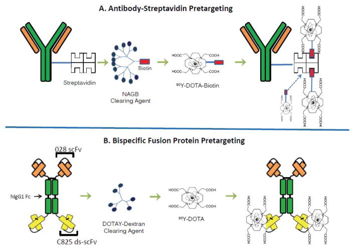
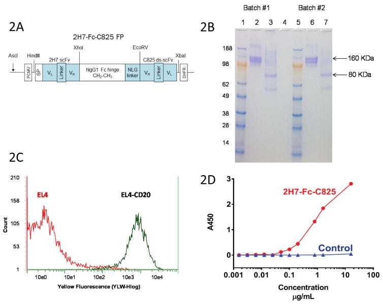
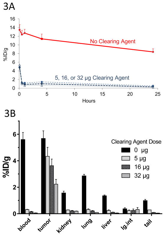
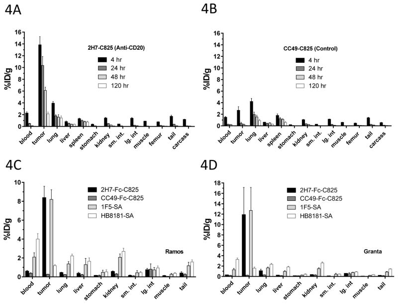
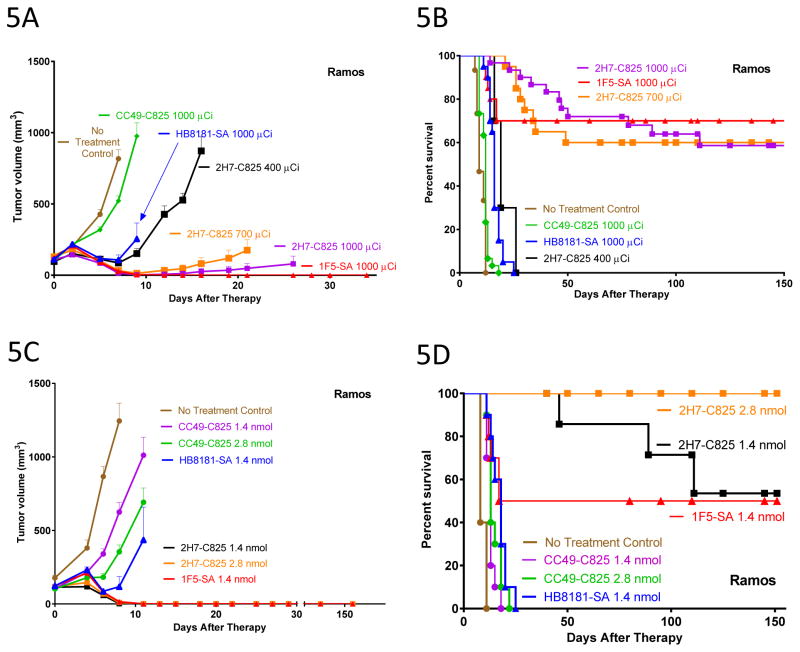
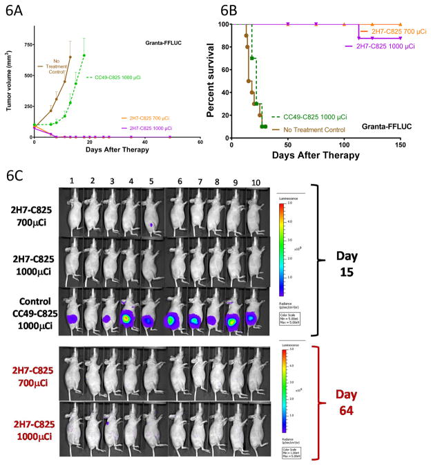
Streptavidin (SA)-biotin pretargeted radioimmunotherapy (PRIT) that targets CD20 in non-Hodgkin lymphoma (NHL) exhibits remarkable efficacy in model systems, but SA immunogenicity and interference by endogenous biotin may complicate clinical translation of this approach. In this study, we engineered a bispecific fusion protein (FP) that evades the limitations imposed by this system. Briefly, one arm of the FP was an anti-human CD20 antibody (2H7), with the other arm of the FP an anti-chelated radiometal trap for a radiolabeled ligand (yttrium[Y]-DOTA) captured by a very high-affinity anti-Y-DOTA scFv antibody (C825). Head-to-head biodistribution experiments comparing SA-biotin and bispecific FP (2H7-Fc-C825) PRIT in murine subjects bearing human lymphoma xenografts demonstrated nearly identical tumor targeting by each modality at 24 hours. However, residual radioactivity in the blood and normal organs was consistently higher following administration of 1F5-SA compared with 2H7-Fc-C825. Consequently, tumor-to-normal tissue ratios of distribution were superior for 2H7-Fc-C825 (P < 0.0001). Therapy studies in subjects bearing either Ramos or Granta subcutaneous lymphomas demonstrated that 2H7-Fc-C825 PRIT is highly effective and significantly less myelosuppressive than 1F5-SA (P < 0.0001). All animals receiving optimal doses of 2H7-Fc-C825 followed by 90Y-DOTA were cured by 150 days, whereas the growth of tumors in control animals progressed rapidly with complete morbidity by 25 days. In addition to demonstrating reduced risk of immunogenicity and an absence of endogenous biotin interference, our findings offer a preclinical proof of concept for the preferred use of bispecific PRIT in future clinical trials, due to a slightly superior biodistribution profile, less myelosuppression, and superior efficacy. Cancer Res; 76(22); 6669-79. ©2016 AACR.


Figures

Figure 1
Figure 1
Figure 1A,B. Schema comparing SA-biotin multistep pretargeted radioimmunotherapy (PRIT) (A) and 2H7-Fc-C825 bispecific FP PRIT (B). Infusion of the anti-CD20 construct is followed by injection of a synthetic CA (N-acetylgalactosamine or DOTAY-dextran) and then infusion of the radiolabeled small molecule.
Figure 2
Figure 2
Figure 2A. Schematic of the 2H7-Fc-C825 (anti-CD20 x anti-Y-DOTA) bispecific Fc fusion antibody gene. An anti-human CD20 2H7 scFv gene and an yttrium-DOTA capturing C825 disulfide-stabilized scFv (ds-scFv) gene were fused to the human IgG1 Fc fragment at the amino and carboxyl ends, respectively. An N-linked glycosylation containing linker (NLG) was incorporated between the Fc and C825 ds-scFv domains, as shown. Figure 2B. SDS-PAGE analysis of the 2H7-Fc-C825 FP. Bispecific 2H7-Fc-C825 fusion polypeptides expressed in CHO-DG44 cells spontaneously formed dimers via the hinge regions. Two batches of 2H7-Fc-C825 FP (5μg) were analyzed by electrophoresis on a 4–20% MES SDS PAGE gel (Invitrogen). Lanes 1 and 5: Seeblue marker proteins; Lanes 2 and 6 show the non-reduced 2H7-Fc-C825 FP (samples boiled); Lanes 3 and 7 show the monomeric 2H7-Fc-C825 FP (samples boiled and reduced with 2-mercaptoethanol); Lane 4 is empty. The gel was stained with Coomassie blue. Figure 2C. Flow Cytometric Analysis of Binding of Purified 2H7-Fc-C825 FP to cells transduced to express hCD20 (EL4-CD20, [black]) and to untransduced (control) EL4 cells (red). EL4 or EL4-CD20 cells (0.5x10⁶ each) were incubated in 100μl of HBSS buffer containing 2% FBS and treated with 1.8μg of the 2H7-Fc-C825 FP for 30 min at 4°C. After washing, the cells were mixed with 2 μl of PE-anti-human Fc antibody in 40μl of HBSS-2% FBS buffer for 30 min at 4°C. After washing 3x,cells were re-suspended in 400μl of PBS buffer containing 1% of formaldehyde and analyzed on a Guava cytometer. Figure 2D. Sandwich ELISA assay demonstrating concentration-dependent binding of the 2H7-Fc-C825 FP to microtiter wells coated with the Y-DOTA ligand. A 96-well plate was coated with 70μl of the BSA-Y-DOTA conjugate (1μg/ml in PBS) and then blocked with 200μl of 2% BSA in PBS buffer. After washing, the wells were treated with 100μl of fusion proteins at a concentration of 16μg/ml followed by serial dilution as indicated. The plate was further treated with HRP-anti-human Fc antibody followed by TMB. A control FP shows no binding to Y-DOTA.
Figure 3
Figure 3. Optimization of DOTAY-Dextran CA Dose
Figure 3A. Blood clearance of circulating C825-Fc-2H7 FP by various doses of DOTAY-Dextran CA. Mice were injected with 1.4nmol of the 2H7-Fc-C825 fusion protein FP followed by 0, 5, 16 or 32μg of DOTAY-Dextran CA 23-hours later. One hour later, 2.4nmol of 90Y-DOTA-Biotin was injected. Blood was collected after 0.08, 0.5, 1, 4, and 24-hours following injection of radioactivity. Results represent the calculated percentages of the injected dose per gram of tissue (%ID/g, ±SD; n=3) after corrections for decay and background subtraction (red square, diluent; filled circle, 5μg DYD; diamond, 16μg DYD; triangle, 32μg DYD). Figure 3B. Biodistribution of 90Y-DOTA-Biotin in organs of mice bearing subcutaneous Ramos xenografts following injection of various doses of DOTAY-Dextran CA. Mice were injected with 1.4nmol of 2H7-Fc-C825 followed by 0, 5, 16, or 32μg of DOTAY-Dextran CA 23-hours later. One hour after injection of DOTAY-Dextran, 2.4nmol of 90Y-DOTA-Biotin was injected. Tissues were harvested 24-hours after the injection of radioactivity. Results represent the calculated mean percentages of the injected dose per gram of tissue (%ID/g, ±SEM; n=3) after corrections for decay and background.
Figure 4
Figure 4
Figure 4A,B. Biodistribution of 90Y-DOTA-Biotin in mice bearing subcutaneous Ramos xenografts. Mice were injected with 1.4nmol of 2H7-Fc-C825 (A) or 1.4nmol of the control, non-binding CC49-Fc-C825 FP (B) followed by 5μg DOTAY-Dextran 23-hours later. One hour after the DOTAY-Dextran, 2.4nmol of 90Y-DOTA-Biotin was injected. Tissues were harvested 4, 24, 48 and 120-hours after injection of radioactivity. Results represent the %ID/g (±SEM; n=5) after corrections for decay and background subtraction. Figure 4C, D. Biodistribution of 90Y-DOTA-Biotin in mice bearing subcutaneous Ramos xenografts (C) or Granta-519 xenografts. Mice were injected with 1.4nmol of 2H7-Fc-C825 (anti-CD20 bispecific), CC49-Fc-C825 (non-binding control bispecific antibody), 1F5-SA (anti-CD20-streptaviding conjugate) or HB8181-SA (non-binding control antibody-streptavidin conjugate), followed 23-hours later by either 5μg DOTAY-Dextran CA (for bispecific antibodies) or 5.8nmol NAGB CA (for antibody-streptavidin conjugates). One hour after injection of the CA, 2.4nmol of 90Y-DOTA-Biotin was administered. Tissues were harvested 4, 24, 48 and 120-hours after injection of radioactivity. Results represent the calculated %ID/g(±SEM; n=5) after corrections for decay and background subtraction.
Figure 5
Figure 5
Figure 5A, B. Comparison of tumor growth (A) and survival (B) in athymic mice bearing subcutaneous Ramos xenografts treated with either bispecific antibody PRIT or streptavidin-biotin PRIT. Mice were injected with 1.4nmol of 2H7-Fc-C825 (anti-CD20 bispecific), CC49-Fc-C825 (non-binding control bispecific antibody), 1F5-SA (anti-CD20-streptaviding conjugate) or HB8181-SA (non-binding control antibody-streptavidin conjugate), followed 23-hours later by either 5 μg DOTAY-Dextran CA (for bispecific antibodies) or 5.8 nmol NAGB CA (for antibody-streptavidin conjugates). One hour later, 2.4nmol DOTA-Biotin radiolabeled with various amounts of 90Y was injected. Tumor growth results represent the mean tumor volume of Ramos xenografts (±SEM; brown, no treatment; green, 1000μCi 90Y following 1.4 mol CC49-Fc-C825 ; blue, 1000μCi 90Y following 1.4nmol HB8181-SA; red, 1000μCi 90Y following 1.4nmol 1F5-SA; black, 400μCi 90Y following 1.4nmol 2H7-Fc-C825 ; orange square, 700μCi 90Y following 1.4nmol 2H7-Fc-C825 ; magenta, 1000μCi 90Y following 1.4nmol 2H7-Fc-C825). Figure 5C. Comparison of Tumor Growth of Ramos xenografts in Athymic mice injected with either 1.4 or 2.8nmol 2H7-Fc-C825 (anti-CD20 Bispecific Ab) or CC49-Fc-C825 (Control Bispecific Ab), 1.4nmol 1F5-SA (Anti-CD20-streptavidin conjugate), or 1.4nmol HB8181-SA (control Ab-streptavidin conjugate), followed 23-hours later by either 5μg DOTAY-Dextran (for bispecific antibodies) or 5.8nmol NAGB (for Ab-streptavidin conjugates). One hour later 2.4nmol of 90Y-DOTA-Biotin radiolabeled with 1000μCi was administered. Results represent the mean tumor volume of Ramos xenografts (±SEM; n=10; brown, no treatment; magenta, 1.4nmol CC49-Fc-C825 ; green, 2.8nmol CC49-FC-C825 ; black, 1.4nmol 2H7-Fc-C825 ; orange, 2.8nmol 2H7-Fc-C825 ; blue, 1.4nmol HB8181-SA; red, 1.4nmol 1F5-SA). Figure 5D. Comparison of survival of athymic mice bearing subcutaneous Ramos xenografts treated with various doses of bispecific antibody PRIT or streptavidin-biotin followed by 1000μCi of 90Y-DOTA-biotin. Mice were treated as described in the legend to figure 5A.
Figure 6
Figure 6
Figure 6A, B. Tumor growth (A) and survival (B) n athymic mice bearing subcutaneous Granta-519luc xenografts treated with bispecific Ab PRIT. Groups of 10 mice each were injected with 1.4nmol of either 2H7-Fc-C825 (Anti-CD20 bispecific) or CC49-Fc-C825 (control bispecific antibody) followed 23-hours later by 5μg of DOTAY-Dextran CA to remove excess circulating FP from the circulation. One hour later, 2.4nmol of 90Y-DOTA-biotin radiolabeled with either 700 or 1000μCi of 90Y was injected. Results represent the mean tumor volume of Granta-519luc xenograft mice (±SEM; n=10; brown, no treatment control; green, 1000μCi 90Y following 1.4nmol CC49-Fc-C825; orange, 700μCi 90Y following 1.4nmol 2H7-Fc-C825 ; magenta, 1000μCi 90Y following 1.4nmol 2H7-Fc-C825). Figure 6C. Whole-body ventral BLI images (from mice treated as described in the legend to Figure 6A) obtained on day 15 (top) demonstrate signal corresponding with measurable disease in the left flank subcutaneous Granta-519luc xenograft tumors of animals receiving nonbinding control FP (CC49-Fc-C825). At the same timepoint, no tumors were identified in mice pretargeted with 2H7-Fc-C825 followed by 1000μCi of 90Y-DOTA-biotin, and one mouse in the treatment group receiving 700μCi of 90Y-DOTA-biotin (animal #5) had measurable disease that was regressing. Day-64 imaging (bottom) of all surviving animals revealed no measurable disease. The imaging data were normalized to the same scale for each timepoint.

References

    1. Siegel RL, Miller KD, Jemal A. Cancer statistics, 2015. CA Cancer J Clin. 2015;65:5–29. - PubMed
    1. Advani RH, Buggy JJ, Sharman JP, Smith SM, Boyd TE, Grant B, et al. Bruton tyrosine kinase inhibitor ibrutinib (PCI-32765) has significant activity in patients with relapsed/refractory B-cell malignancies. Journal of clinical oncology : official journal of the American Society of Clinical Oncology. 2013;31:88–94. - PMC - PubMed
    1. Gopal AK, Kahl BS, de Vos S, Wagner-Johnston ND, Schuster SJ, Jurczak WJ, et al. PI3Kdelta inhibition by idelalisib in patients with relapsed indolent lymphoma. The New England journal of medicine. 2014;370:1008–18. - PMC - PubMed
    1. Larson SM, Carrasquillo JA, Cheung NK, Press OW. Radioimmunotherapy of human tumours. Nature reviews Cancer. 2015;15:347–60. - PMC - PubMed
    1. Palanca-Wessels MC, Czuczman M, Salles G, Assouline S, Sehn LH, Flinn I, et al. Safety and activity of the anti-CD79B antibody-drug conjugate polatuzumab vedotin in relapsed or refractory B-cell non-Hodgkin lymphoma and chronic lymphocytic leukaemia: a phase 1 study. Lancet Oncol. 2015;16:704–15. - PubMed

Publication types