. 2016 Nov;13(11):1152-1165.
doi: 10.1080/15476286.2016.1229734. Epub 2016 Sep 3.

Interplay between RNA-binding protein HuR and microRNA-125b regulates p53 mRNA translation in response to genotoxic stress

Affiliations

Interplay between RNA-binding protein HuR and microRNA-125b regulates p53 mRNA translation in response to genotoxic stress

Deepika Ahuja et al. RNA Biol. 2016 Nov.

Abstract

Tumor suppressor protein p53 plays a crucial role in maintaining genomic integrity in response to DNA damage. Regulation of translation of p53 mRNA is a major mode of regulation of p53 expression under genotoxic stress. The AU/U-rich element-binding protein HuR has been shown to bind to p53 mRNA 3'UTR and enhance translation in response to DNA-damaging UVC radiation. On the other hand, the microRNA miR-125b is reported to repress p53 expression and stress-induced apoptosis. Here, we show that UVC radiation causes an increase in miR-125b level in a biphasic manner, as well as nuclear cytoplasmic translocation of HuR. Binding of HuR to the p53 mRNA 3'UTR, especially at a site adjacent to the miR-125b target site, causes dissociation of the p53 mRNA from the RNA-induced silencing complex (RISC) and inhibits the miR-125b-mediated translation repression of p53. HuR prevents the oncogenic effect of miR-125b by reversing the decrease in apoptosis and increase in cell proliferation caused by the overexpression of miR-125b. The antagonistic interplay between miR-125b and HuR might play an important role in fine-tuning p53 gene expression at the post-transcriptional level, and thereby regulate the cellular response to genotoxic stress.

Keywords: Cancer; DNA damage; HuR; RNA-binding protein; miR-125b; microRNA; p53; translation regulation.


Figures

Figure 1.
Figure 1.
HuR reverses miR-125b-mediated translation repression of p53 mRNA. (A) Partial sequence of p53 3′UTR (nt. 512 to 1190) indicating the miR-125b binding site and the 3 HuR binding sites. (B) Immunoblots of lysates of MCF7 cells transfected with 3 increasing concentrations of pSUPER-miR-125b and cotransfected with 2 increasing concentrations of pCI-neo-myc-HuR probed with p53, myc, and GAPDH antibodies. (C) Immunoblots of lysates of MCF7 cells transfected with 2 increasing concentrations of pCI-neo-myc-HuR probed with p53, myc, and GAPDH antibodies. (D) Cells transfected with Fluc-p53-3′UTR and Fluc reporter gene constructs and pCMV-Rluc were cotransfected with 2 increasing concentrations of pSUPER-miR-125b and 2 concentrations of pCI-neo-myc-HuR. Fluc values are normalized to Rluc values as transfection control. (E) MCF7 cells were either untransfected (−miR-125b/−HuR) or transfected with pSUPER-miR-125b together with (+miR-125b/+HuR) or without pCI-neo-myc-HuR (+miR-125b/−HuR) or with pCI-neo-myc-HuR alone (−miR-125b/+HuR). Cell lysates were immunoprecipitated with HuR and Ago2 antibody and control IgG. RNA associated with the immunoprecipitates was subjected to qRT-PCR using p53 and GAPDH primers, and p53 mRNA levels were normalized to GAPDH mRNA levels. (F) Ribosomal fractions from MCF7 cells, either mock transfected or transfected with pSUPER-miR-125b or pSUPER-miR-125b and pCI-neo-myc-HuR, or with pCI-neo-myc-HuR alone were analyzed by sucrose density gradient fractionation. rRNA content, measured at 254 nm, is plotted against fraction numbers. RNA isolated from selected fractions was analyzed by semi-quantative RT-PCR using p53 and GAPDH primers. The level of mRNA content in each fraction, represented as band intensity of PCR products of each lane as % of total band intensity of all lanes, are plotted against respective fraction numbers below.
Figure 2.
Figure 2.
UVC irradiation causes nuclear-cytoplasmic translocation of HuR and prevents miR-125b-mediated translation repression of p53. (A) Immunoblots of lysates of MCF7 cells exposed to UVC radiation and collected at indicated time points post UVC-exposure, probed with p53 and GAPDH antibodies. (B) qRT-PCR of total RNA isolated from UVC-treated MCF7 cells collected at indicated time points using miR-125b specific primers. miR-125b RNA levels were normalized to U6 snRNA levels. Data represents fold change of normalized miR-125b RNA level in UVC-treated cells over UVC-untreated cells. (C) Immunoblots of lysates of cells exposed to UVC radiation and collected at indicated time points post UVC-exposure, probed with HuR and GAPDH antibodies. (D) Lysates of MCF7 cell collected post UVC exposure were immunoprecipitated with HuR antibody or control IgG. RNA associated with the immunoprecipitate was subjected to RT-PCR using p53 or GAPDH primers. Input represents total RNA isolated from cell lysate. (E) MCF7 cells were UVC treated and immunofluorescence of cells collected at various time points post UVC treatment was observed using anti-HuR primary and AlexaFluor568-conjugated secondary antibodies (red). Nucleus was visualized using DAPI staining (blue). (F) MCF7 cells were UVC irradiated following which cells collected at various time points were fractionated into nuclear and cytoplasmic fractions. Fractions were immunoblotted with anti-HuR, anti-lamin (nuclear marker) and anti- β actin (cytoplasmic marker) antibodies.
Figure 3.
Figure 3.
HuR competes with miR-125b to cause the UVC-mediated enhancement of p53 mRNA translation. (A) MCF7 cells were transfected with HuR siRNA or control siRNA and either exposed or not exposed to UVC. Lysates of cells collected at indicated time points were immunoblotted with HuR, p53 and GAPDH antibodies. (B) MCF7 cells were transfected with HuR siRNA or control siRNA and UVC irradiated. Lysates of cells collected at indicated time points were immunoprecipitated with control IgG, HuR and Ago2 antibodies. RNA isolated from immunoprecipitates was reverse transcribed and p53 mRNA level was estimated by qPCR. (C) MCF7 cells were transfected with miR-125b antagomiR or a control oligonucleotide and UVC irradiated. Lysates of cells collected at indicated time points were immunoprecipitated with control IgG, HuR and Ago2 antibodies. RNA isolated from immunoprecipitates was reverse transcribed and p53 mRNA level was estimated by qPCR. (D) MCF7 cells were transfected with pSUPER-miR-125b plasmid or a control plasmid and UVC irradiated. Lysates of cells collected at indicated time points were immunoprecipitated with control IgG, HuR and Ago2 antibodies. RNA isolated from immunoprecipitates was reverse transcribed and p53 mRNA level was estimated by qPCR.
Figure 4.
Figure 4.
The binding site of HuR proximal to the miR-125b target site is necessary and sufficient for preventing translation repression. (A) Schematic diagram of wild type (WT) p53 3′UTR indicating miR-125b and HuR-binding sites and various deletions mutants. (B) 32P-UTP labeled full length and various deletion mutants of p53 3′UTR RNA were incubated with purified HuR protein, UV-crosslinked, digested with RNase A and resolved on 10% SDS-PAGE. (C) Reporter gene constructs containing wild type and various deletion mutants of p53 3′UTR were transfected into MCF7 cells together with pSUPER-miR-125b or pCI-Neo-myc-HuR or both.
Figure 5.
Figure 5.
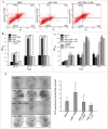
HuR reverses the anti-apoptotic and cell proliferative effect of miR-125b. (A) MCF7 cells were either mock transfected, transfected with pSUPER-miR-125b alone or together with pCI-neo-myc-HuR and then serum starved for 48 hours to induce apoptosis. Cells were stained with AnnexinV-FITC and PI to detect apoptosis by flow cytometry. The x-axis and y-axis represents PI and Annexin staining respectively. The percentage of cells in each quadrant is indicated. (B) MCF7 cells mock transfected, or transfected with pSUPER-miR-125b or pCI-neo-myc-HuR alone or in with both pSUPER-miR-125b and pCI-neo-myc-HuR or UVC treatment were allowed to grow post-transfection and MTT assay was performed at indicated time points. (C) MCF7 cells mock transfected, or UVC irradiated or transfected with pSUPER-miR-125b alone or in combination with UVC and with UVC + HuR siRNA were allowed to grow post-transfection and MTT assay was performed at 24, 48 and 72 hours. (D) MCF7 cells were mock transfected, or transfected with pSUPER-miR-125b alone or in combination with pCI-neo-myc-HuR or UVC treatment. 103 cells were seeded and colonies were counted after 14 d for GFP expression and by crystal violet staining. Ratios of GFP-expressing colonies/total number of colonies from 3 experiments are plotted.
Figure 6.
Figure 6.
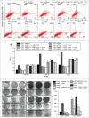
The antagonistic effect of HuR and miR-125b on cell proliferation and apoptosis is mediated via p53. (A) MCF7 cells were either transfected with control siRNA or p53 siRNA, in combination with miR-125b, HuR, miR-125b and HuR and miR-125b and UVC. Cells were subjected to apoptotic stimulus (48 hour serum starvation) following which cells were stained with AlexaFluor 488-Annexin V and PtdIns to detect apoptosis by flow cytometry. The x-axis and y-axis represents PI and Annexin staining respectively. The percentage of cells in each quadrant of representative flow cytograms is indicated. (B) MCF7 cells were either transfected with control siRNA or p53 siRNA, in combination with miR-125b, HuR, mir-125b and HuR and miR-125b and UVC. Cells were allowed to grow post-transfection or UVC exposure and MTT assay was performed at indicated time points. (C) MCF7 cells were either transfected with control siRNA or p53 siRNA, in combination with miR-125b, HuR, mir-125b and HuR and miR-125b and UVC. 103 cells were seeded and colonies were counted after 14 d by crystal violet staining. Average number of colonies in each treatment from 2 independent experiments is plotted.
Figure 7.
Figure 7.
Antagonistic interplay between HuR and miR-125b regulates p53 expression in response to UVC irradiation. Proposed model showing nuclear-cytoplasmic translocation of HuR on UVC irradiation resulting in HuR binding to p53 3′UTR and causing dissociation of miR-125b-RISC complex from p53 RNA allowing rescue of translation.

References

    1. Giaccia AJ, Kastan MB. The complexity of p53 modulation: emerging patterns from divergent signals. Genes Dev 1998; 12:2973-83; PMID:9765199; http://dx.doi.org/10.1101/gad.12.19.2973 - DOI - PubMed
    1. Horn HF, Vousden KH. Coping with stress: multiple ways to activate p53. Oncogene 2007; 26:1306-16; PMID:17322916; http://dx.doi.org/10.1038/sj.onc.1210263 - DOI - PubMed
    1. Ray PS, Grover R, Das S. Two internal ribosome entry sites mediate the translation of p53 isoforms. EMBO Rep 2006; 7:404-10; PMID:16440000; http://dx.doi.org/10.1038/sj.embor.7400623 - DOI - PMC - PubMed
    1. Vilborg A, Wilhelm MT, Wiman KG. Regulation of tumor suppressor p53 at the RNA level. J Mol Med (Berl) 2010; 88:645-52; PMID:20306257; http://dx.doi.org/10.1007/s00109-010-0609-2 - DOI - PubMed
    1. Takagi M, Absalon MJ, McLure KG, Kastan MB. Regulation of p53 translation and induction after DNA damage by ribosomal protein L26 and nucleolin. Cell 2005; 123:49-63; PMID:16213212; http://dx.doi.org/10.1016/j.cell.2005.07.034 - DOI - PubMed