. 2016 Sep 21;91(6):1356-1373.
doi: 10.1016/j.neuron.2016.08.009. Epub 2016 Sep 1.

Modulating Neuronal Competition Dynamics in the Dentate Gyrus to Rejuvenate Aging Memory Circuits

Affiliations

Modulating Neuronal Competition Dynamics in the Dentate Gyrus to Rejuvenate Aging Memory Circuits

Kathleen M McAvoy et al. Neuron. .

Abstract

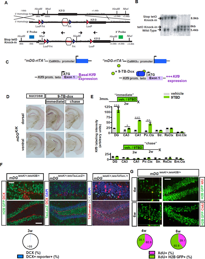
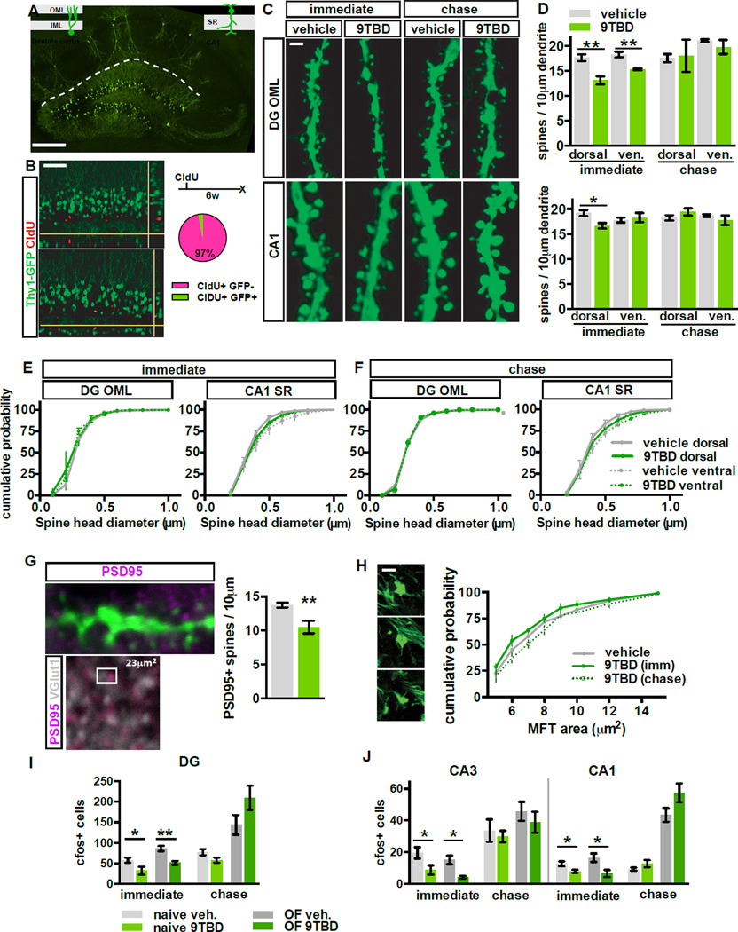
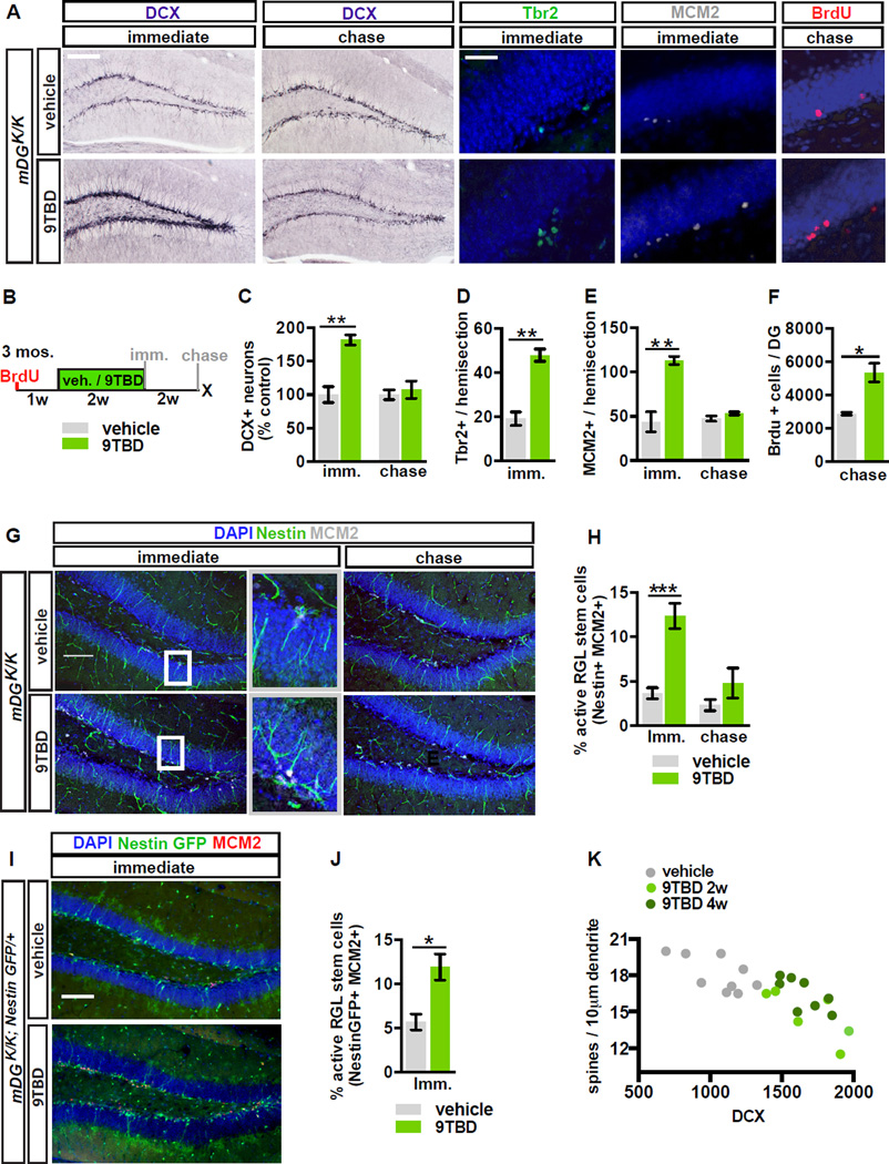
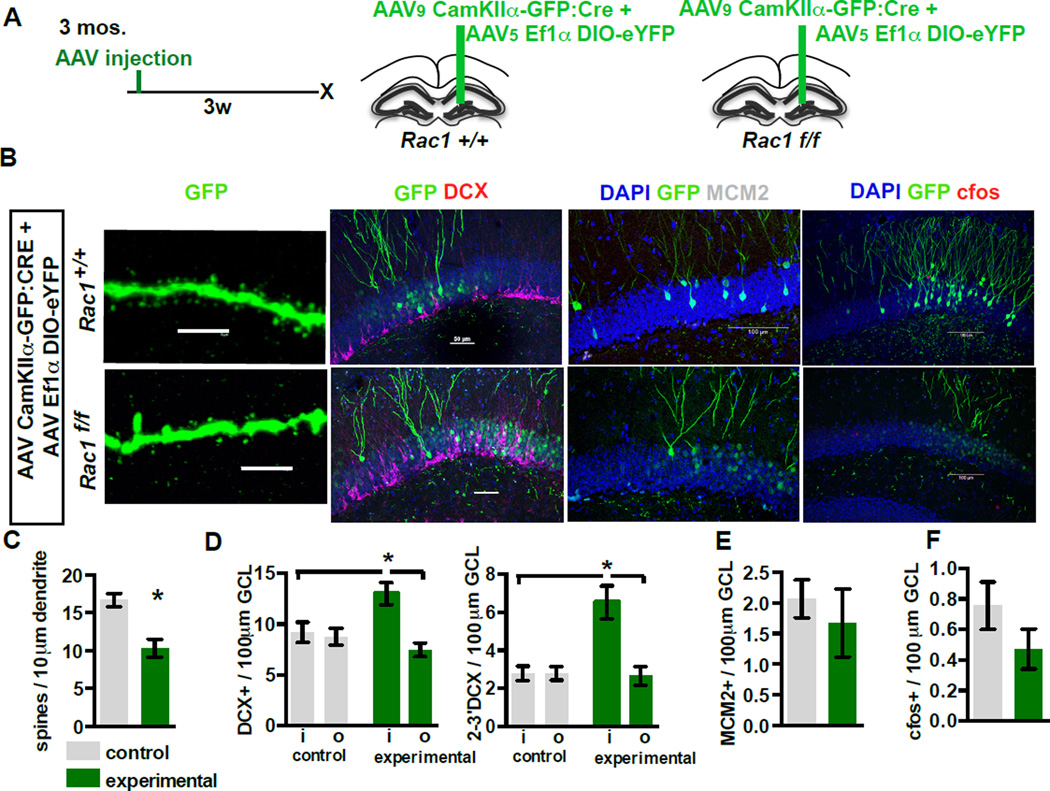
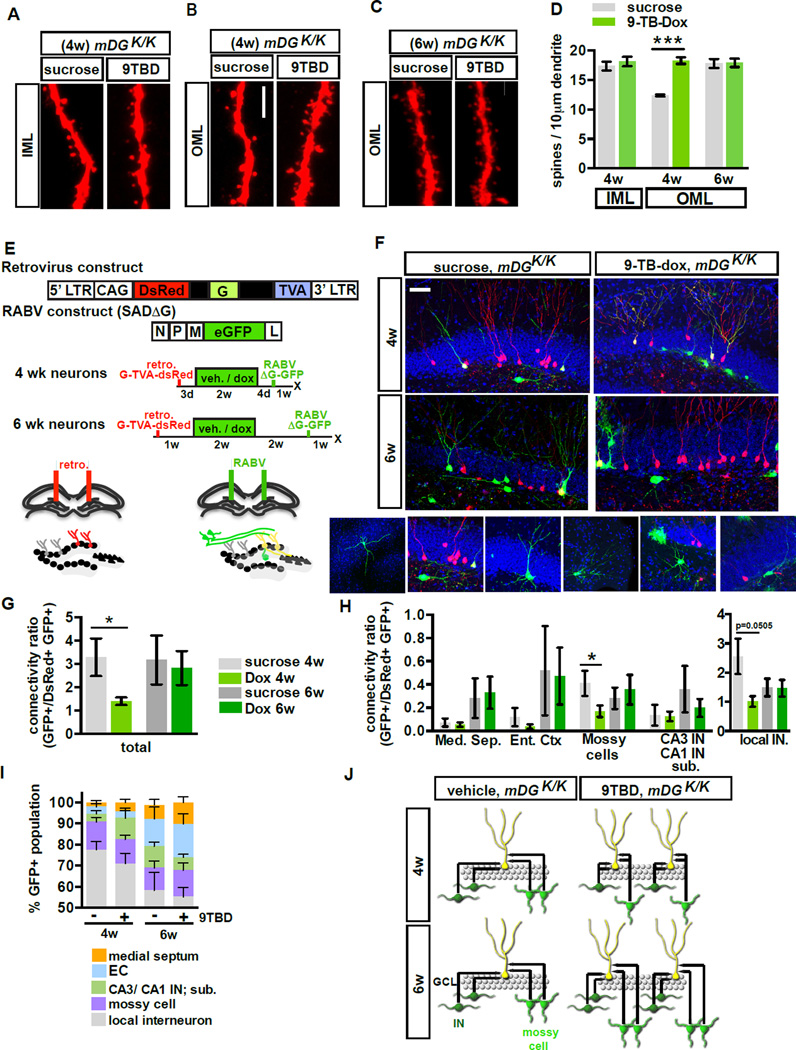
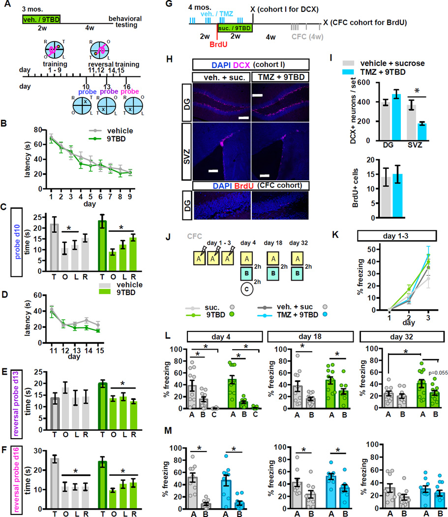
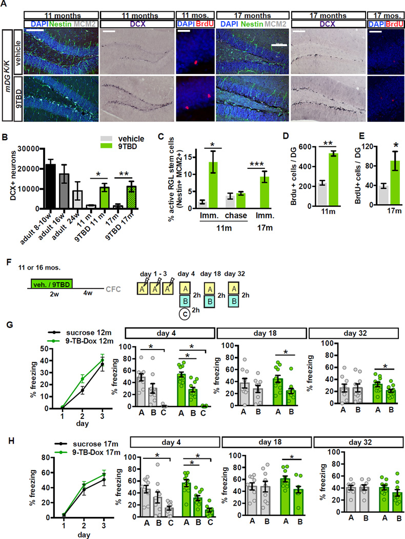
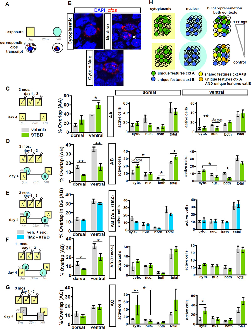
The neural circuit mechanisms underlying the integration and functions of adult-born dentate granule cell (DGCs) are poorly understood. Adult-born DGCs are thought to compete with mature DGCs for inputs to integrate. Transient genetic overexpression of a negative regulator of dendritic spines, Kruppel-like factor 9 (Klf9), in mature DGCs enhanced integration of adult-born DGCs and increased NSC activation. Reversal of Klf9 overexpression in mature DGCs restored spines and activity and reset neuronal competition dynamics and NSC activation, leaving the DG modified by a functionally integrated, expanded cohort of age-matched adult-born DGCs. Spine elimination by inducible deletion of Rac1 in mature DGCs increased survival of adult-born DGCs without affecting proliferation or DGC activity. Enhanced integration of adult-born DGCs transiently reorganized adult-born DGC local afferent connectivity and promoted global remapping in the DG. Rejuvenation of the DG by enhancing integration of adult-born DGCs in adulthood, middle age, and aging enhanced memory precision.


Figures

Figure 1
Figure 1. A genetic system for inducible and reversible overexpression of Klf9 in mature DGCs
A) Strategy for targeting Klf9 locus to generate tetO-Klf9 knock-in mouse line. B) Southern blot on F2 offspring DNA digested with NheI and probed with the 3’ probe showing all 3 alleles (WT, Stop tetO-Knock-in, and TetO-Knock-in). C-E) Inducible and reversible overexpression of Klf-9. (C) Schematic of tet-on genetic system. (D) In situ hybridization shows Klf9 transcripts in mDGK/K mice are increased following two weeks of 9TBD treatment in drinking water (immediate) and are restored after a two-week chase period. (E) Quantification of D (arbitrary units) at the immediate (top, n=5,3) and chase timepoint (n=3,3; bottom). F) The mDG rtTA line drives expression in DGCs older than 3 weeks of age. Confocal images show absence of DCX and teto-reporter overlap in DGCs. G) rtTA transactivates the tetO-H2B GFP transgene in DGCs 4 weeks of age and older. (n=3 mice). Scale bar: 500µm (D), 20µm top, 100µm below (F), 20µm top, 75µm below (G). For all figures data represent mean ± SEM unless otherwise noted and ***p<0.001, **p<0.01, *p<0.05.
Figure 2
Figure 2. Reversible overexpression of Klf9 transiently decreases dendritic spine density and activity of mature DGCs
A) GFP is expressed in a subset of DGCs and CA1 pyramidal neurons in mDGK/K;Thy1-GFP(M)/+ mice. B) GFP+ DGCs in mDGK/K;Thy1-GF(M)/+ are 6 weeks of age or older. Confocal scans of the GCL showing 6 week-old DGCs (CldU+) expressing GFP (arrowhead indicates overlap, n=3). C,D) Inducible Klf9 overexpression decreases dendritic spine density. (C) Maximum intensity projection confocal images of individual dendritic segments from OML (top) and CA1-SR (below) at the immediate and chase timepoint in mDGK/K;Thy1-GFP/+ mice. (D) Quantification of C (n=4,3). E,F) The distribution of spine head diameter is unchanged in OML or SR of mDGK/K;Thy1-GFP/+ mice (E) at the immediate (F) and chase timepoints (n=4,3 immediate, n=3,3 chase). G) Inducible Klf9 overexpression decreases the density of PSD95+ dendritic spines. Left, top: overlap between PSD95 and GFP-expressing dendrite. Left, below: representative image from DG OML showing adjacent opposing puncta of PSD95 and Vesicular glutamate transporter-1 (vGlut1). Right: quantification of PSD95+ dendritic spine density in the OML (n=3,5). H) Klf9 overexpression in mature DGCs does not affect MFT size. Graph displays the percentage distribution of MFT area (n=7[vehicle], 4[immediate], 3[chase]). I, J) Klf9 overexpression in mature DGCs transiently decreases the cfos+ population at (I) the immediate timepoint (home cage (n=7,7), 30 minutes of open field exploration (n=6,3)) but not the chase timepoint (home cage n=6,6, open field n=3,3). Scale bar: 500µm (A), 20µm (B), 2µm (C,G,H).
Figure 3
Figure 3. Reversible overexpression of Klf9 in mature DGCs modulates activation of NSCs and neuronal competition dynamics
A - F) Adult hippocampal neurogenesis is reversibly enhanced in mDGK/K mice. (A) Images show labeling for DCX, Tbr2, MCM2, and BrdU. (B) Schematic indicates experimental design for Fig. 3A, C-J. (C) Reversible expansion of the DCX+ population in 9TBD treated mDG K/K mice (n=5,3 immediate, n=6,7 chase). (D) Increased number of progenitors (Tbr2+), (E) reversible enhancement of dividing cells (MCM2+) (n=3,3), and (F) enhanced survival of 5 week-old adult-born cells in GCL of 9TBD-treated mDGK/K mice (n=3,3). G - J) NSC activation is reversibly enhanced in mDGK/K mice. (G) Representative images of activation of NSCs (arrowheads, Nestin+ MCM2+) with boxes to indicate magnified region. (H) Quantification of G (n=7,7). (I) Representative images of activated NSCs in vehicle and 9TBD-treated mDGK/K;NestinGFP/+mice. (J) Quantification of I (n=3,4). K) Dendritic spine density of mature DGCs is inversely correlated with the number of DCX+ cells in dorsal DG. Dots represent individual animals. Scale bar: 100µm, 20 µm (A), 100µm (G,I).
Figure 4
Figure 4. Conditional elimination of Rac1 in mature DGCs decreases spine density and increases the survival of adult-born DGCs
A-D) Elimination of Rac1 expands the DCX+ population of adult-born DGCs. (A) Timeline and viral system. (B) Representative images of (left to right): OML dendritic segments, DCX, MCM2, and c-fos from Rac1 WT (top, AAV9-CaMKIIα-Cre:GFP / AAV8-EF1α-DIO-eYFP injected Rac1+/+ animals) and Rac1 negative neurons (below, Rac1f/f animals) three weeks after virus infection (scale bars, left to right: 5µm, 50µm, 100µm, 100µm). (C) Inducible loss of Rac1 reduces dendritic spine density (n=7,5). Control group also includes AAV9-CaMKIIα-GFP / AAV8-EF1α-DIO-eYFP injected Rac1f/f animals to control for potential genotype effects (n=4 of 7). (D) Inducible loss of Rac1 increases the total DCX+ population (left) and the more mature DCX+ population (right) only in the area in which Cre virus is expressed in Rac1f/f animals. E,F) Inducible loss of Rac1 does not affect the MCM2+ population (E), or c-fos expression (F) in the virally transduced neurons (n=7,5)
Figure 5
Figure 5. Genetic enhancement of adult hippocampal neurogenesis transiently reorganizes local afferent connectivity of maturing adult-born DGCs
A - D) 4 but not 6-week-old adult-born DGCs of 9TBD-treated mDGK/K mice have greater spine density than vehicle treated mDGK/K mice. (A, C) Maximum intensity projections of confocal z-stack scans of OML and (B) IML dendritic segments 4 weeks (A,B) or 6 weeks (C) after injection of DsRed-expressing retrovirus. 9TBD was administered as shown in Fig. 5E. (D) Quantification of A-C (4wk n=5,4, 6w n=3,3). E - J) Decreased local monosynaptic inputs onto 4-week-old, but not 6-week-old, adult-born DGCs in 9TBD treated mDGK/K mice. (E) Schematic showing rabies virus constructs and injection paradigm for F-J. (F) Representative images of dorsal DG sections showing retrovirally labeled cells (red), starter cells (yellow), and presynaptic partners (green). Below, images of specific presynaptic cell types. (G) Connectivity ratio of 4-week, but not 6-week-old, adult-born DGCs is decreased in 9TBD-treated mDGK/Kmice relative to controls (n=4 for all groups) with (H) specific reductions in connectivity to mossy cells and interneurons (n=4 DG INs p=0.0505). (I) 4 and 6-week-old adult-born DGCs of vehicle and 9TBD treated mDGK/K show similar distributions of pre-synaptic cells. (J) Population-level model conveying how enhancing adult hippocampal neurogenesis reorganizes inputs from mossy cells and DG INs. Scale bar: 5µm (A,C), 20 µm (F).
Figure 6
Figure 6. Genetic expansion of a cohort of 5–8 week-old adult-born DGCs decreases spatial interference and enhances long-term memory strength and precision
A - F) (A) Schematic of MWM paradigm used in B-F. (B) Vehicle and 9TBD treated mDGK/K mice exhibit similar latencies to locate the hidden platform across training days (n=10,10). (C) Both groups preferred the target quadrant during the probe trial on day 10. (D) Vehicle and 9TBD-treated mDGK/K mice exhibit similar latencies to locate the hidden platform during training at the reversal location. (E) In the first reversal probe (day 13 prior to training) 9TBD-treated mDGK/K mice, but not controls, spent significantly more time in the target quadrant whereas (F) both groups spent similar amounts of time swimming in the target quadrant during the probe trial on day 16. G-I) Concurrent TMZ treatment prevents the KLF9 overexpression-induced expansion of the adult-born neuron population. (G) Schematic of TMZ and 9TBD dosing used for H,I, K, M (H) Representative images show no change in the DCX+ population of adult-born DGCs, but a reduction in DCX+ adult-born neurons in the SVZ in TMZ/9TBD treated mDGK/K mice. Scale bars 200mm. Below, representative images of BrdU labeling as described in I. (I) Above: In the DG, TMZ treatment prevents the KLF9 overexpression-induced increase in neurogenesis (n=4), and in the SVZ, where Klf9 overexpression does not affect neurogenesis, TMZ treatment significantly reduced neurogenesis (n=4,5). Below: Quantification of DG BrdU+ cells shows no change. BrdU was given at the start of 9TBD or sucrose treatment (i.e. halfway through the vehicle or TMZ treatment paradigm) and animals were sacrificed after CFC testing was completed (n=4,4). J - M) Expansion of the 5–8-week-old adult-born DGC population improves memory precision. (J) Schematic of the contextual fear conditioning paradigm. (K) Vehicle and 9TBD-treated (n=10,11), and vehicle/sucrose and TMZ/9TBD treated (n=8,8) mDGK/K mice exhibited comparable acquisition of contextual fear memory over 3 days. (L) 9TBD-treated mDGK/K mice exhibited modestly improved discrimination of similar contexts at day 32 (t-test, A vs. B, 9TBD: p=0.0546) and enhanced long-term contextual fear memory at day 32 (t-test, vehicle A vs. 9TBD A: p=0.0495). (M) Vehicle/sucrose and TMZ/9TBD treated mDGK/K mice showed no differences in discrimination at day 4, 18, or 32. For CFC data, graphs show mean ± SEM with individual animals shown as circles.
Figure 7
Figure 7. Contextual memory precision is improved in middle-aged and aged mice with expanded populations of 5–8 week-old adult-born DGCs
A - D) Enhancement of adult hippocampal neurogenesis in middle-aged and aged mDGK/K mice. (A) Representative hippocampal images from 11 and 17-month-old vehicle or 9TBD-treated mDGK/K mice showing labeling for DCX, Nestin and MCM2 (activated NSCs), and BrdU. (B) The DCX+ population is increased in 9TBD-treated mDGK/K mice at 11 months (10667±2011) and 17 months old (11160±1286) to levels comparable to that of 5–6-month-old mice (9000±2266)(11m n=3,4, 17m: n=4,4) (C, D). Enhanced (C) activation of NSCs and (D) survival of 3 week-old adult-born cells in 9TBD-treated 11 and 17-month-old mDGK/K mice (11m n=3,4, 17m n=3,3 for survival n=4,4). F - H) 12 and 17-month-old mice with expanded populations of adult-born neurons show modest enhancements in a contextual fear conditioning task. (F) Schematic of the CFC paradigm used in G, H (12m n=8,10, 17m n=9,9). (G) 12 month-old mDGK/K mice with an expanded population of 5–8 week-old adult-born DGCs exhibit discrimination of similar contexts at day 4, day 18 and day 32. (H) 17-month-old mDGK/K mice with expanded population of 5–8 week-old adult-born DGCs exhibit discrimination of similar contexts at day 4 and day 18 but not day 32. Scale bar: 20 µm (BrdU) 100µm (remainder) (A).
Figure 8
Figure 8. Expansion of a cohort of 5–8 week-old adult-born DGCs enhances global remapping in the DG
A) Schematic outlining catFISH concept using c-fos intronic and full-length cDNA probes as utilized in B– G. B) DGCs exhibiting cytoplasmic, nuclear, or nuclear and cytoplasmic localization of c-fos transcripts. C) Expanding the cohort of 5–8 week-old adult-born DGCs enhances reactivation of DG cellular assemblies in ventral DG following repeat exposure to the same context (ventral A-A, n=6,4). Both groups of mice showed similar numbers of active cells (Right). D - F) Expansion of a cohort of 5–8 week-old adult-born DGCs (E, n=6,6), significantly decreases reactivation of cellular assemblies in dorsal and ventral DG following exposure to similar contexts (A–B) in adult (D) (n=7,4) and middle-aged (F) (n=4,5) mice. In adult mice, this expansion increases active cells following first exposure and increases sparseness following second exposure (D, Right). G) Genetic expansion of a cohort of 5–8 week-old adult-born DGCs does not affect global remapping in DG in response to a distinct context (n=6,3) although the population of active cells following the first exposure is increased (Right). H) Model: Increasing neurogenesis increases the pool of adult-born DGCs re-activated by features common to both contexts (yellow circles). This in turn decreases the likelihood of re-activating DGCs used to encode unique features of previously experienced contexts (orange circles), thereby decreasing overlap between engrams of contexts A and B.

Comment in

References

    1. Aimone JB, Deng W, Gage FH. Resolving new memories: a critical look at the dentate gyrus, adult neurogenesis, and pattern separation. Neuron. 2011;70:589–596. - PMC - PubMed
    1. Akers KG, Martinez-Canabal A, Restivo L, Yiu AP, De Cristofaro A, Hsiang HL, Wheeler AL, Guskjolen A, Niibori Y, Shoji H, et al. Hippocampal neurogenesis regulates forgetting during adulthood and infancy. Science. 2014;344:598–602. - PubMed
    1. Altman J, Das GD. Autoradiographic and histological evidence of postnatal hippocampal neurogenesis in rats. J Comp Neurol. 1965;124:319–335. - PubMed
    1. Bakker A, Kirwan CB, Miller M, Stark CE. Pattern separation in the human hippocampal CA3 and dentate gyrus. Science. 2008;319:1640–1642. - PMC - PubMed
    1. Bakker A, Krauss GL, Albert MS, Speck CL, Jones LR, Stark CE, Yassa MA, Bassett SS, Shelton AL, Gallagher M. Reduction of hippocampal hyperactivity improves cognition in amnestic mild cognitive impairment. Neuron. 2012;74:467–474. - PMC - PubMed

MeSH terms