

Clinical PET-MR Imaging in Breast Cancer and Lung Cancer
- PMID: 27593245
- PMCID: PMC5538357
- DOI: 10.1016/j.cpet.2016.05.008
Clinical PET-MR Imaging in Breast Cancer and Lung Cancer
Abstract
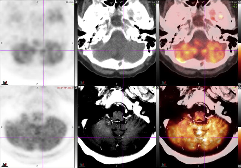
Hybrid imaging systems have dramatically improved thoracic oncology patient care over the past 2 decades. PET-MR imaging systems have the potential to further improve imaging of thoracic neoplasms, resulting in diagnostic and therapeutic advantages compared with current MR imaging and PET-computed tomography systems. Increasing soft tissue contrast and lesion sensitivity, improved image registration, reduced radiation exposure, and improved patient convenience are immediate clinical advantages. Multiparametric quantitative imaging capabilities of PET-MR imaging have the potential to improve understanding of the molecular mechanisms of cancer and treatment effects, potentially guiding improvements in diagnosis and therapy.
Keywords: Breast cancer; Hybrid imaging; Lung cancer; Oncology; PET-MR imaging; Thoracic imaging.
Copyright © 2016 Elsevier Inc. All rights reserved.
Figures

Similar articles
-
Breast PET/MR Imaging.Radiol Clin North Am. 2017 May;55(3):579-589. doi: 10.1016/j.rcl.2016.12.011. Epub 2017 Feb 1. Radiol Clin North Am. 2017. PMID: 28411681 Free PMC article. Review.
-
PET/MR Imaging for Chest Diseases: Review of Initial Studies on Pulmonary Nodules and Lung Cancers.Magn Reson Imaging Clin N Am. 2015 May;23(2):245-59. doi: 10.1016/j.mric.2015.01.008. Magn Reson Imaging Clin N Am. 2015. PMID: 25952518 Review.
-
Improved Detection of Small Pulmonary Nodules Through Simultaneous MR/PET Imaging.PET Clin. 2018 Jan;13(1):89-95. doi: 10.1016/j.cpet.2017.09.001. PET Clin. 2018. PMID: 29157389 Review.
-
PET/MR imaging in the detection and characterization of pulmonary lesions: technical and diagnostic evaluation in comparison to PET/CT.J Nucl Med. 2014 May;55(5):724-9. doi: 10.2967/jnumed.113.129247. Epub 2014 Mar 20. J Nucl Med. 2014. PMID: 24652827
-
Quantitative PET/MR imaging of lung cancer in the presence of artifacts in the MR-based attenuation correction maps.Acta Radiol. 2020 Jan;61(1):11-20. doi: 10.1177/0284185119848118. Epub 2019 May 15. Acta Radiol. 2020. PMID: 31091969 No abstract available.
Cited by
-
Development and validation of a novel diagnostic model for assessing lung cancer metastasis in a Chinese population based on multicenter real-world data.Cancer Manag Res. 2019 Oct 29;11:9213-9223. doi: 10.2147/CMAR.S217970. eCollection 2019. Cancer Manag Res. 2019. PMID: 31807063 Free PMC article.
-
Amyloid precursor protein promotes the migration and invasion of breast cancer cells by regulating the MAPK signaling pathway.Int J Mol Med. 2020 Jan;45(1):162-174. doi: 10.3892/ijmm.2019.4404. Epub 2019 Nov 13. Int J Mol Med. 2020. PMID: 31746365 Free PMC article.
-
A Novel Sonification Approach to Support the Diagnosis of Alzheimer's Dementia.Front Neurol. 2017 Dec 7;8:647. doi: 10.3389/fneur.2017.00647. eCollection 2017. Front Neurol. 2017. PMID: 29270150 Free PMC article.
-
Application of serum Raman spectroscopy combined with classification model for rapid breast cancer screening.Front Oncol. 2023 Oct 26;13:1258436. doi: 10.3389/fonc.2023.1258436. eCollection 2023. Front Oncol. 2023. PMID: 37965448 Free PMC article.
-
Recent Advances in Multimodal Molecular Imaging of Cancer Mediated by Hybrid Magnetic Nanoparticles.Polymers (Basel). 2021 Sep 3;13(17):2989. doi: 10.3390/polym13172989. Polymers (Basel). 2021. PMID: 34503029 Free PMC article. Review.
References
Publication types
MeSH terms
Grants and funding
LinkOut - more resources
Full Text Sources
Other Literature Sources
Medical
Miscellaneous
