

Decreased Progesterone Receptor B/A Ratio in Endometrial Cells by Tumor Necrosis Factor-Alpha and Peritoneal Fluid from Patients with Endometriosis
- PMID: 27593876
- PMCID: PMC5011280
- DOI: 10.3349/ymj.2016.57.6.1468
Decreased Progesterone Receptor B/A Ratio in Endometrial Cells by Tumor Necrosis Factor-Alpha and Peritoneal Fluid from Patients with Endometriosis
Abstract
Purpose: Progesterone resistance is thought to be a major factor that contributes to progression of endometriosis. However, it is not clear what causes progesterone resistance in endometriosis. This study aimed to assess whether cytokines or peritoneal fluid can affect progesterone receptor (PR) expression in endometrial cells and to verify whether PR expression is reduced in endometriosis.
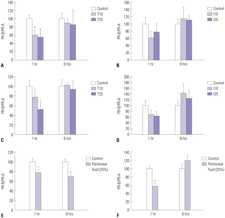
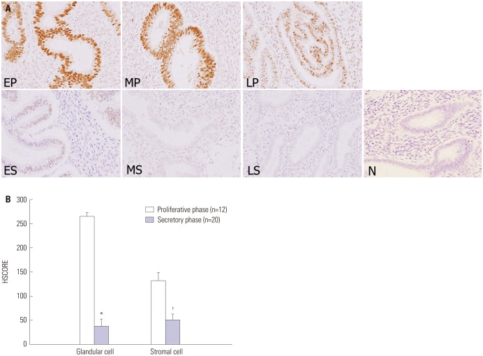
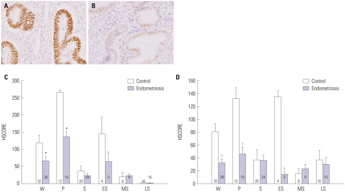
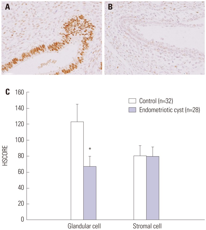
Materials and methods: The PR-B/A ratio was measured via real-time polymerase chain reaction after in vitro culture, in which endometrial cells were treated with either tumor necrosis factor-alpha (TNF-α), interleukin-1 beta, or peritoneal fluid obtained from women with advanced-stage endometriosis. Immunohistochemistry was performed to compare PR-B expression between eutopic and ectopic endometrial tissues from women with and without advanced-stage endometriosis.
Results: The PR-B/A ratio was significantly decreased by treatment with either TNF-α (p=0.011) or peritoneal fluid from women with advanced-stage endometriosis (p=0.027). Immunoreactivity of PR-B expression was significantly lower during the secretory phase than during the proliferative phase in endometrial tissues from control subjects (p<0.001). PR-B expression was significantly reduced in the eutopic endometrium (p=0.031) and ovarian endometrioma (p=0.036) from women with advanced-stage endometriosis compared with eutopic endometrium tissues from control subjects.
Conclusion: Progesterone resistance in endometriosis may be caused by proinflammatory conditions in the pelvic peritoneal microenvironment.
Keywords: Endometriosis; cytokine; endometrium; peritoneal fluid; progesterone receptor.
Conflict of interest statement
The authors have no financial conflicts of interest.
Figures
References
-
- Eskenazi B, Warner ML. Epidemiology of endometriosis. Obstet Gynecol Clin North Am. 1997;24:235–258. - PubMed
-
- Giudice LC, Kao LC. Endometriosis. Lancet. 2004;364:1789–1799. - PubMed
-
- Eisermann J, Gast MJ, Pineda J, Odem RR, Collins JL. Tumor necrosis factor in peritoneal fluid of women undergoing laparoscopic surgery. Fertil Steril. 1988;50:573–579. - PubMed
-
- Hill JA, Anderson DJ. Lymphocyte activity in the presence of peritoneal fluid from fertile women and infertile women with and without endometriosis. Am J Obstet Gynecol. 1989;161:861–864. - PubMed
MeSH terms
Substances
Supplementary concepts
LinkOut - more resources
Full Text Sources
Other Literature Sources
Medical
Research Materials
