

Osteochondral Autograft from the Ipsilateral Femoral Head by Surgical Dislocation for Treatment of Femoral Head Fracture Dislocation: A Case Report
- PMID: 27593886
- PMCID: PMC5011290
- DOI: 10.3349/ymj.2016.57.6.1527
Osteochondral Autograft from the Ipsilateral Femoral Head by Surgical Dislocation for Treatment of Femoral Head Fracture Dislocation: A Case Report
Abstract
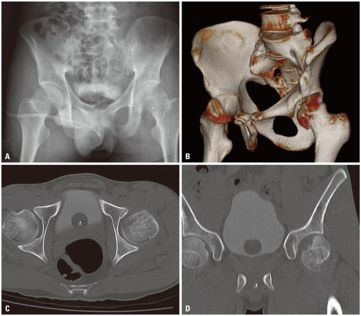
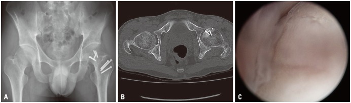
As anatomical reduction of the articular surface of femoral head fractures and restoration of damaged cartilage are essential for good long-term results, many treatment options have been suggested, including fixation of the fracture using various surgical exposures and implants, as well as arthroscopic irrigation and debridement, bone marrow stimulating techniques, osteochondral allograft, autograft, and autogenous chondrocyte implantation. We report a case of osteochondral autograft harvested from its own femoral articular surface through surgical hip dislocation. The osteochondral graft was harvested from the inferior non-weight-bearing articular surface and grafted to the osteochondral defect. One year later, the clinical and radiological results were good, without the collapse of the femoral head or arthritic change. This procedure introduced in our case is considered convenient and able to lessen surgical time without morbidity of the donor site associated with the harvest.
Keywords: Femoral head; autograft transplant; fractures cartilage; hip dislocation.
Conflict of interest statement
The authors have no financial conflicts of interest.
Figures

References
-
- Pipkin G. Treatment of grade IV fracture-dislocation of the hip. J Bone Joint Surg Am. 1957;39-A:1027–1042 passim. - PubMed
-
- Davis JB. Simultaneous femoral head fracture and traumatic hip dislocation. Am J Surg. 1950;80:893–895. - PubMed
-
- Butler JE. Pipkin Type-II fractures of the femoral head. J Bone Joint Surg Am. 1981;63:1292–1296. - PubMed
-
- Henle P, Kloen P, Siebenrock KA. Femoral head injuries: which treatment strategy can be recommended? Injury. 2007;38:478–488. - PubMed
-
- Hougaard K, Thomsen PB. Traumatic posterior fracture-dislocation of the hip with fracture of the femoral head or neck, or both. J Bone Joint Surg Am. 1988;70:233–239. - PubMed
Publication types
MeSH terms
LinkOut - more resources
Full Text Sources
Other Literature Sources
Medical
Research Materials
