

Biliary architecture of livers exhibiting right-sided ligamentum teres: an indication for preoperative cholangiography prior to major hepatectomy
- PMID: 27594117
- PMCID: PMC5094486
- DOI: 10.1016/j.hpb.2016.08.002
Biliary architecture of livers exhibiting right-sided ligamentum teres: an indication for preoperative cholangiography prior to major hepatectomy
Abstract
Objective: To obtain information about the basic biliary anatomy of livers with right-sided ligamentum teres (RSLT).
Summary of background data: RSLT is a relatively rare anomaly with a reported incidence of 0.2-1.2%. Although the portal/hepatic venous and arterial anatomy of livers with RSLT has already been established, the biliary architecture of such livers remains unclear.
Methods: RSLT was detected in 48 patients during 12,071 consecutive image readings (0.4%). Of these patients, the cholangiograms of 46 patients were analyzed, and their intrahepatic biliary tree confluence patterns were classified.
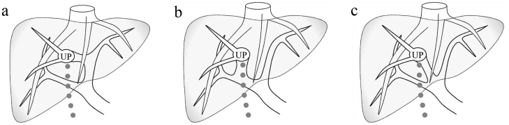
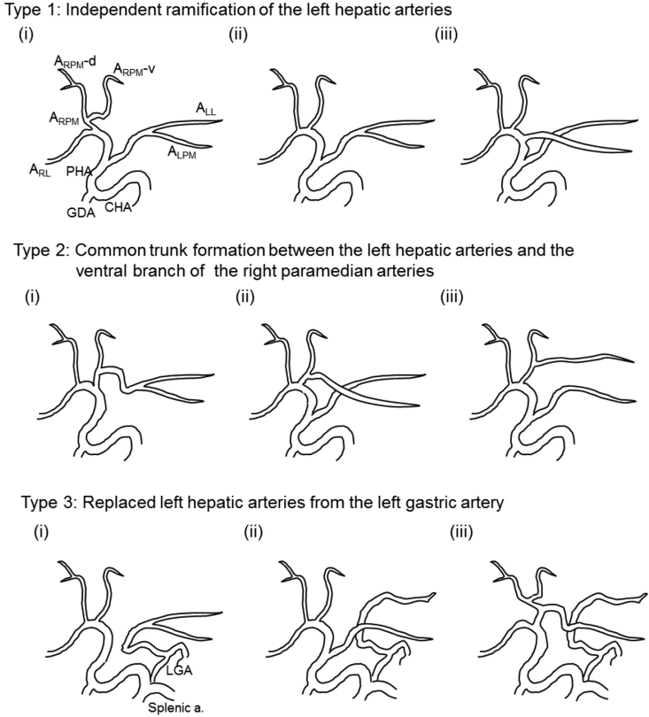
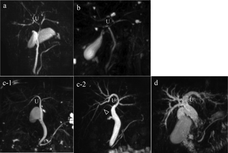
Results: The following four unique biliary confluence patterns were identified in livers with RSLT: the symmetrical type (23/46), independent right lateral type (13/46), total left type (6/46), and total right type (1/46). Analyses of the portal and arterial branching patterns of these livers showed that there were no correlations between their biliary confluence patterns and their portal or arterial ramification patterns.
Conclusion: The basic biliary architecture of livers with RSLT was clarified. As the RSLT patients' anomalous biliary confluences differed from those seen in normal livers and were difficult to predict, preoperative cholangiography should be performed prior to complex hepatobiliary surgery involving livers with RSLT to ensure patient safety.
Copyright © 2016 The Author(s). Published by Elsevier Ltd.. All rights reserved.
Figures


References
-
- Kawai K., Koizumi M., Honma S., Tokiyoshi A., Kodama K. Right ligamentum teres joining to the right branch of the portal vein. Anat Sci Int. 2008;83:49–54. - PubMed
-
- Maetani Y., Itoh K., Kojima N., Tabuchi T., Shibata T., Asonuma K. Portal vein anomaly associated with deviation of the ligamentum teres to the right and malposition of the gallbladder. Radiology. 1998;207:723–728. - PubMed
-
- Shindoh J., Satou S., Aoki T., Beck Y., Hasegawa K., Sugawara Y. Hidden symmetry in asymmetric morphology: significance of Hjortsjo's anatomical model in liver surgery. Hepatogastroenterology. 2012;59:519–525. - PubMed
MeSH terms
LinkOut - more resources
Full Text Sources
Other Literature Sources
Medical
Research Materials
