

Computed tomography features of supracardiac total anomalous pulmonary venous connection in an infant
- PMID: 27594934
- PMCID: PMC4996912
- DOI: 10.1016/j.radcr.2016.04.005
Computed tomography features of supracardiac total anomalous pulmonary venous connection in an infant
Abstract
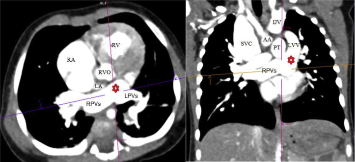
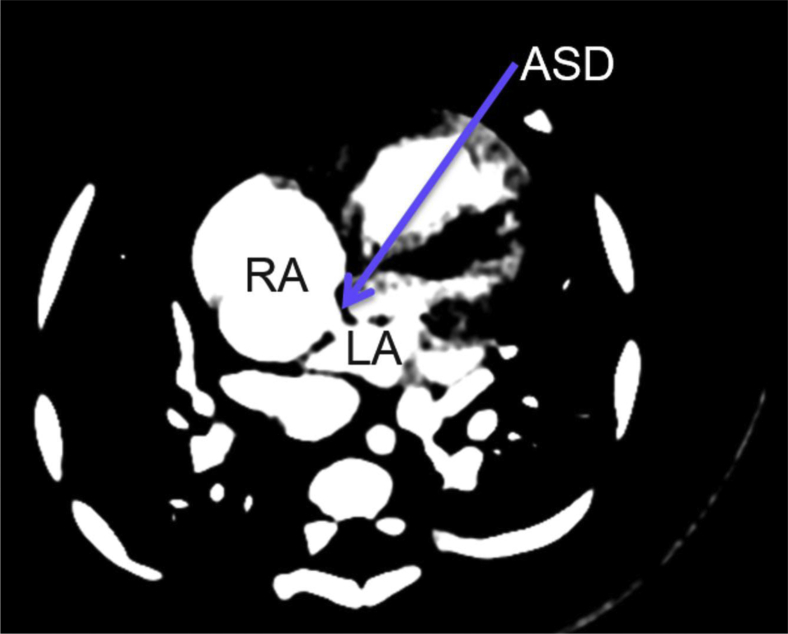
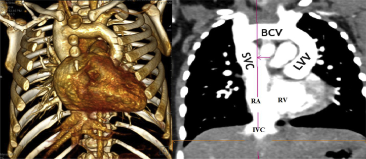
Total anomalous pulmonary venous connection (TAPVC) is a rare congenital anomaly of the pulmonary veins drainage. In this entity, the pulmonary veins, instead of draining to left atrium, connect abnormally to the systemic venous circulation. A right-to-left shunt is obligatory for survival. Based on its type and degree of pulmonary venous obstruction, TAPVC may result in pulmonary hypertension and congestive heart failure. In severe cases, urgent diagnosis and surgical correction is essential to reduce morbidity and mortality. Echocardiography as the first and safest imaging modality for cardiovascular abnormalities may fail in complete depiction of some complex feature of TAPVC. Computed tomography angiography is then a noninvasive and sensitive choice for mapping the pulmonary veins without the need for invasive cardiac catheterization. Contrast-enhanced MR angiography can be a radiation-free alternative. Authors present a computed tomography-detected supracardiac TAPVC with small patent ductus arteriosus in a 2 months cyanotic infant.
Keywords: anomalous pulmonary venous connection; congenital heart disease; cyanosis.
Figures
References
-
- Ogawa M., Nakagawa M., Hara M., Ito M., Goto T., Ohte N. Total anomalous pulmonary venous connection in a 64-year-old man: a case report. Ann Thorac Cardiovasc Surg. 2013;19(1):46–48. - PubMed
-
- Lakshminrusimha S., Wynn R.J., Youssfi M., Pabalan M.J., Bommaraju M., Kirmani K. Use of CT angiography in the diagnosis of total anomalous venous return. J Perinatol. 2009;29(6):458–461. - PubMed
-
- Dillman J.R., Yarram S.G., Hernandez R.J. Imaging of pulmonary venous developmental anomalies. AJR Am J Roentgenol. 2009;192(5):1272–1285. - PubMed
-
- Hines M.H., Hammon J.W. Anatomy of total anomalous pulmonary venous connection. Oper Tech Thorac Cardiovasc Surg. 2001;6(1):2–7.
-
- DeLeon S.Y., Gidding S.S., Ilbawi M.N., Idriss F.S., Muster A.J., Cole R.B. Surgical management of infants with complex cardiac anomalies associated with reduced pulmonary blood flow and total anomalous pulmonary venous drainage. Ann Thorac Surg. 1987;43(2):207–211. - PubMed
Publication types
LinkOut - more resources
Full Text Sources
Other Literature Sources
