. 2016 Aug 4;1(12):e87012.
doi: 10.1172/jci.insight.87012.

Classical dendritic cells mediate fibrosis directly via the retinoic acid pathway in severe eye allergy

Affiliations

Classical dendritic cells mediate fibrosis directly via the retinoic acid pathway in severe eye allergy

Sarah D Ahadome et al. JCI Insight. .

Abstract

Fibrosis is a shared end-stage pathway to lung, liver, and heart failure. In the ocular mucosa (conjunctiva), fibrosis leads to blindness in trachoma, pemphigoid, and allergy. The indirect fibrogenic role of DCs via T cell activation and inflammatory cell recruitment is well documented. However, here we demonstrate that DCs can directly induce fibrosis. In the mouse model of allergic eye disease (AED), classical CD11b+ DCs in the ocular mucosa showed increased activity of aldehyde dehydrogenase (ALDH), the enzyme required for retinoic acid synthesis. In vitro, CD11b+ DC-derived ALDH was associated with 9-cis-retinoic acid ligation to retinoid x receptor (RXR), which induced conjunctival fibroblast activation. In vivo, stimulating RXR led to rapid onset of ocular mucosal fibrosis, whereas inhibiting ALDH activity in DCs or selectively depleting DCs markedly reduced fibrosis. Collectively, these data reveal a profibrotic ALDH-dependent pathway by DCs and uncover a role for DC retinoid metabolism.


Figures
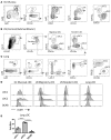
Figure 1
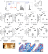
Figure 1. Uniquely high aldehyde dehydrogenase (ALDH) activity in steady-state type-2 classical DCs (cDC2) is conserved in multiple tissues.
(AC) Gating strategies for analyzing steady-state cDC1 and cDC2 in ocular (Oc) mucosa, draining submandibular/cervical lymph node (LN), and lung from normal C57BL/6 mice (PMN, neutrophils; Eo, eosinophils; Mo/MF, classical monocytes and macrophages). (D and E) Comparison of ALDH activity in cDC1s and cDC2s. Cell suspensions were stained with ALDEFLUOR to assess ALDH activity. Negative controls are shown for the respective cDC2 populations, which were ALDEFLUOR-stained samples treated with the ALDH inhibitor diethylaminobenzaldehyde (DEAB). Data are representative of duplicate data points from 2 separate experiments on pooled conjunctiva from n = 10 mice. Lung data is representative of triplicate data points from 3 separate samples from n = 20 mice (error bars represent ±SEM, ***P < 0.0005, ****P < 0.00005, 1-way ANOVA with Bonferroni correction). Each experiment was carried out at least twice.
Figure 2
Figure 2. Fibrogenic role for CD11b+ BMDC-derived ALDH on ocular mucosal fibroblasts.
(A and B) Uniquely high aldehyde dehydrogenase (ALDH) activity of CD11b+ (11b) BM-derived DCs (BMDC), relative to CD103+ (103) BMDCs. (C) CD11b+ BMDC-derived ALDH impairs normal fibroblast proliferation and contractility in vitro. Primary conjunctival fibroblasts were plated in media for the proliferation assay or a free-floating collagen matrix for the contractility assay. Added to the wells were complete media only, conditioned media from CD11b+ BMDCs treated with or without diethylaminobenzaldehyde (DEAB), or conditioned media from CD103+ BMDCs as indicated. Gel contraction and proliferation was measured 1 d later. (D) Retinoid x receptor (RXR) signaling is responsible for CD11b+ BMDC-derived ALDH impairment of normal fibroblast contractility and proliferation. Fibroblasts were plated in the presence of CD11b+ BMDC-conditioned media or not and treated with RXR or retinoic acid receptor (RAR) inhibitor as indicated. (E) 9-cis-RA (9c) mediates RXR signaling that causes impairment of normal fibroblast contractility and proliferation. Primary conjunctival fibroblasts were plated, and some samples received 9c or all-trans-RA (AT), in addition to RXR inhibitor as indicated. (F) RXR agonist, bexarotene (Bex), impairs normal fibroblast contractility and proliferation (Veh, vehicle). (G) Local administration of Bex in vivo induces ocular mucosal fibrosis. Subconjunctival injection of Bex (10 ng) was administered to one eye, whereas the vehicle control was injected into the fellow eye. Clinical images were obtained, and Masson’s trichrome–stained sections of the conjunctiva were assessed (white arrows indicate diseased eye; black arrows indicate subepithelial collagen deposition). Triplicate data points represent fibroblast data from 3 separate fibroblast cultures from 3 different mice (error bars represent ±SEM, ***P < 0.0005, ****P < 0.00005, 1-way ANOVA with Bonferroni correction). In vivo data are representative of n = 5 mice. Each experiment was carried out at least twice.
Figure 3
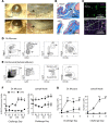
Figure 3. Development of ocular mucosal fibrosis during allergic eye disease (AED) is associated with augmented cDC2-derived ALDH activity.
Mice were immunized with OVA/adjuvant and challenged topically once daily with an OVA instillation to induce AED. (A and B) Ocular mucosal fibrosis develops in AED (black arrows). Data from whole eyes sectioned and stained with Masson’s trichrome are representative of n = 3 per group (inset shows fibrosis/opaque white lesion in AED, not seen in naive mice). (CF) Ocular (Oc) mucosal type-2 classical DC (cDC2) confer increased aldehyde dehydrogenase (ALDH) activity in AED. (C) Whole mount immunofluorescence of the ocular mucosa is representative of n = 5 per group. (D and E) Ocular mucosa and draining lymph node (LN) in AED were collected for flow cytometry analysis. Representative flow cytometry plots are shown from tissues collected at 6 hr after challenge on day 3. (F) Quantitation was performed on tissues collected at 20 min (20) and 6 hr (6) after OVA challenge on days (d) 0 through 3 to measure ALDH activity by flow cytometry. (G) Ocular mucosal cDC2 increase in number during AED. Flow cytometry was carried out for enumeration of cDCs. Data shown for the ocular mucosal cDCs are representative of duplicate data points from 2 separate experiments. Cell suspensions were pooled samples from n = 10 naive mice or n = 6 mice for each experiment (error bars represent ±SEM; *P < 0.05, **P < 0.005; 1-way ANOVA with Bonferroni correction). Each experiment was carried out at least twice.
Figure 4
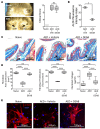
Figure 4. Local inhibition of cDC2-derived aldehyde dehydrogenase (ALDH) protects mice from ocular mucosal fibrosis during allergic inflammation.
Mice induced with allergic eye disease (AED) were treated with topical diethylaminobenzaldehyde (DEAB) in one eye and vehicle control in the fellow/contralateral eye. (A and B) Equivalent levels of allergic inflammation in AED is achieved in the setting of unilateral DEAB instillation (black arrows). Clinical image and scores are derived from n = 12 mice. (B) Unilateral DEAB instillation in AED leads to reduced ALDH activity in type-2 classical DC (cDC2), as compared with those from the vehicle-treated contralateral eye. Flow cytometry data are derived from triplicate data points of pooled conjunctiva from n = 12 mice. (C) Unilateral DEAB instillation during AED leads to reduced collagen deposition in the ocular mucosa of the treated eye (black arrows). Images represent n = 6 mice. (D and E) Unilateral DEAB instillation restores normal levels of proliferation, contractility, collagen production, and α smooth muscle actin (α) of AED conjunctival fibroblasts from the treated eye. Fibroblast data are represented by triplicate data points from 3 separate fibroblast cultures from 3 different mice (error bars represent ±SEM; *P < 0.05, ****P < 0.00005; 1-way ANOVA with Bonferroni correction). Each experiment was carried out at least twice.
Figure 5
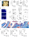
Figure 5. Local DC depletion protects CD11c-eGFP/DTR mice from developing ocular mucosal fibrosis during allergic inflammation.
Ipsilateral diphtheria toxin (DTx) instillation was given topically (3.125 ng) on days –1, 3, and 5 of challenge, whereas the contralateral eye received PBS control. (A) Equivalent levels of allergic inflammation in allergic eye disease (AED) is achieved despite unilateral DTx instillation to CD11c-eGFP/DTR mice (black arrows). Clinical scores and enumeration of ocular mucosal eosinophils (Eos) and CD4+ T cells (flow cytometry) compared DTx-treated eyes vs. PBS-treated contralateral eyes on day 7. (B) Deletion efficacy of unilateral DTx instillation. Intravital multiphoton microscopy of DTx-treated eye and vehicle-treated contralateral eyes was carried out in AED mice to verify DC deletion in the DTx-treated eye (SHG, second harmonic generation). Lymph nodes (LNs) were collected for flow cytometry to verify deletion of ipsilateral migratory type-2 classical DCs (cDC2). Data is derived from triplicate data points from n = 5 mice. (C) Unilateral DTx instillation during AED leads to reduced collagen deposition in the ocular mucosa of the treated eye (black arrows). Micrographs represent n = 5 mice. (D) Unilateral DTx instillation restores normal levels of proliferation, contractility, and collagen production of AED conjunctival fibroblasts from the treated eye. Fibroblast data are represented by triplicate data points from 3 separate fibroblast cultures from 3 different mice (error bars represent ±SEM; **P < 0.005, ****P < 0.00005; 1-way ANOVA with Bonferroni correction). Each experiment was carried out at least twice.

References

    1. Lee CG, et al. Interleukin-13 induces tissue fibrosis by selectively stimulating and activating transforming growth factor beta(1) J Exp Med. 2001;194(6):809–821. doi: 10.1084/jem.194.6.809. - DOI - PMC - PubMed
    1. Wynn TA. Common and unique mechanisms regulate fibrosis in various fibroproliferative diseases. J Clin Invest. 2007;117(3):524–529. doi: 10.1172/JCI31487. - DOI - PMC - PubMed
    1. Chiaramonte MG, Donaldson DD, Cheever AW, Wynn TA. An IL-13 inhibitor blocks the development of hepatic fibrosis during a T-helper type 2-dominated inflammatory response. J Clin Invest. 1999;104(6):777–785. doi: 10.1172/JCI7325. - DOI - PMC - PubMed
    1. Leonardi A, Cortivo R, Fregona I, Plebani M, Secchi AG, Abatangelo G. Effects of Th2 cytokines on expression of collagen, MMP-1, and TIMP-1 in conjunctival fibroblasts. Invest Ophthalmol Vis Sci. 2003;44(1):183–189. doi: 10.1167/iovs.02-0420. - DOI - PubMed
    1. Saw VP, et al. Conjunctival interleukin-13 expression in mucous membrane pemphigoid and functional effects of interleukin-13 on conjunctival fibroblasts in vitro. Am J Pathol. 2009;175(6):2406–2415. doi: 10.2353/ajpath.2009.090579. - DOI - PMC - PubMed