. 2016 Oct;22(10):1094-1100.
doi: 10.1038/nm.4177. Epub 2016 Sep 5.

Persisting positron emission tomography lesion activity and Mycobacterium tuberculosis mRNA after tuberculosis cure

Affiliations

Persisting positron emission tomography lesion activity and Mycobacterium tuberculosis mRNA after tuberculosis cure

Stephanus T Malherbe et al. Nat Med. 2016 Oct.

Erratum in

Abstract

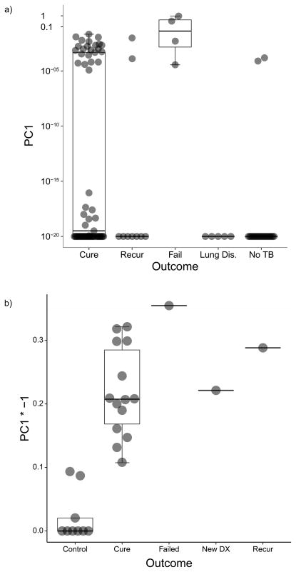
The absence of a gold standard to determine when antibiotics induce a sterilizing cure has confounded the development of new approaches to treat pulmonary tuberculosis (PTB). We detected positron emission tomography and computerized tomography (PET-CT) imaging response patterns consistent with active disease, along with the presence of Mycobacterium tuberculosis (MTB) mRNA in sputum and bronchoalveolar lavage samples, in a substantial proportion of adult, HIV-negative patients with PTB after a standard 6-month treatment plus 1 year follow-up, including patients with a durable cure and others who later developed recurrent disease. The presence of MTB mRNA in the context of nonresolving and intensifying lesions on PET-CT images might indicate ongoing transcription, suggesting that even apparently curative treatment for PTB may not eradicate all of the MTB bacteria in most patients. This suggests an important complementary role for the immune response in maintaining a disease-free state. Sterilizing drugs or host-directed therapies, and better treatment response markers, are probably needed for the successful development of improved and shortened PTB-treatment strategies.


Conflict of interest statement

statement. We have no competing financial interest to declare.

Figures

Figure 1
Figure 1
Flow chart of study design. (a) In, South-Africa, 99 sputum culture positive Pulmonary Tuberculosis (PTB) patients underwent 18F-FDG PET-CT scans at diagnosis (Dx), month 1 (M1) and month 6 (M6) of treatment. Fifty patients also had PET-CT scans 1 year after the end of treatment (EOT + 1y). QRT-PCR assays for MTB mRNA were performed on month 6 sputum from 75 patients and end of treatment bronchoalveolar lavage (BAL) samples of 15 patients. (b) QRT-PCR assays were repeated on sputum from 20 community controls, and five controls with lung disease other than PTB; as well as BAL samples of 10 controls undergoing diagnostic bronchoscopies for suspected lung cancer. (c) PET-CT scans were also performed on 14 sputum culture positive PTB patients from South-Korea at Dx, M1 and M6.
Figure 2
Figure 2
Representative Dx, M1 and M6 PET-CT images for cured patients after 6 months of standard treatment. 3-dimensional anterior (top panels) and transverse views, at the level of blue lines, (lower panels). (a) Resolved scan, without residual abnormal FDG uptake, some structural abnormalities remain on CT scan. (b) Improved scan, where all lesions have improved since diagnosis, but abnormal CT lesions and increased uptake persist. This example includes thick-walled cavities with high FDG uptake. (c) Mixed scan response with either new FDG-avid lesions or increased intensity of some lesions. This example shows a large new FDG-avid nodule with high intensity uptake in the left lung upper lobe.
Figure 3
Figure 3
Clinical outcome and associated PET-CT findings (due to rounding of percentage values totals may equal to 99% or 101%; Lesion intensity rankings summary in Supplementary Dataset 5). (a) South-African: Clinical outcome and associated M6 scan response pattern. Mixed scan response was more likely to have an unfavourable outcome (Fisher exact test, two-sided: P = 0.002). Source data in Supplementary Dataset 6. (b) South-Korea: Patients grouped into those diagnosed with MDR strains or with drug-sensitive or rifampicin mono-resistant (Rif-mono) strains. All MDR cases culture converted within 6 months, while the DS/Rif-mono group includes nine cured, one unevaluable and one recurrent patient. Source data in Supplementary Dataset 2. (c) South-Africa: Clinical outcome and associated EOT + 1y scan response pattern in comparison to M6. Source data in Supplementary Dataset 7.
Figure 4
Figure 4
Representative Dx, M6 and EOT + 1y, PET-CT images for patients after 6 months of standard treatment. 3-dimensional anterior (top) and transverse views (lower) at the level of horizontal blue line. (a) A large new FDG-avid nodule in right lung apex lesion develops by M6, but improves over the next year, with some residual increased FDG uptake. (b) Bilateral upper lobe cavities improve during treatment, but nodules with normal FDG-uptake at M6 develop new cavitation and increased uptake by EOT + 1y. (c) Multiple lesions with continuing reduction in FDG-avidity at M6 and EOT + 1y but with a large new area of consolidation at EOT + 1y. This patient was Gene Xpert positive, but culture negative at the time of EOT + 1y scan, but diagnosed with recurrent disease 6 months later (18 months after treatment completion), while patients (a) and (b) maintained cure.
Figure 5
Figure 5
Transformed (arcsinh) principal component one of mRNA transcript PCR data, demonstrating separation by detectable MTB mRNA as captured in PC1. Box plots represent median (middle), 75th percentile (top) and 25th percentile (bottom); whiskers represent range. (a) M6 sputum from 75 PTB cases by clinical category; 22 of 60 cured cases, all four failed treatment cases and two of the nine recurrent cases in the group have detectable MTB mRNA in their sputum. None of the other lung disease participants and two of 20 healthy community controls have detectable MTB mRNA (Fisher Exact Test, two-sided: P < 0.01). Source data in Supplementary Dataset 3. (b) EOT bronchoalveolar lavage samples by clinical category. The new PTB case and 15 EOT cases (one failed treatment, one recurrent and 13 cases that maintained cure), all still have significantly detectable mRNA. Three of the six Quantiferon® positive controls show inconclusive levels of MTB mRNA, whereas the Quantiferon® negative controls have no detectable MTB mRNA (Fisher Exact test, two-sided: P < 0.001). Source data in Supplementary Dataset 4.

Comment in

  • A broader spectrum of tuberculosis.
    Schnappinger D, Ehrt S. Schnappinger D, et al. Nat Med. 2016 Oct 6;22(10):1076-1077. doi: 10.1038/nm.4186. Nat Med. 2016. PMID: 27711059 No abstract available.

References

    1. Cox HS, Morrow M, Deutschmann PW. Long term efficacy of DOTS regimens for tuberculosis: systematic review. BMJ. 2008;336:484–487. - PMC - PubMed
    1. WHO. Global Tuberculosis Report. 2015;20
    1. Gillespie SH, et al. Four-Month Moxifloxacin-Based Regimens for Drug-Sensitive Tuberculosis. N Engl J Med. 2014;371:1577–1587. - PMC - PubMed
    1. Jindani A, et al. High-Dose Rifapentine with Moxifloxacin for Pulmonary Tuberculosis. N Engl J Med. 2014;371:1599–1608. - PMC - PubMed
    1. Luzze H, et al. Relapse more common than reinfection in recurrent tuberculosis 1–2 years post treatment in urban Uganda. Int J Tuberc Lung Dis. 2013;17:361–367. - PMC - PubMed

Publication types

MeSH terms