

Detection of Isoforms Differing by a Single Charge Unit in Individual Cells
- PMID: 27595864
- PMCID: PMC5201312
- DOI: 10.1002/anie.201606039
Detection of Isoforms Differing by a Single Charge Unit in Individual Cells
Erratum in
-
Corrigendum: Detection of Isoforms Differing by a Single Charge Unit in Individual Cells.Angew Chem Int Ed Engl. 2016 Dec 5;55(49):15200. doi: 10.1002/anie.201609785. Angew Chem Int Ed Engl. 2016. PMID: 27897425 No abstract available.
Abstract
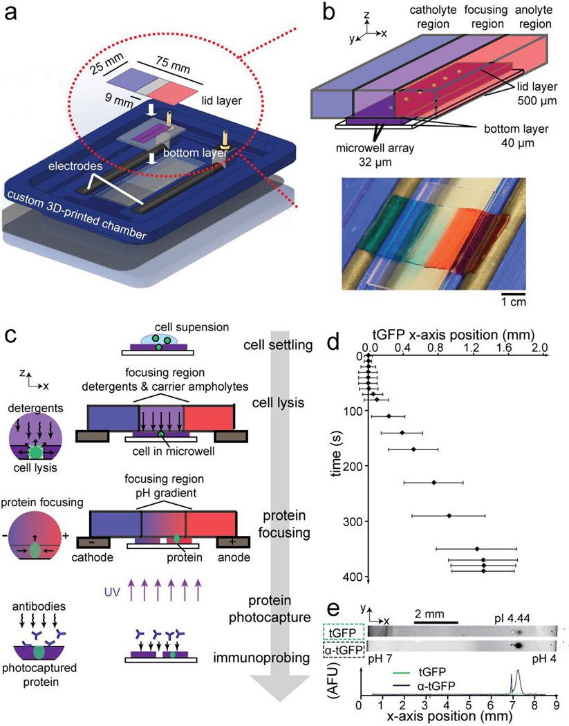
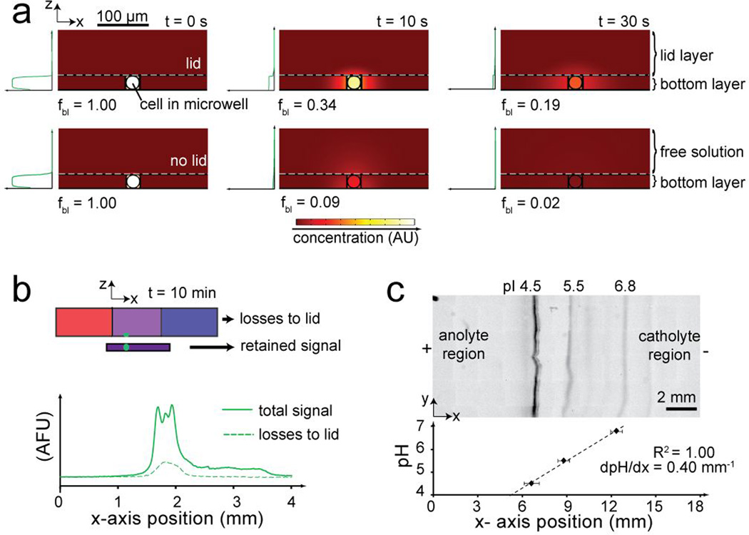
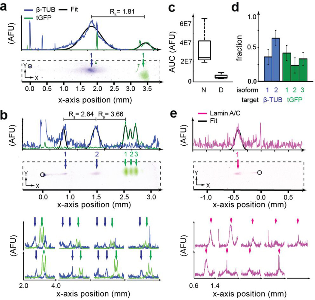
To measure protein isoforms in individual mammalian cells, we report single-cell resolution isoelectric focusing (scIEF) and high-selectivity immunoprobing. Microfluidic design and photoactivatable materials establish the tunable pH gradients required by IEF and precisely control the transport and handling of each 17-pL cell lysate during analysis. The scIEF assay resolves protein isoforms with resolution down to a single-charge unit, including both endogenous cytoplasmic and nuclear proteins from individual mammalian cells.
Keywords: immunoassays; isoelectric focusing; lab-on-a-chip; proteomics; single-cell analysis.
© 2016 WILEY-VCH Verlag GmbH & Co. KGaA, Weinheim.
Figures
Similar articles
-
Reducing Cathodic Drift during Isoelectric Focusing Using Microscale Immobilized pH Gradient Gels.Anal Chem. 2024 May 28;96(21):8648-8656. doi: 10.1021/acs.analchem.4c00788. Epub 2024 May 8. Anal Chem. 2024. PMID: 38716690 Free PMC article.
-
Single-Cell High-Resolution Detection and Quantification of Protein Isoforms Differing by a Single Charge Unit.Methods Mol Biol. 2019;1855:501-509. doi: 10.1007/978-1-4939-8793-1_44. Methods Mol Biol. 2019. PMID: 30426445
-
Microfluidic Free-Flow Isoelectric Focusing with Real-Time pI Determination.Methods Mol Biol. 2019;1906:113-124. doi: 10.1007/978-1-4939-8964-5_8. Methods Mol Biol. 2019. PMID: 30488389
-
Current trends in capillary isoelectric focusing of proteins.J Chromatogr B Biomed Sci Appl. 1997 Oct 10;699(1-2):91-104. doi: 10.1016/s0378-4347(96)00208-3. J Chromatogr B Biomed Sci Appl. 1997. PMID: 9392370 Review.
-
Immobilized pH gradient isoelectric focusing as a first-dimension separation in shotgun proteomics.J Biomol Tech. 2005 Sep;16(3):181-9. J Biomol Tech. 2005. PMID: 16461941 Free PMC article. Review.
Cited by
-
Single cell protein analysis for systems biology.Essays Biochem. 2018 Oct 26;62(4):595-605. doi: 10.1042/EBC20180014. Print 2018 Oct 26. Essays Biochem. 2018. PMID: 30072488 Free PMC article. Review.
-
Strategies for Development of a Next-Generation Protein Sequencing Platform.Trends Biochem Sci. 2020 Jan;45(1):76-89. doi: 10.1016/j.tibs.2019.09.005. Epub 2019 Oct 30. Trends Biochem Sci. 2020. PMID: 31676211 Free PMC article. Review.
-
Reducing Cathodic Drift during Isoelectric Focusing Using Microscale Immobilized pH Gradient Gels.Anal Chem. 2024 May 28;96(21):8648-8656. doi: 10.1021/acs.analchem.4c00788. Epub 2024 May 8. Anal Chem. 2024. PMID: 38716690 Free PMC article.
-
Controlling Dispersion during Single-Cell Polyacrylamide-Gel Electrophoresis in Open Microfluidic Devices.Anal Chem. 2018 Nov 20;90(22):13419-13426. doi: 10.1021/acs.analchem.8b03233. Epub 2018 Nov 2. Anal Chem. 2018. PMID: 30346747 Free PMC article.
-
Multiplexed in-gel microfluidic immunoassays: characterizing protein target loss during reprobing of benzophenone-modified hydrogels.Sci Rep. 2019 Oct 28;9(1):15389. doi: 10.1038/s41598-019-51849-8. Sci Rep. 2019. PMID: 31659305 Free PMC article.
References
-
- Alfaro JA, Sinha A, Kislinger T, Boutros PC. Nat. Methods. 2014;11:1107–1113. - PubMed
Publication types
MeSH terms
Substances
Grants and funding
LinkOut - more resources
Full Text Sources
Other Literature Sources
Miscellaneous
