

The X-Ray Crystal Structure of the Keratin 1-Keratin 10 Helix 2B Heterodimer Reveals Molecular Surface Properties and Biochemical Insights into Human Skin Disease
- PMID: 27595935
- PMCID: PMC5514376
- DOI: 10.1016/j.jid.2016.08.018
The X-Ray Crystal Structure of the Keratin 1-Keratin 10 Helix 2B Heterodimer Reveals Molecular Surface Properties and Biochemical Insights into Human Skin Disease
Abstract
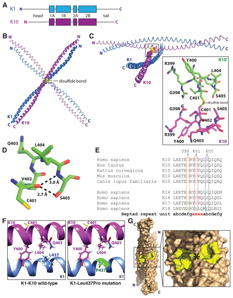
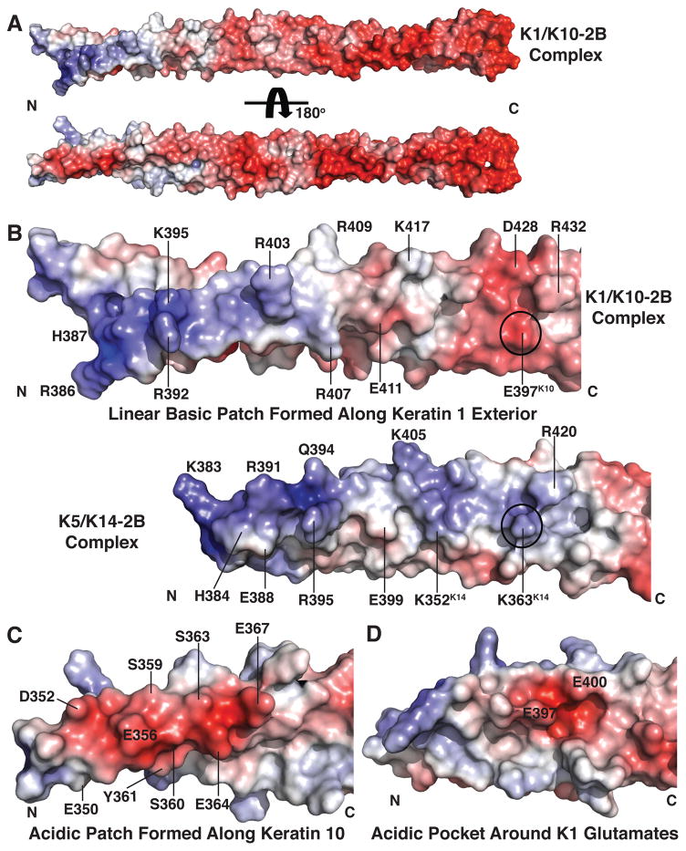
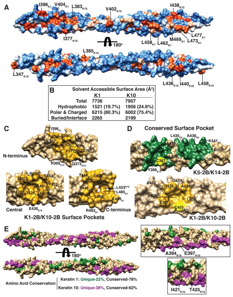
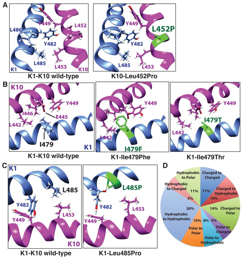
Keratins 1 (K1) and 10 (K10) are the primary keratins expressed in differentiated epidermis. Mutations in K1/K10 are associated with human skin diseases. We determined the crystal structure of the complex between the distal (2B) helices of K1 and K10 to better understand how human keratin structure correlates with function. The 3.3 Å resolution structure confirms many features inferred by previous biochemical analyses, but adds unexpected insights. It demonstrates a parallel, coiled-coil heterodimer with a predominantly hydrophobic intermolecular interface; this heterodimer formed a higher order complex with a second K1-K10-2B heterodimer via a Cys401K10 disulfide link, although the bond angle is unanticipated. The molecular surface analysis of K1-K10-2B identified several pockets, one adjacent to the disulfide linkage and conserved in K5-K14. The solvent accessible surface area of the K1-K10 structure is 20-25% hydrophobic. The 2B region contains mixed acidic and basic patches proximally (N-terminal), whereas it is largely acidic distally (C-terminal). Mapping of conserved and nonconserved residues between K1-K10 and K5-K14 onto the structure demonstrated the majority of unique residues align along the outer helical ridge. Finally, the structure permitted a fresh analysis of the deleterious effects caused by K1/K10 missense mutations found in patients with phenotypic skin disease.
Copyright © 2016 The Authors. Published by Elsevier Inc. All rights reserved.
Conflict of interest statement
The authors state no conflict of interest.
Figures
Similar articles
-
Crystal Structure of Keratin 1/10(C401A) 2B Heterodimer Demonstrates a Proclivity for the C-Terminus of Helix 2B to Form Higher Order Molecular Contacts.Yale J Biol Med. 2020 Mar 27;93(1):3-17. eCollection 2020 Mar. Yale J Biol Med. 2020. PMID: 32226330 Free PMC article.
-
In silico predicted structural and functional insights of all missense mutations on 2B domain of K1/K10 causing genodermatoses.Oncotarget. 2016 Aug 16;7(33):52766-52780. doi: 10.18632/oncotarget.10599. Oncotarget. 2016. PMID: 27421141 Free PMC article.
-
Human keratin 1/10-1B tetramer structures reveal a knob-pocket mechanism in intermediate filament assembly.EMBO J. 2019 Jun 3;38(11):e100741. doi: 10.15252/embj.2018100741. Epub 2019 Apr 29. EMBO J. 2019. PMID: 31036554 Free PMC article.
-
Keratins and skin disorders.Cell Biol Int. 1996 Apr;20(4):261-74. Cell Biol Int. 1996. PMID: 8664850 Review.
-
Lessons from disorders of epidermal differentiation-associated keratins.Histol Histopathol. 2002 Jan;17(1):331-8. doi: 10.14670/HH-17.331. Histol Histopathol. 2002. PMID: 11813882 Review.
Cited by
-
Keratin 6, 16 and 17-Critical Barrier Alarmin Molecules in Skin Wounds and Psoriasis.Cells. 2019 Aug 1;8(8):807. doi: 10.3390/cells8080807. Cells. 2019. PMID: 31374826 Free PMC article. Review.
-
Binding of the periplakin linker requires vimentin acidic residues D176 and E187.Commun Biol. 2020 Feb 21;3(1):83. doi: 10.1038/s42003-020-0810-y. Commun Biol. 2020. PMID: 32081916 Free PMC article.
-
Structural heterogeneity of cellular K5/K14 filaments as revealed by cryo-electron microscopy.Elife. 2021 Jul 29;10:e70307. doi: 10.7554/eLife.70307. Elife. 2021. PMID: 34323216 Free PMC article.
-
Role of the keratin 1 and keratin 10 tails in the pathogenesis of ichthyosis hystrix of Curth Macklin.PLoS One. 2018 Apr 24;13(4):e0195792. doi: 10.1371/journal.pone.0195792. eCollection 2018. PLoS One. 2018. PMID: 29689068 Free PMC article.
-
Genotype‒Structurotype‒Phenotype Correlations in Patients with Pachyonychia Congenita.J Invest Dermatol. 2021 Dec;141(12):2876-2884.e4. doi: 10.1016/j.jid.2021.03.035. Epub 2021 Jun 8. J Invest Dermatol. 2021. PMID: 34116063 Free PMC article.
References
-
- Bunick CG. X-ray crystal structure of the keratin 1-keratin 10 heterodimer reveals a molecular basis for associated keratinopathies. J Invest Dermatol; Society for Investigative Dermatology 74th Annual Meeting; Atlanta, GA. 2015. pp. S58–S69. - PubMed
MeSH terms
Substances
Grants and funding
LinkOut - more resources
Full Text Sources
Other Literature Sources
Medical
Molecular Biology Databases
Research Materials
