. 2016 Sep 5;16(1):713.
doi: 10.1186/s12885-016-2756-5.

CD44 and RHAMM are essential for rapid growth of bladder cancer driven by loss of Glycogen Debranching Enzyme (AGL)

Affiliations

CD44 and RHAMM are essential for rapid growth of bladder cancer driven by loss of Glycogen Debranching Enzyme (AGL)

Darby Oldenburg et al. BMC Cancer. .

Abstract

Background: Loss of Amylo-alpha-1-6-glucosidase-4-alpha-glucanotransferase (AGL) drives rapid proliferation of bladder cancer cells by upregulating Hyaluronic acid(HA) Synthase (HAS2) mediated HA synthesis. However the role of HA receptors CD44 and Hyaluronan Mediated Motility Receptor (RHAMM) in regulating the growth of bladder cancer cells driven by loss of AGL has not been studied.

Methods: Western blot analysis and Terminal deoxynucleotidyl transferase (TdT) dUTP Nick-End Labeling (TUNEL) assay was carried out to study cellular apoptosis with HAS2, CD44 and RHAMM loss in bladder cancer cells with and without AGL expression. Proliferation and softagar assays were carried out to study cellular anchorage dependent and independent growth. Clinicopathologic analysis was carried out on bladder cancer patient datasets.

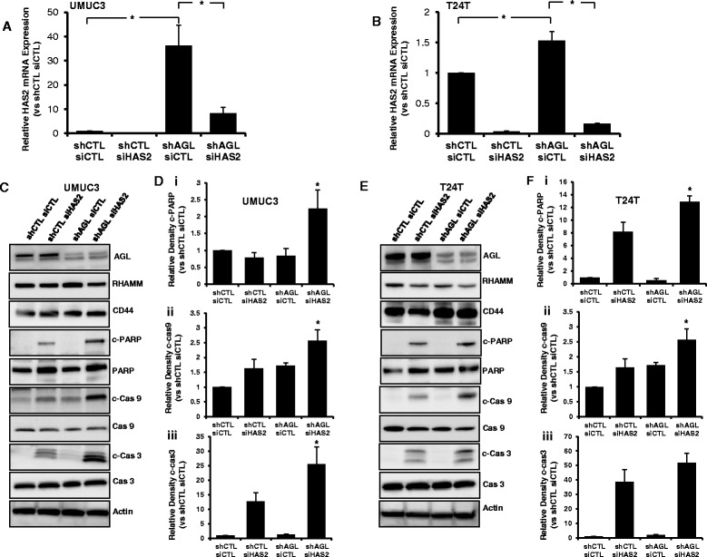
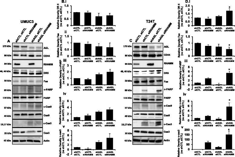
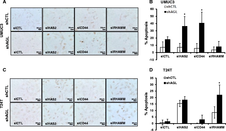
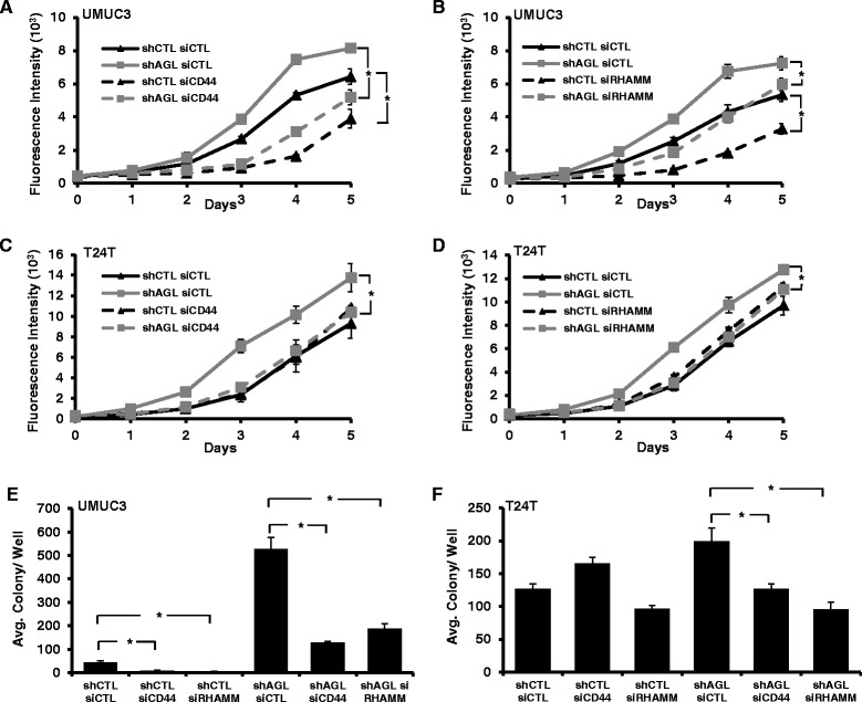
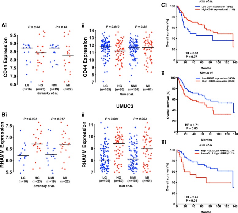
Results: Higher amounts of cleaved Cas3, Cas9 and PARP was observed in AGL low bladder cancer cell with loss of HAS2, CD44 or RHAMM. TUNEL staining showed more apoptotic cells with loss of HAS2, CD44 or RHAMM in AGL low bladder cancer cells. This revealed that bladder cancer cells whose aggressive growth is mediated by loss of AGL are susceptible to apoptosis with loss of HAS2, CD44 or RHAMM. Interestingly loss of either CD44 or RHAMM induces apoptosis in different low AGL expressing bladder cancer cell lines. Growth assays showed that loss of CD44 and RHAMM predominantly inhibit anchorage dependent and independent growth of AGL low bladder cancer cells. Clinicopathologic analysis revealed that high RHAMM mRNA expression is a marker of poor patient outcome in bladder cancer and patients with high RHAMM and low AGL tumor mRNA expression have poor survival.

Conclusion: Our findings strongly point to the importance of the HAS2-HA-CD44/RHAMM pathway for rapid growth of bladder cancer cells with loss of AGL and provides rational for targeting this pathway at various steps for "personalized" treatment of bladder cancer patients based of their AGL expression status.

Keywords: AGL; Bladder cancer; CD44; HAS2; RHAMM.


Figures

Fig. 1
Fig. 1
HAS2 loss and apoptosis in bladder cancer cells +/− AGL. a, b qRT-PCR demonstrating efficacy of HAS2 depletion in UMUC3 and T24T control (shCTL) and AGL knockdown (shAGL) cells. Cells were plated and 24 h later transfected with scrambled (siCTL) or directed siRNA against HAS2 (siHAS2). Details of siRNA used are in Materials and Methods. Cells were harvested at 48 h for mRNA followed by qRT-PCR analysis (n = 3). c 48 h after UMUC3 shCTL and shAGL cells were transfected with scrambled siRNA (siCTL) or siRNA against HAS2 (siHAS2), cells were lysed and expression of CD44, RHAMM and the proteins involved in the apoptotic pathway were detected by Western blot. d Densitometric analysis of cleaved apoptotic proteins normalized to total protein and the UMUC3 shCTL siCTL sample (n = 3). e 48 h after T24T shCTL and shAGL were transfected with scrambled siRNA (siCTL) or siRNA against HAS2 (siHAS2), cells were lysed and expression of CD44, RHAMM and the proteins involved in the apoptotic pathway were detected by Western blot. f Densitometric analysis of cleaved apoptotic proteins normalized to total protein and the T24T shCTL siCTL sample (n = 3). Results are shown as mean ± SD, *P < 0.05
Fig. 2
Fig. 2
CD44 loss and apoptosis in bladder cancer cells +/− AGL. a UMUC3 shCTL and shAGL cells were plated and 24 h later transfected with scrambled siRNA (siCTL) or siGENOME SMARTpool siRNA against CD44(siCD44). Details of siRNA are in Material and Methods. Cells were lysed 48 h after transfection and Western blot was carried out for proteins involved in apoptosis. b Densitometric analysis of cleaved apoptotic proteins normalized to total protein and the UMUC3 shCTL siCTL sample (n = 3). DR5 and Fas normalized to Actin and the UMUC3 shCTL siCTL sample (n = 3). Results are shown as mean ± SD, *P < 0.05. c T24T shCTL and shAGL cells were plated and 24 h later transfected with scrambled siRNA (siCTL) or siGENOME SMARTpool siRNA against CD44 (siCD44). Details of siRNA are in Material and Methods. Cells were lysed 48 h after transfection and Western blot was carried out for proteins involved in apoptosis. d Densitometric analysis of cleaved apoptotic proteins normalized to total protein and the T24T shCTL siCTL sample (n = 3). DR5 and Fas normalized to Actin and the shCTL siCTL sample (n = 3). Results are shown as mean ± SD, *P < 0.05
Fig. 3
Fig. 3
RHAMM loss and apoptosis in bladder cancer cells +/− AGL. a UMUC3 shCTL and shAGL cells were plated and 24 h later transfected with scrambled siRNA (siCTL) or siGENOME SMARTpool siRNA against RHAMM (siRHAMM). Details of siRNA are in Material and Methods. Cells were lysed 48 h after transfection and Western blot was carried out for proteins involved in apoptosis. b Densitometric analysis of cleaved apoptotic proteins normalized to total protein and the UMUC3 shCTL siCTL sample (n = 3). DR5 and Fas normalized to Actin and the UMUC3 shCTL siCTL sample (n = 3). Results are shown as mean ± SD, *P < 0.05. c T24T shCTL and shAGL cells were plated and 24 h later transfected with scrambled siRNA (siCTL) or siGENOME SMARTpool siRNA against RHAMM (siRHAMM). Details of siRNA are in Material and Methods. Cells were lysed 48 h after transfection and Western blot was carried out for proteins involved in apoptosis. d Densitometric analysis of cleaved apoptotic proteins normalized to total protein and the T24T shCTL siCTL sample (n = 3). DR5 and Fas normalized to Actin and the UMUC3 shCTL siCTL sample (n = 3). Results are shown as mean ± SD, *P < 0.05
Fig. 4
Fig. 4
TUNEL assay to detect apoptosis with HAS2, CD44 or RHAMM loss in bladder cancer cells +/− AGL. a, b UMUC3 and T24T shCTL and shAGL cells were plated in chambered slides and 24 h later transfected with scrambled siRNA or siRNA against HAS2 (siHAS2), CD44 (siCD44) or RHAMM (siRHAMM). Details of siRNA are in Material and Methods. 48 h after transfection cells were subjected to TUNEL assay according to manufacturer protocol as mentioned in Material and Methods. Images were taken at a 40X magnification using Olympus IX71 microscope. c, d Quantification of TUNEL staining in UMUC3 and T24T shCTL and shAGL cells with the different gene knockdowns (n = 3). TUNEL staining quantified using ImageJ. Results are shown as mean ± SD, *P < 0.05
Fig. 5
Fig. 5
Anchorage dependent and independent growth with CD44 or RHAMM loss in bladder cancer cells +/− AGL. ad 72 h after UMUC3 or T24T shCTL and shAGL were transfected with siCTL, siCD44 or siRHAMM, they were plated for monolayer growth (n = 6) in 96-welled plate (103 cells/well) for 5 days followed by CyQUANT assay. e, f 72 h after UMUC3 or T24T shCTL and shAGL were transfected with siRNA against CD44 (siCD44) or RHAMM (siRHAMM) they were plated in agar for evaluation of anchorage independent growth (15x103 cells/well) in 6 well plate (n = 3). Results are shown as mean ± SD, *P < 0.05
Fig. 6
Fig. 6
Relationship of CD44, RHAMM and AGL mRNA to clinicopathologic variables in human bladder cancer. a, b CD44 and RHAMM mRNA expression in high grade (HG) and muscle invasive (MI) bladder tumors compared to low grade (LG) and non-muscle invasive (NMI) bladder tumors in two independent patient datasets (i) Stransky et al. [11] and (ii) Kim et al. [10] (Additional file 1: Table S1). c Kaplan Meier analysis of categorized (high/low) mRNA levels of i) CD44; ii) RHAMM and iii) AGL and RHAMM and overall survival in the Kim et al. [10] bladder patient dataset. Hazard Ratios (HR) and logrank P values are shown. High- and low-expression groups were determined by an optimal cutoff that gave the best p-value and was selected from nine different percentiles (from 10th to 90th). The optimal cutoff was 20th percentile for CD44, 60th percentile for RHAMM and 30th percentile for AGL

References

    1. Murray RK, Granner DK, Mayes PA, Rodwell VW. Harper's Illustrated Biochemistry. 26. New York: McGraw Hill; 2003. p. 180.
    1. Kishnani PS, Austin SL, Arn P, Bali DS, Boney A, Case LE, Chung WK, Desai DM, El-Gharbawy A, Haller R, Smit GP, Smith AD, Hobson-Webb LD, Wechsler SB, Weinstein DA, Watson MS. Glycogen storage disease type III diagnosis and management guidelines. Genet Med. 2010;12(7):446–63. doi: 10.1097/GIM.0b013e3181e655b6. - DOI - PubMed
    1. Shen J, Bao Y, Liu HM, Lee P, Leonard JV, Chen YT. Mutations in exon 3 of the glycogen debranching enzyme gene are associated with glycogen storage disease type III that is differentially expressed in liver and muscle. J Clin Invest. 1996;98(2):352–7. doi: 10.1172/JCI118799. - DOI - PMC - PubMed
    1. Guin S, Pollard C, Ru Y, Ritterson Lew C, Duex JE, Dancik G, Owens C, Spencer A, Knight S, Holemon H, Gupta S, Hansel D, Hellerstein M, Lorkiewicz P, Lane AN, Fan TW, Theodorescu D. Role in tumor growth of a glycogen debranching enzyme lost in glycogen storage disease. J Natl Cancer Inst. 2014;106:5. doi: 10.1093/jnci/dju062. - DOI - PMC - PubMed
    1. Guin S, Ru Y, Agarwal N, Lew CR, Owens C, Comi GP, Theodorescu D. Loss of Glycogen Debranching Enzyme AGL Drives Bladder Tumor Growth via Induction of Hyaluronic Acid Synthesis. Clin Cancer Res. 2016;22(5):1274–83. doi: 10.1158/1078-0432.CCR-15-1706. - DOI - PMC - PubMed

Publication types

MeSH terms

LinkOut - more resources