Review
. 2017 Jan;72(1):83-93.
doi: 10.1136/thoraxjnl-2016-208357. Epub 2016 Sep 5.

Chest electrical impedance tomography examination, data analysis, terminology, clinical use and recommendations: consensus statement of the TRanslational EIT developmeNt stuDy group

Collaborators, Affiliations
Review

Chest electrical impedance tomography examination, data analysis, terminology, clinical use and recommendations: consensus statement of the TRanslational EIT developmeNt stuDy group

Inéz Frerichs et al. Thorax. 2017 Jan.

Abstract

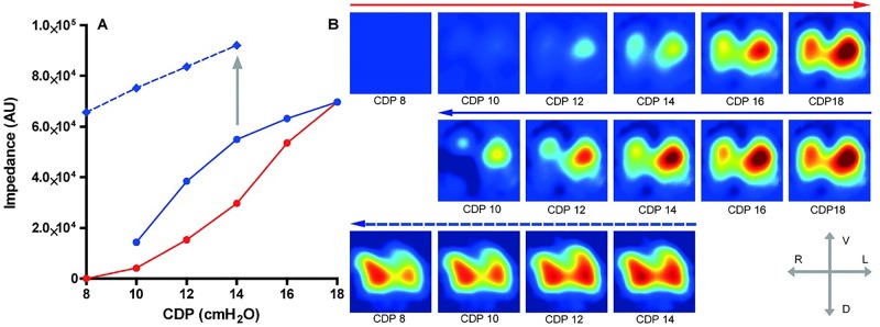
Electrical impedance tomography (EIT) has undergone 30 years of development. Functional chest examinations with this technology are considered clinically relevant, especially for monitoring regional lung ventilation in mechanically ventilated patients and for regional pulmonary function testing in patients with chronic lung diseases. As EIT becomes an established medical technology, it requires consensus examination, nomenclature, data analysis and interpretation schemes. Such consensus is needed to compare, understand and reproduce study findings from and among different research groups, to enable large clinical trials and, ultimately, routine clinical use. Recommendations of how EIT findings can be applied to generate diagnoses and impact clinical decision-making and therapy planning are required. This consensus paper was prepared by an international working group, collaborating on the clinical promotion of EIT called TRanslational EIT developmeNt stuDy group. It addresses the stated needs by providing (1) a new classification of core processes involved in chest EIT examinations and data analysis, (2) focus on clinical applications with structured reviews and outlooks (separately for adult and neonatal/paediatric patients), (3) a structured framework to categorise and understand the relationships among analysis approaches and their clinical roles, (4) consensus, unified terminology with clinical user-friendly definitions and explanations, (5) a review of all major work in thoracic EIT and (6) recommendations for future development (193 pages of online supplements systematically linked with the chief sections of the main document). We expect this information to be useful for clinicians and researchers working with EIT, as well as for industry producers of this technology.

Keywords: ARDS; Assisted Ventilation; Imaging/CT MRI etc; Paediatric Lung Disaese.


Conflict of interest statement

Competing interests: IF: Grants from The European Union's 7th Framework Programme for Research and Technological Development (WELCOME, Grant No. 611223) and from The European Union's Framework Programme for Research and Innovation Horizon 2020 (CRADL, Grant No. 668259), personal fees from Dräger, outside the submitted work. MBPA: Grants from Dixtal/Philips and Timpel SA, outside the submitted work. AHvK: non-financial support from CareFusion, outside the submitted work. DGT: Goe-MF II EIT system provided by CareFusion for unrestricted research use, two Swisstom Pioneer EIT systems and consumables fully purchased from the manufacturer, unrestricted assistance in customising research software for specific research needs by Swisstom. ZZ: Grant from the German Federal Ministry of Education and Research (MOSES, Grant No. 03FH038I3), personal fees from Dräger Medical, outside the submitted work. BG: Personal fees from Swisstom, outside the submitted work. SHB: Co-founder, employee and Chief Medical Officer of Swisstom AG, inventor of several EIT-related patents and patent applications owned by Swisstom AG and Timpel SA. ET: Employee of Dräger; several EIT-related patents pending and issued to Dräger. OS: Personal fees and non-financial support from Dräger Medical, outside the submitted work; owner of an issued patent EP 228009A1. VT: Personal fees from Timpel, outside the submitted work. DG: Personal fees from Dräger Medical, outside the submitted work. SL: Grants, personal fees and non-financial support from Dräger Medical, grants and non-financial support from Philips Research, grants and non-financial support from BMBF (private/public partnership project with Weinmann GmbH), grants and non-financial support from BMWi (public/private partnership ZIM project with Fritz Stephan GmbH), outside the submitted work; several patents pending, licensed and issued to Dräger Medical AG & Co KGaA.

Figures

Figure 1
Figure 1
Schematic presentation of the chest EIT examination and data analysis. The drawings, examples of EIT images and EIT measures illustrate the different steps involved. The images were generated using the GREIT image reconstruction algorithm from data acquired in a healthy adult subject with the Goe-MF II EIT device (CareFusion, Höchberg, Germany). (These images as well as the data shown in subsequent figures originate from examinations approved by the institutional ethics committees and acquired with written informed consent.) ARDS, acute respiratory distress syndrome; EIT, electrical impedance tomography; rel. ΔZ, relative impedance change; GI, global inhomogeneity index; CoV, centre of ventilation; PEEP, positive end-expiratory pressure; ROP, regional opening pressure; RVD, regional ventilation delay.
Figure 2
Figure 2
Electrical impedance tomography (EIT) waveforms simultaneously registered in one image pixel in the dorsal region of the dependent (left) and in another pixel in the dorsal region of the non-dependent lungs (right) in a healthy man aged 43 years lying on his right side. The raw data were obtained using the Goe-MF II EIT device (CareFusion, Höchberg, Germany) and reconstructed with the GREIT algorithm. During the first 30 s, the subject was breathing quietly, then he was instructed to hold his breath for 20 s after tidal inspiration, finally he took three deep breaths. The periodic ventilation-related changes of the EIT signal, given as relative impedance change (rel. ΔZ), are higher than those associated with the cardiac action. (The latter are best discernible during the apnoea phase.) The tidal changes in rel. ΔZ during the first and the third phases of this measurement are higher in the dependent than in the non-dependent lung reflecting the physiologically known higher ventilation of the dependent lung regions in adult subjects spontaneously breathing at the functional residual capacity level. During the apnoeic phase, the EIT signal falls continuously in the dependent lung due to local gas volume loss caused by the continuing gas exchange. This is not observed in the non-dependent lung. The changes in rel. ΔZ synchronous with the heartbeat are of comparable amplitude in both pixels. The breathing rate (BR) and heart rate (HR) in each of the three examination phases given in the lower part of the figure were derived from frequency filtering of the EIT signal.
Figure 3
Figure 3
An adult mechanically ventilated patient examined by electrical impedance tomography (EIT) (Enlight, Timpel, Sao Paulo, Brazil; with a finite element method (FEM)-based Newton Raphson image reconstruction algorithm) during a decremental positive end-expiratory pressure (PEEP) trial. The patient suffered from severe acute respiratory distress syndrome as detected in the CT scan (top left) obtained at the same chest plane where the EIT electrode belt was located during the EIT examination. The panels 1–10 show regional lung hyperdistension and collapse at each PEEP step in white and blue colours, respectively. The percentages of collapse and overdistension are provided at the right side of each panel and in the diagram (bottom left). With decreasing PEEP, hyperdistension fell (green curve) and collapse rose (blue curve). The crossover point between the curves is highlighted by the red arrow. The corresponding PEEP step preceding the crossover shows the value of 17 cm H2O in red in panel 4.
Figure 4
Figure 4
Ventilation distribution in a supine adult patient in the course of one tidal inflation during controlled and assisted mechanical ventilation in the pressure-controlled mode. The data were acquired with the Enlight device (Timpel, Sao Paulo, Brazil) using a finite element method (FEM)-based Newton Raphson reconstruction algorithm. The electrical impedance tomography (EIT) images show the impedance variation compared with the beginning of the respiratory cycle at five time points highlighted by dashed lines (A–E). In controlled ventilation, anterior and posterior regions inflated synchronously, as seen in the blue and red waveforms. In contrast, when the same patient was allowed to perform spontaneous efforts, the pendelluft phenomenon was detected by EIT with a volume shift from the anterior to the posterior regions attributed to differences in local driving forces and lung mechanics. The stronger posterior diaphragm excursion ‘sucked’ air from the anterior regions, which deflated at the beginning of inspiration, causing a transient local overdistension in the posterior regions. This excessive volume shift can be seen in the blue waveform as an overshoot (simultaneously with an undershoot in the red anterior lung waveform) before decreasing to the mechanical equilibrium with the ventilator, followed by relatively synchronous expiration. Red and blue regions in the EIT images imply a decrease and an increase in regional impedance, respectively, when compared with the beginning of inspiration.
Figure 5
Figure 5
An adult patient with a mediastinal mass admitted to the intensive care unit with severe respiratory failure and difficult mechanical ventilation (peak inspiratory pressures >40 cmH2O). The CT image suggested severe airway obstruction caused by tumour invasion, with probable obstruction of right pulmonary artery (top left). Black arrows in the top left panel show the severe obstruction of the main bronchi, more pronounced on the left. This supported the surgical plan of right-sided pneumonectomy along with tumour excision. As part of the detailed patient evaluation, an electrical impedance tomography (EIT) examination (Enlight, Timpel, Sao Paulo, Brazil; with a finite element method (FEM)-based Newton Raphson algorithm) was performed to estimate regional ventilation and perfusion. Regional EIT waveforms showed smaller ventilation amplitude in the left lung (red) in comparison to the right lung (blue). During a prolonged expiratory pause, intrinsic positive end-expiratory pressure (PEEPi) was detected in the proximal airway pressure (Pprox) signal. At the same time, an evident redistribution of air occurred: the left lung exhibited a decrease and the right lung an increase in air content. When ventilation returned, the opposite occurred, with cumulative air trapping in the left and concomitant deflation of the right lungs. A CT image, colour-coded to enhance the low lung density, demonstrated the marked air trapping in the left lung (bottom left). The functional EIT images showed the right lung to exhibit higher ventilation (bottom middle) and perfusion (bottom right) than the left lung. These findings resulted in changed surgical plans and preservation of the right lung. After the removal of the compressing mass (rhabdomyosarcoma), which was ultimately not invading the right pulmonary artery, the patient was successfully extubated 2 days later.
Figure 6
Figure 6
Electrical impedance tomography (EIT) examination of a patient aged 67 years with COPD performed during forced full expiration (Goe-MF II EIT device (CareFusion, Höchberg, Germany) with the GREIT image reconstruction). The regional EIT waveforms (bottom left) originate from four image pixels highlighted in the functional EIT ventilation image of this patient (top). The waveforms were normalised to better visualise the regional dissimilarities in lung emptying. The histogram (bottom right) shows the heterogeneity of EIT-derived pixel ratios of FEV1 and FVC. rel. ΔZ, relative impedance change.
Figure 7
Figure 7
End-expiratory lung volume changes measured by electrical impedance tomography (EIT) (Goe-MF II EIT device (CareFusion, Höchberg, Germany) with the GREIT image reconstruction) in a preterm infant (weight 800 g) with respiratory distress syndrome subjected to an oxygenation-guided lung recruitment procedure during high-frequency ventilation before and after exogenous surfactant administration. Using the impedance changes at a continuous distending pressure of 8 cmH2O as the reference value, both the inflation (solid red line) and the deflation limbs (solid blue line) before and the deflation limb (dashed blue line) after surfactant treatment (grey arrow) are mapped (A). In addition, the regional impedance changes in the cross-sectional slice of the chest during each incremental and decremental pressure step are presented as functional EIT images (B). Red colour indicates a large variation and blue a small variation in impedance. Note the presence of lung hysteresis before surfactant treatment (A) and the dissimilar regional changes in lung aeration (B). Also note the increase in lung aeration and its distribution after surfactant treatment and the stabilising effect on the deflation limb. AU, arbitrary unit; CDP, continuous distending pressure; R, right; L, left; V, ventral; D, dorsal.

References

    1. Barber D, Brown B. Applied potential tomography. J Phys E Sci Instrum 1984;17:723–33. 10.1088/0022-3735/17/9/002 - DOI
    1. Bodenstein M, David M, Markstaller K. Principles of electrical impedance tomography and its clinical application. Crit Care Med 2009;37:713–24. 10.1097/CCM.0b013e3181958d2f - DOI - PubMed
    1. Costa EL, Lima RG, Amato MB. Electrical impedance tomography. Curr Opin Crit Care 2009;15:18–24. 10.1097/MCC.0b013e3283220e8c - DOI - PubMed
    1. Frerichs I. Electrical impedance tomography (EIT) in applications related to lung and ventilation: a review of experimental and clinical activities. Physiol Meas 2000;21:R1–21. 10.1088/0967-3334/21/2/201 - DOI - PubMed
    1. Frerichs I, Becher T, Weiler N. Electrical impedance tomography imaging of the cardiopulmonary system. Curr Opin Crit Care 2014;20:323–32. 10.1097/MCC.0000000000000088 - DOI - PubMed

Publication types