. 2017 Apr 1;34(7):1291-1301.
doi: 10.1089/neu.2016.4628. Epub 2016 Oct 13.

Synaptic Mitochondria Sustain More Damage than Non-Synaptic Mitochondria after Traumatic Brain Injury and Are Protected by Cyclosporine A

Affiliations

Synaptic Mitochondria Sustain More Damage than Non-Synaptic Mitochondria after Traumatic Brain Injury and Are Protected by Cyclosporine A

Jacqueline R Kulbe et al. J Neurotrauma. .

Abstract

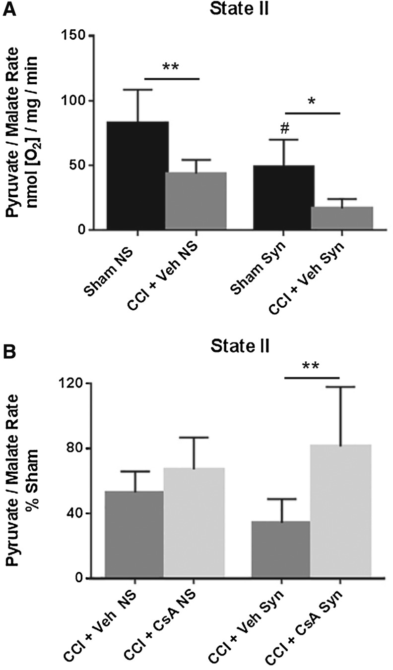
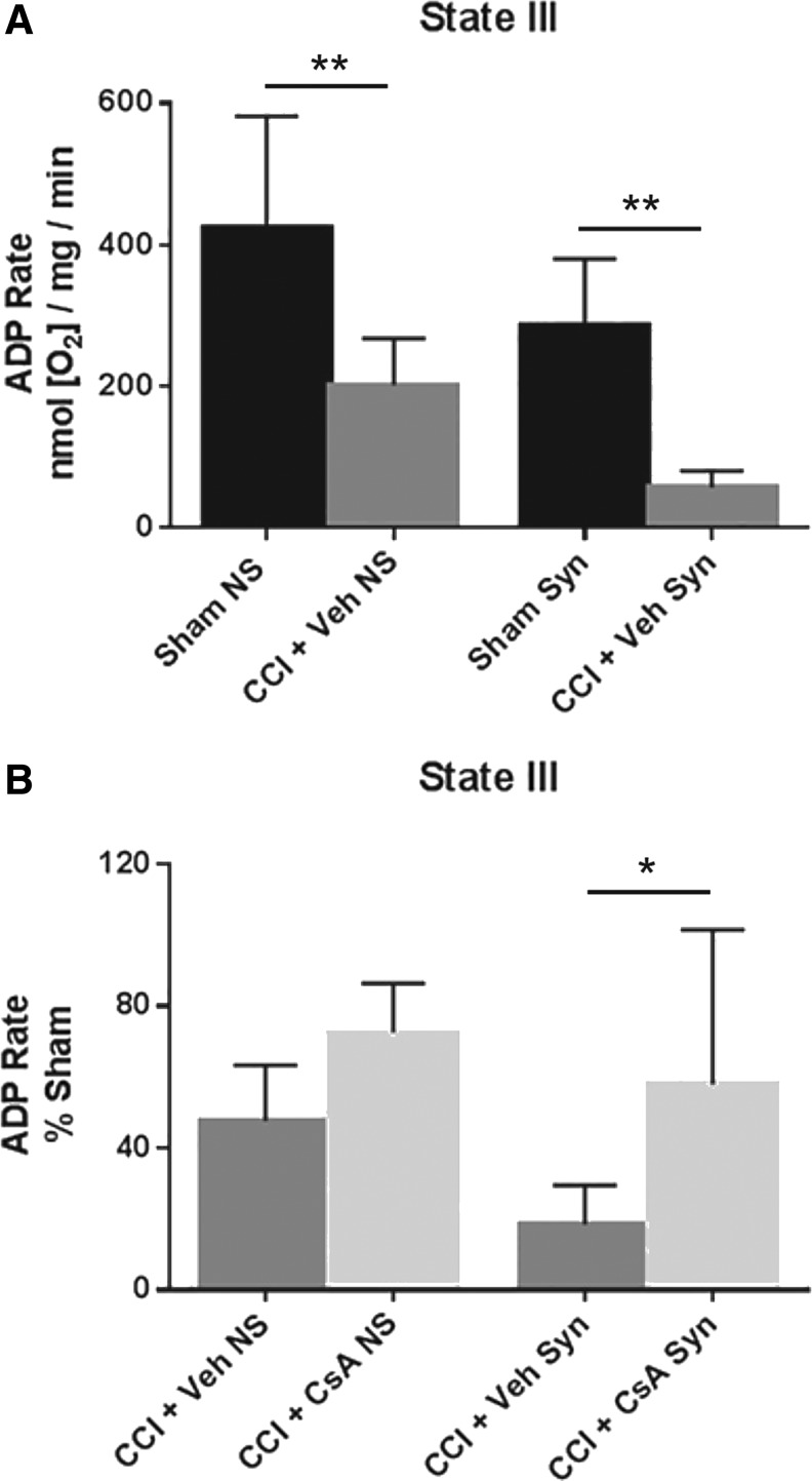
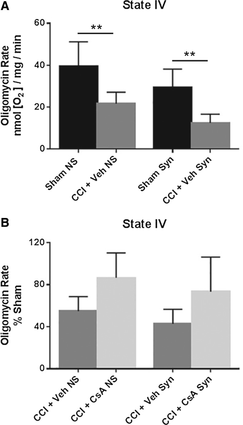
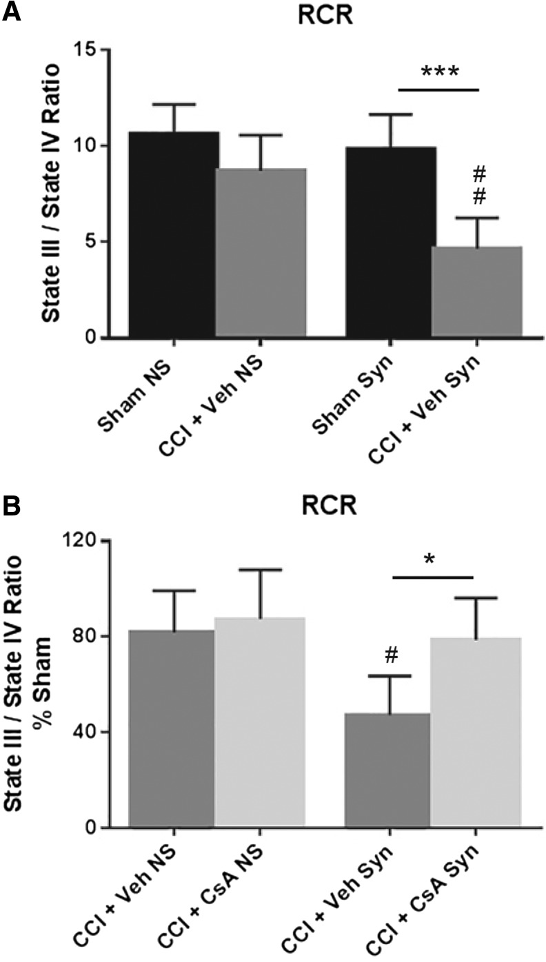
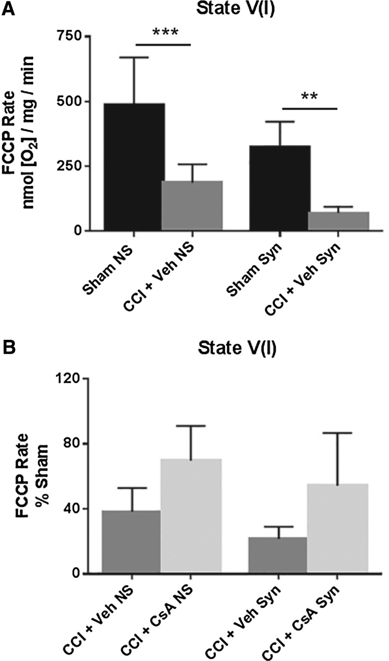
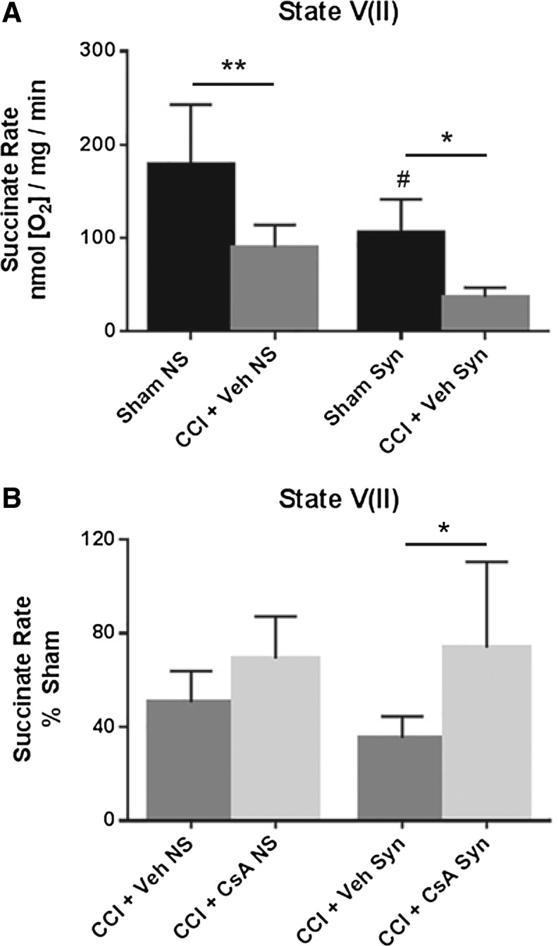
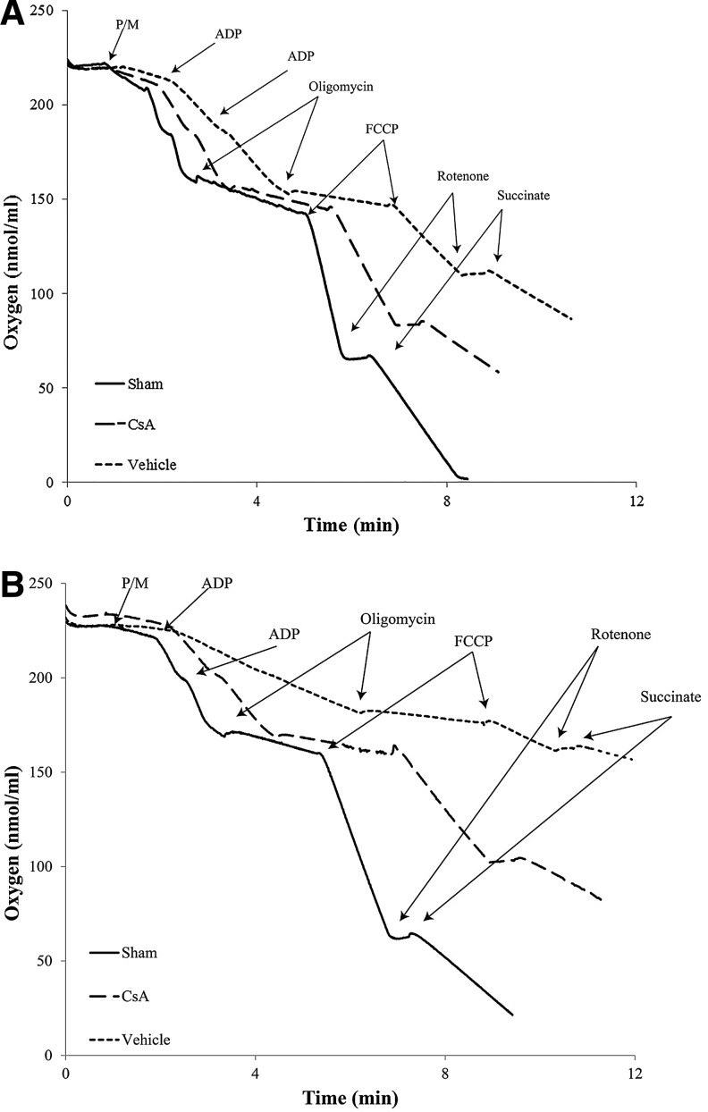
Currently, there are no Food and Drug Administration (FDA)-approved pharmacotherapies for the treatment of those with traumatic brain injury (TBI). As central mediators of the secondary injury cascade, mitochondria are promising therapeutic targets for prevention of cellular death and dysfunction after TBI. One of the most promising and extensively studied mitochondrial targeted TBI therapies is inhibition of the mitochondrial permeability transition pore (mPTP) by the FDA-approved drug, cyclosporine A (CsA). A number of studies have evaluated the effects of CsA on total brain mitochondria after TBI; however, no study has investigated the effects of CsA on isolated synaptic and non-synaptic mitochondria. Synaptic mitochondria are considered essential for proper neurotransmission and synaptic plasticity, and their dysfunction has been implicated in neurodegeneration. Synaptic and non-synaptic mitochondria have heterogeneous characteristics, but their heterogeneity can be masked in total mitochondrial (synaptic and non-synaptic) preparations. Therefore, it is essential that mitochondria targeted pharmacotherapies, such as CsA, be evaluated in both populations. This is the first study to examine the effects of CsA on isolated synaptic and non-synaptic mitochondria after experimental TBI. We conclude that synaptic mitochondria sustain more damage than non-synaptic mitochondria 24 h after severe controlled cortical impact injury (CCI), and that intraperitoneal administration of CsA (20 mg/kg) 15 min after injury improves synaptic and non-synaptic respiration, with a significant improvement being seen in the more severely impaired synaptic population. As such, CsA remains a promising neuroprotective candidate for the treatment of those with TBI.

Keywords: cyclosporine A; mitochondria; neuroprotection; synaptic and non-synaptic; traumatic brain injury.


Conflict of interest statement

No competing financial interests exist.

Figures

<b>FIG. 1.</b>
FIG. 1.
(A) Effect of injury on non-synaptic and synaptic mitochondria for state II respiration (pyruvate/malate) 24 h after severe controlled cortical impact (CCI). (B) Effect of early post-injury (15 min) intraperitoneal administration of cyclosporine A (CsA) (20 mg/kg) on non-synaptic and synaptic mitochondria for state II respiration (pyruvate/malate) 24 h after severe CCI, calculated as % sham. Sham NS, sham non-synaptic (n = 6), Sham Syn, sham synaptic (n = 6), CCI + Veh NS, CCI + vehicle non-synaptic (n = 6), CCI + Veh Syn, CCI + vehicle synaptic (n = 6), CCI + CsA NS, CCI + CsA non-synaptic (n = 8), CCI + CsA Syn, CCI + CsA synaptic (n = 8); values, mean ± standard deviation; two-way analysis of variance followed by Tukey post hoc; *p < 0.05, **p < 0.01, #p < 0.05 compared with non-synaptic sham.
<b>FIG. 2.</b>
FIG. 2.
(A) Effect of injury on non-synaptic and synaptic mitochondria for state III respiration (pyruvate/malate/adenosine diphosphate [ADP]) 24 h after severe controlled cortical impact (CCI). (B) Effect of early post-injury (15 min) intraperitoneal administration of cyclosporine A (CsA) (20 mg/kg) on non-synaptic and synaptic mitochondria for state III respiration (pyruvate/malate/ADP) 24 h after severe CCI, calculated as % sham. Sham NS, sham non-synaptic (n = 6), Sham Syn, sham synaptic (n = 6); CCI + Veh NS, CCI + vehicle non-synaptic (n = 6); CCI + Veh Syn, CCI + vehicle synaptic (n = 6); CCI + CsA NS, CCI + CsA non-synaptic (n = 8); CCI + CsA Syn, CCI + CsA synaptic (n = 8); values = mean ± standard deviation; two-way analysis of variance followed by Tukey post hoc; *p < 0.05, **p < 0.01.
<b>FIG. 3.</b>
FIG. 3.
(A) Effect of injury on non-synaptic and synaptic mitochondria for state IV respiration (oligomycin) 24 h after severe controlled cortical impact (CCI). (B) Effect of early post-injury (15 min) intraperitoneal administration of cyclosporine A (CsA) (20 mg/kg), on non-synaptic and synaptic mitochondria for state IV respiration (oligomycin) 24 h after severe CCI, calculated as % sham. Sham NS, sham non-synaptic (n = 6); Sham Syn, sham synaptic (n = 6); CCI + Veh NS, CCI + vehicle non-synaptic (n = 6); CCI + Veh Syn, CCI + vehicle synaptic (n = 6); CCI + CsA NS, CCI + CsA non-synaptic (n = 8); CCI + CsA Syn, CCI + CsA synaptic (n = 8); values = mean ± standard deviation; two-way analysis of variance followed by Tukey post hoc; **p < 0.01.
<b>FIG. 4.</b>
FIG. 4.
(A) Effect of injury on non-synaptic and synaptic mitochondria for respiratory control ratio (RCR) (state III/state IV) 24 h after severe controlled cortical impact (CCI). (B) Effect of early post-injury (15 min) intraperitoneal administration of cyclosporine A (CsA) (20 mg/kg), on non-synaptic and synaptic mitochondria for RCR (state III/state IV) 24 h after severe CCI, calculated as % sham. Sham NS, sham non-synaptic (n = 6); Sham Syn, sham synaptic (n = 6); CCI + Veh NS, CCI + vehicle non-synaptic (n = 6); CCI + Veh Syn, CCI + vehicle synaptic (n = 6); CCI + CsA NS, CCI + CsA non-synaptic (n = 8); CCI + CsA Syn, CCI + CsA synaptic (n = 8); values = mean ± standard deviation; two-way analysis of variance followed by Tukey post hoc; *p < 0.05, **p < 0.001, #p < 0.05 vs non-synaptic vehicle, ##p < 0.01 vs. non-synaptic vehicle.
<b>FIG. 5.</b>
FIG. 5.
(A) Effect of injury on non-synaptic and synaptic mitochondria for state V(I) respiration (FCCP) 24 h after severe controlled cortical impact (CCI). (B) Effect of early post-injury (15 min) intraperitoneal administration of cyclosporine A (CsA) (20 mg/kg) on non-synaptic and synaptic mitochondria for state V(I) respiration (FCCP) 24 h after severe CCI, calculated as % sham. Sham NS, sham non-synaptic (n = 6); Sham Syn, sham synaptic (n = 6); CCI + Veh NS, CCI + vehicle non-synaptic (n = 6); CCI + Veh Syn, CCI + vehicle synaptic (n = 6); CCI + CsA NS, CCI + CsA non-synaptic (n = 8); CCI + CsA Syn, CCI + CsA synaptic (n = 8); values = mean ± standard deviation; two-way analysis of variance followed by Tukey post hoc; **p < 0.01, ***p < 0.001.
<b>FIG. 6.</b>
FIG. 6.
(A) Effect of injury on non-synaptic and synaptic mitochondria for state V(II) respiration (rotenone/succinate) 24 h after severe controlled cortical impact (CCI). (B) Effect of early post-injury (15 min) intraperitoneal administration of CsA (20 mg/kg) on non-synaptic and synaptic mitochondria for state V(II) respiration (rotenone/succinate) 24 h after severe CCI, calculated as % sham. Sham NS, sham non-synaptic (n = 6); Sham Syn, sham synaptic (n = 6); CCI + Veh NS, CCI + vehicle non-synaptic (n = 6); CCI + Veh Syn, CCI + vehicle synaptic (n = 6); CCI + CsA NS, CCI + CsA non-synaptic (n = 8); CCI + CsA Syn, CCI + CsA synaptic (n = 8); values = mean ± standard deviation; two-way analysis of variance followed by Tukey post hoc; *p < 0.05, **p < 0.01, #p < 0.05 vs. non-synaptic sham.
<b>FIG. 7.</b>
FIG. 7.
Representative oxymetric traces indicating rates of oxygen consumption of sham, vehicle, and cyclosporine A (CsA) (20 mg/kg, intraperitoneally, 15 min post-injury) for (A) non-synaptic and (B) synaptic mitochondria isolated from ipsilateral cortex 24 h after severe controlled cortical impact (CCI). Purified mitochondrial protein (>30 μg) was suspended in respiration buffer (125 mmol/L KCl, 2 mmol/L MgCl2, 2.5 mmol/L KH2PO4, 0.1% BSA, 20 mmol/L HEPES, pH 7.2) in a final volume of 250 μl, and oxygen consumption rates were measured using a Clark-type oxygen electrode in the presence of 5 mmol/L pyruvate and 2.5 mmol/L malate (state II), two boluses of 150 μmol/L ATP (state III), 2 μmol/L oligomycin (state IV), 2 μmol/L FCCP (state VI), and 100 nmol/L rotenone and 10 mmol/L succinate (state VII). Sham, sham; CsA, CCI + CsA; vehicle, CCI + vehicle; ADP, adenosine diphosphate.

Similar articles

Cited by

References

    1. Thurman D.J., Alverson C., Dunn K.A., Guerrero J., and Sniezek J.E. (1999). Traumatic brain injury in the United States: a public health perspective. J. Head Trauma Rehabil. 14, 602–615 - PubMed
    1. Finkelstein E.A, Corso P.S., and Miller T.R. (2006). The Incidence and Economic Burden of Injuries in the United States. Oxford University Press: New York
    1. Coronado V.G., McGuire L.C., Faul M.F., Sugerman D.E., and Pearson W.S. (2012). Traumatic brain injury epidemiology and public health issues. in: Brain Injury Medicine: Principles and Practice. Zasler N.D., Katz D.I., and Zafonte R.D. (eds). Demos Medical Publishing: New York, pps. 84–100
    1. Maas A.I., Stocchetti N., and Bullock R. (2008). Moderate and severe traumatic brain injury in adults. Lancet Neurol. 7, 728–741 - PubMed
    1. Faden A.I., Demediuk P., Panter S.S., and Vink R. (1989). The role of excitatory amino acids and NMDA receptors in traumatic brain injury. Science 244, 798–800 - PubMed

Publication types

MeSH terms