. 2016 Sep 6:6:32977.
doi: 10.1038/srep32977.

Gamma radiation at a human relevant low dose rate is genotoxic in mice

Affiliations

Gamma radiation at a human relevant low dose rate is genotoxic in mice

Anne Graupner et al. Sci Rep. .

Abstract

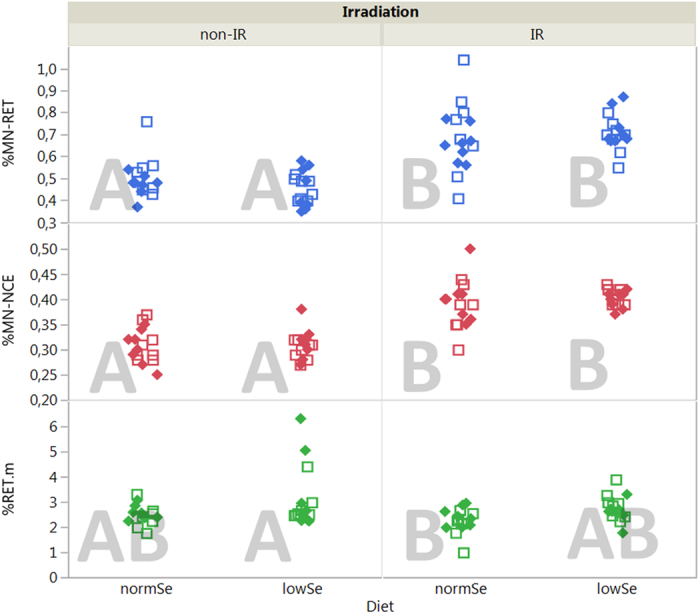
Even today, 70 years after Hiroshima and accidents like in Chernobyl and Fukushima, we still have limited knowledge about the health effects of low dose rate (LDR) radiation. Despite their human relevance after occupational and accidental exposure, only few animal studies on the genotoxic effects of chronic LDR radiation have been performed. Selenium (Se) is involved in oxidative stress defence, protecting DNA and other biomolecules from reactive oxygen species (ROS). It is hypothesised that Se deficiency, as it occurs in several parts of the world, may aggravate harmful effects of ROS-inducing stressors such as ionising radiation. We performed a study in the newly established LDR-facility Figaro on the combined effects of Se deprivation and LDR γ exposure in DNA repair knockout mice (Ogg1(-/-)) and control animals (Ogg1(+/-)). Genotoxic effects were seen after continuous radiation (1.4 mGy/h) for 45 days. Chromosomal damage (micronucleus), phenotypic mutations (Pig-a gene mutation of RBC(CD24-)) and DNA lesions (single strand breaks/alkali labile sites) were significantly increased in blood cells of irradiated animals, covering three types of genotoxic activity. This study demonstrates that chronic LDR γ radiation is genotoxic in an exposure scenario realistic for humans, supporting the hypothesis that even LDR γ radiation may induce cancer.


Conflict of interest statement

S.D.D. is an employee of Litron Laboratories; Litron holds patents covering flow cytometric methods for scoring GPI anchor-deficient erythrocytes and sells kits based on this technology (In Vivo MutaFlow); Litron holds patents covering flow cytometric methods for scoring micronucleated erythrocytes and sells kits based on this technology (In Vivo MicroFlow).

Figures

Figure 1
Figure 1. Differences of Least Square Means (LSM) of untransformed data between groups after AIC.
cf. Statistical analysis in the method section. P-values for the factors irradiation and diet and their interaction (irradiation*diet) are given in brackets (in bold for p < 0.05). LSM are indicated in red for endpoints where irradiation or low Se diet had a stronger effect; LSM are indicated in green for endpoints where non-irradiation or normal Se diet had a stronger effect. aDifference of LSM between irradiated and non-irradiated mice (LSMIR – LSMnonIR). Positive values indicate a higher response in the investigated endpoint in the irradiated groups compared with the non-irradiated groups. bDifference of LSM between low and adequate Se diet (LSMlowSe – LSMnormSe). Positive values indicate a higher response in the investigated endpoint in the groups fed with low Se diet compared with groups fed with normal Se diet. cSingle strand breaks and alkali labile sites. dFpg-sensitive sites, i.e. oxidized DNA lesions.
Figure 2
Figure 2. Micronucleus assay.
Upper panel: Mean percentage of micronucleated blood reticulocytes (% MN-RET). Middle panel: micronucleated normochromic erythrocytes (% MN-NCE). Lower panel: percentage reticulocytes (% RET) of unexposed (non-IR) and chronic LDR irradiated (IR) mice given two different diets (normSe and lowSe). Solid diamonds (Ogg1+/−) and hollow squares (Ogg1−/−) represent individuals (8 mice per group). Similar letters indicate that there is no significant difference between groups (Tukey’s HSD).
Figure 3
Figure 3. Pig-a gene mutation assay.
Mutant phenotype frequencies of RETCD24− (upper panel), RBCCD24− (middle panel) and % RET (lower panel) of unexposed (non-IR) and chronic LDR irradiated (IR) mice given two different diets (normSe and lowSe). Solid diamonds (Ogg1+/−) and hollow squares (Ogg1−/−) represent individuals (6–8 mice per group). Similar letters indicate that there is no significant difference between groups (Tukey’s HSD).
Figure 4
Figure 4. SCGE assay, ssb/als.
Single strand breaks and alkali labile sites (ssb/als) detected by the alkaline SCGE assay in whole blood of mice after 45 days (upper panel) of chronic LDR γ radiation and at day 90, i.e. after 45 days recovery (lower panel). Solid diamonds (Ogg1+/−) and hollow squares (Ogg1−/−) represent individuals (8 mice per group). Similar letters indicate that there is no significant difference between groups (Tukey’s HSD).
Figure 5
Figure 5. SCGE assay, oxidised DNA lesions.
Fpg-sensitive sites (Fpg-ss) detected by the alkaline SCGE assay in whole blood of mice for each time point and genotype separately. Panel A: Ogg1+/− at day 45; Panel B: Ogg1+/− at day 90; Panel C: Ogg1−/− at day 45; Panel D: Ogg1−/− at day 90. Solid diamonds (Ogg1+/−) and hollow squares (Ogg1−/−) represent individuals (4–8 mice per group). Similar letters indicate that there is no significant difference between groups (Tukey’s HSD).
Figure 6
Figure 6. Study design with respect to irradiation and endpoint analysis.
The mice were continuously exposed to LDR γ radiation for 45 days. At day 45, blood samples for MN assay and SCGE were taken from 64 mice before killing and harvest of organs. The remaining 64 mice were kept for another 45 days without irradiation (recovery). Blood samples for Pig-a gene mutation assay and SCGE were taken on day 59 and day 90, respectively. These mice were killed at day 90 and organs were harvested.

Similar articles

Cited by

References

    1. Suzuki K. & Yamashita S. Low-dose radiation exposure and carcinogenesis. Japanese journal of clinical oncology 42, 563–568 (2012). - PubMed
    1. Scott B. R. It’s time for a new low-dose-radiation risk assessment paradigm--one that acknowledges hormesis. Dose-response: a publication of International Hormesis Society 6, 333–351 (2008). - PMC - PubMed
    1. UNSCEAR. Report of the United Nations Scientific Committee on the Effects of Atomic Radiation 2010: 57th session: summary of low-dose radiation effects on health. (2010).
    1. UNSCEAR. Sources, effects and risks of ionizing radiation. Annex A: Levels and effects of radiation exposure due to the nuclear accident after the 2011 great east-Japan earthquake and tsunami. (United Nations, New York, 2014).
    1. Olipitz W. et al.. Integrated molecular analysis indicates undetectable change in DNA damage in mice after continuous irradiation at ~400-fold natural background radiation. Environmental health perspectives 120, 1130–1136 (2012). - PMC - PubMed

Publication types

LinkOut - more resources