. 2016 Sep 5;12(1):182.
doi: 10.1186/s12917-016-0821-6.

Normal computed tomographic features and reference values for the coelomic cavity in pet parrots

Affiliations

Normal computed tomographic features and reference values for the coelomic cavity in pet parrots

Irene A Veladiano et al. BMC Vet Res. .

Abstract

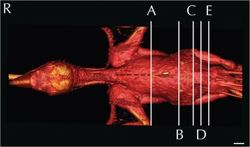
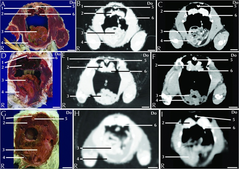
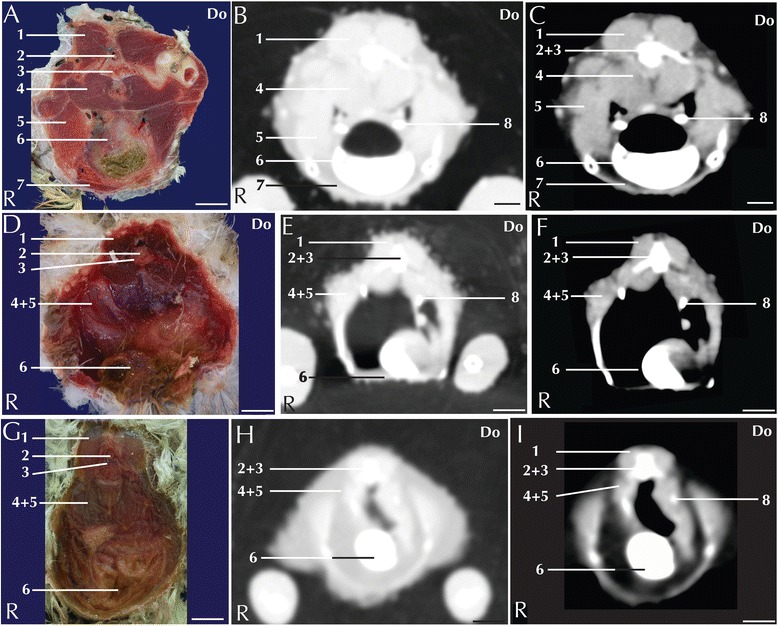
Background: The increasing popularity gained by pet birds over recent decades has highlighted the role of avian medicine and surgery in the global veterinary scenario; such a need for speciality avian medical practice reflects the rising expectation for high-standard diagnostic imaging procedures. The aim of this study is to provide an atlas of matched anatomical cross-sections and contrast-enhanced CT images of the coelomic cavity in three highly diffused psittacine species.

Results: Contrast-enhanced computed tomographic studies of the coelomic cavity were performed in 5 blue-and-gold macaws, 4 African grey parrots and 6 monk parakeets by means of a 4-multidetector-row CT scanner. Both pre- and post-contrast scans were acquired. Anatomical reference cross-sections were obtained from 5 blue-and-gold macaw, 7 African grey parrot, and 9 monk parakeet cadavers. The specimens were stored in a -20 °C freezer until completely frozen and then sliced at 5-mm intervals by means of a band saw. All the slices were photographed on both sides. Individual anatomical structures were identified by means of the available literature. Pre- and post-contrast attenuation reference values for the main coelomic organs are reported in Hounsfield units (HU).

Conclusions: The results provide an atlas of matched anatomical cross-sections and contrast-enhanced CT images of the coelomic cavity in three highly diffused psittacine species.

Keywords: African grey parrot; Blue-and-gold macaw; Coelomic cavity; Computed tomography; Monk parakeet.


Figures

Fig. 1
Fig. 1
Three-dimensional reconstruction of the body of a blue-and-gold macaw. Lines A-E indicate the level of the matched cross-sections and CT images displayed in Figs. 2, 3, 4, 5, 6 and 7
Fig. 2
Fig. 2
Matched cross-sections and CT images at the level of the heart corresponding to line A in Fig. 1. Matched cross-sections and CT images of blue-and-gold macaw (a-c), African grey parrot (d-f) and monk parakeet (g-i). The CT images have been reconstructed with a soft tissue kernel and displayed in pulmonary (b-e-h) and abdomen (c-f-i) window. Do is dorsal and R are right. Bar = 1 cm. 1. Lung; 2. Scapulohumeralis muscle; 3. Intrapulmonary primary bronchus; 4. Scapulohumeral caudal muscle; 5. Right atrium; 6. Pectoral muscle (thoracobrachialis portion); 7. Right ventricle; 8. Pectoral muscle (sternobrachialis portion); 9. Vertebra; 10. Spinal cord; 11. Aorta; 12. Oesophagus; 13. Pulmonary trunk; 14. Cranial thoracic air sac; 15. Carina sterni; 16. Cardiac muscle, left ventricle; 17. Supracoracoid muscle
Fig. 3
Fig. 3
Matched cross-sections and CT images at the level of the liver corresponding to line B in Fig. 1. Matched cross-sections and CT images of blue-and-gold macaw (a-c), African grey parrot (d-f) and monk parakeet (g-i). The CT images have been reconstructed with a soft tissue kernel and displayed in pulmonary (b-e-h) and abdomen (c-f-i) window. Do is dorsal and R are right. Bar = 1 cm. 1. Vertebra; 2. Kidney (cranial lobe); 3. Common iliac vein; 4. Spleen; 5. Caudal vena cava; 6. Liver, right lobe; 7. Carina Sterni; 8. Longissimus dorsi muscle; 9. Spinal cord; 10. Gonads; 11. Air sac; 12. Proventriculus; 13. Liver, medial part of the left lobe; 14. Liver, lateral part of the left lobe; 15. Pectoral muscle; 16. Supracoracoid muscle
Fig. 4
Fig. 4
Matched cross-sections and CT images at the level of the ventriculus corresponding to line C in Fig. 1. Matched cross-sections and CT images of blue-and-gold macaw (a-c), African grey parrot (d-f) and monk parakeet (g-i). The CT images have been reconstructed with a soft tissue kernel and displayed in pulmonary (b-e-h) and abdomen (c-f-i) window. Do is dorsal and R are right. Bar = 1 cm. 1. Spinal cord; 2. Medial lobe of the right kidney; 3. Air sac wall; 4. Small intestine; 5. Pancreas. 6. Ingesta; 7. Liver; 8. Vertebra; 9. Left ureter; 10. Ventriculus, muscular wall
Fig. 5
Fig. 5
Matched cross-sections and CT images at the level of the kidneys corresponding to line D in Fig. 1. Matched cross-sections and CT images of blue-and-gold macaw (a-c), African grey parrot (d-f) and monk parakeet (g-i). The CT images have been reconstructed with a soft tissue kernel and displayed in pulmonary (b-e-h) and abdomen (c-f-i) window. Do is dorsal and R are right. Bar = 1 cm. 1. Vertebra; 2. Medial lobe of the right kidney; 3. Intestine; 4. Pectoral muscle (thoracic portion); 5. Spinal cord; 6. Left ureter
Fig. 6
Fig. 6
Matched cross-sections and CT images at the level of the cloaca corresponding to line E in Fig. 1. Matched cross-sections and CT images of blue-and-gold macaw (a-c), African grey parrot (d-f) and monk paraket (g-i). The CT images have been reconstructed with a soft tissue kernel and displayed in pulmonary (b-e-h) and abdomen (c-f-i) window. Do is dorsal and R are right. Bar = 1 cm. 1. Musculus levator caudae; 2. Spinal cord; 3. Vertebra; 4. Musculus levator cloacae; 5. Musculus transversus cloacae; 6. Cloaca; 7. Pectoral muscle (abdominal portion); 8. Left ureter
Fig. 7
Fig. 7
Matched cross-sections and CT images at the level of the gonads corresponding to line E in Fig. 1. Matched cross-sections and CT images of female blue-and-gold macaw (a-c), and male blue-and-gold macaw (d-f). The CT images have been reconstructed with a soft tissue kernel and displayed in pulmonary (b-e) and abdomen (c-f) window. Do is dorsal and R are right. Bar = 1 cm. 1. Vertebra; 2. Spinal cord; 3. Kidney; 4. Ovary; 5. Spleen; 6. Liver; 7. Pectoral muscle; 8. Longissimus dorsi muscle; 9. Proventriculus; 10. Coracoid bone; 11. Testicles

Similar articles

Cited by

References

    1. Groskin R. Prologue. In: Ritchie BW, Harrison GJ, Harrison LR, editors. Avian medicine: principle and application. 1. Lake Worth: Wingers publishing inc; 1994. pp. 17–25.
    1. Raftery A. The initial presentation: triage and critical care. In: Harcourt-Brown N, Chitty J, editors. BSAVA Manual of Psittacine Birds. 2. Gloucester: British small animal veterinary association; 2005. pp. 35–49.
    1. Alonso-Farré JM, Gonzalo-Orden M, Barreiro-Vázquez JD, Ajenjio JM, Barreiro-Lois A, Llarena-Reino M, Degollada E. Cross-sectional anatomy, computed tomography and magnetic resonance imaging of the thoracic region of common dolphin (Delphinus delphis) and striped dolphin (Stenella coeruleoalba) Anat Histol Embryol. 2014;43(3):221–229. doi: 10.1111/ahe.12065. - DOI - PubMed
    1. Endo H, Sasaki H, Hayashi Y, Petrov EA, Amano M, Suzuki N, Miyazaki N. CT examination of the head of the Baikal seal (Phoca sibirica) J Anat. 1999;194(1):119–126. doi: 10.1046/j.1469-7580.1999.19410119.x. - DOI - PMC - PubMed
    1. Zotti A, Banzato T, Cozzi B. Cross-sectional anatomy of the rabbit neck and trunk: Comparison of computed tomography and cadaver anatomy. Res Vet Sci. 2009;87(2):171–176. doi: 10.1016/j.rvsc.2009.02.003. - DOI - PubMed