. 2017 Mar;76(3):612-619.
doi: 10.1136/annrheumdis-2016-209424. Epub 2016 Sep 5.

Treatment of inflammatory arthritis via targeting of tristetraprolin, a master regulator of pro-inflammatory gene expression

Affiliations

Treatment of inflammatory arthritis via targeting of tristetraprolin, a master regulator of pro-inflammatory gene expression

E A Ross et al. Ann Rheum Dis. 2017 Mar.

Abstract

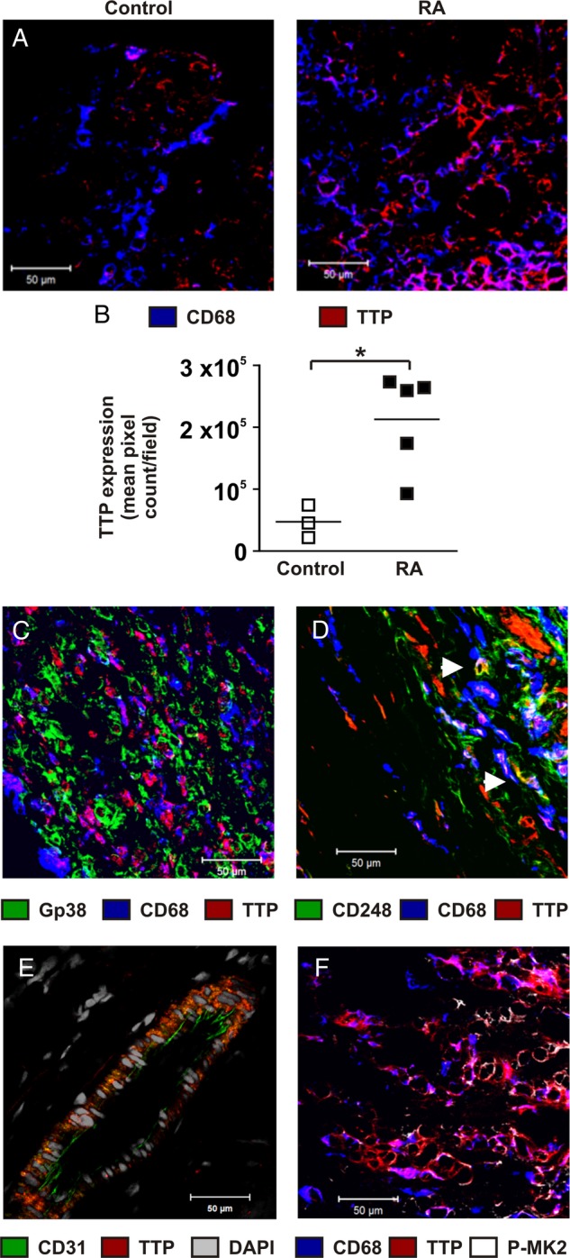
Objectives: Tristetraprolin (TTP), a negative regulator of many pro-inflammatory genes, is strongly expressed in rheumatoid synovial cells. The mitogen-activated protein kinase (MAPK) p38 pathway mediates the inactivation of TTP via phosphorylation of two serine residues. We wished to test the hypothesis that these phosphorylations contribute to the development of inflammatory arthritis, and that, conversely, joint inflammation may be inhibited by promoting the dephosphorylation and activation of TTP.

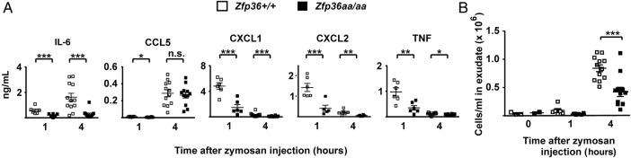
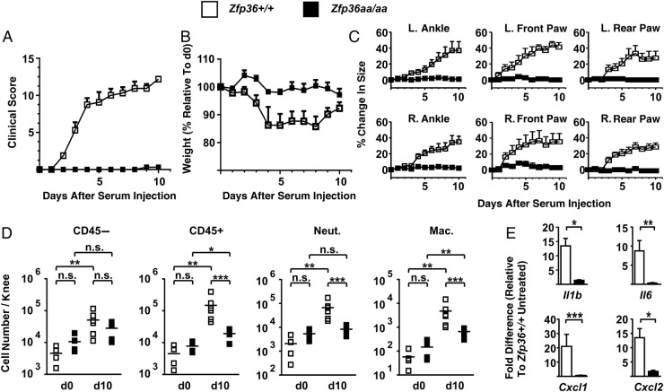
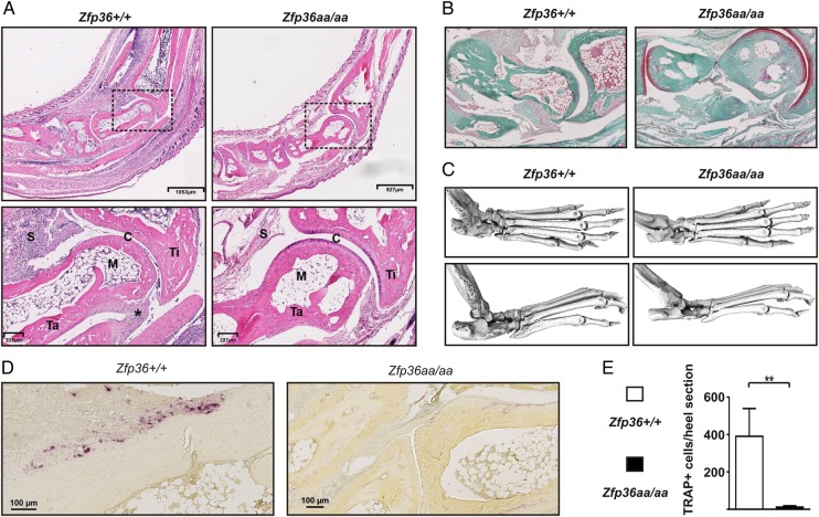
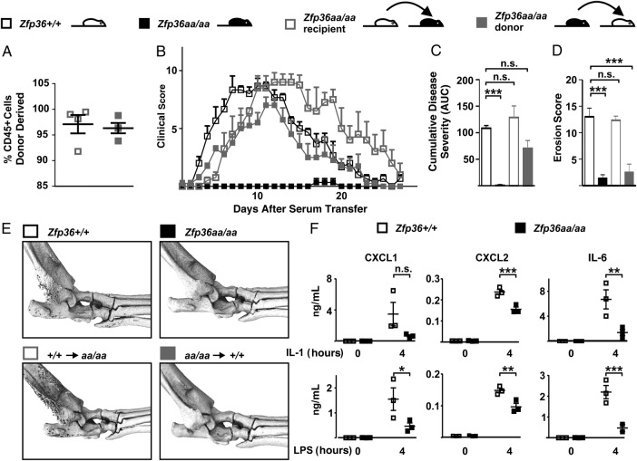
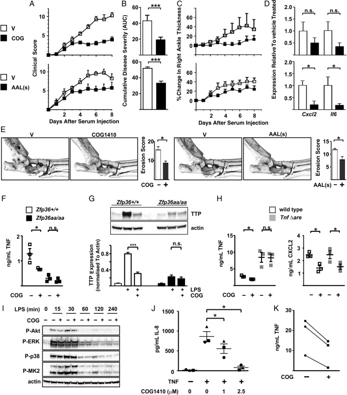
Methods: The expression of TTP and its relationship with MAPK p38 activity were examined in non-inflamed and rheumatoid arthritis (RA) synovial tissue. Experimental arthritis was induced in a genetically modified mouse strain, in which endogenous TTP cannot be phosphorylated and inactivated. In vitro and in vivo experiments were performed to test anti-inflammatory effects of compounds that activate the protein phosphatase 2A (PP2A) and promote dephosphorylation of TTP.

Results: TTP expression was significantly higher in RA than non-inflamed synovium, detected in macrophages, vascular endothelial cells and some fibroblasts and co-localised with MAPK p38 activation. Substitution of TTP phosphorylation sites conferred dramatic protection against inflammatory arthritis in mice. Two distinct PP2A agonists also reduced inflammation and prevented bone erosion. In vitro anti-inflammatory effects of PP2A agonism were mediated by TTP activation.

Conclusions: The phosphorylation state of TTP is a critical determinant of inflammatory responses, and a tractable target for novel anti-inflammatory treatments.

Keywords: Cytokines; Fibroblasts; Inflammation; Rheumatoid Arthritis; TNF-alpha.


Conflict of interest statement

Competing interests: MPV is a founder, owner and employee of Cognosci, Inc., which holds patents on apoE-mimetics including COG1410.

Figures

Figure 1
Figure 1
Expression of tristetraprolin (TTP) in the rheumatoid synovium. Biopsies from non-inflamed (A, left) or rheumatoid arthritis (RA) synovium (A, right, C-F) were stained using antibodies with the specificities indicated, and with DAPI (4'6-diamidino-2'-phenylindole) nuclear stain. The nuclear stain is shown only in (D), to identify the topology of the vessel wall. Antibody isotype-matched control stains were essentially blank (see online supplementary figure S1). (B) Sections of non-inflamed or RA synovial tissue were stained with antibody against TTP, and staining was quantified using ImageJ, by pixel counting of three fields per section, selected and scanned in blinded manner. *p<0.05 (Mann-Whitney U test).
Figure 2
Figure 2
Zfp36aa/aa mice are protected from zymosan-induced local inflammation. Dorsal air pouches were created and localised inflammation was induced by injection of 7 μL of 1 mg/mL zymosan. After 1 or 4 hours mice were humanely culled. (A) Cytokines and chemokines were measured in air pouch exudate fluid. Graphs represent means±SEM from 6 mice (1 hour) or 12 mice (4 hours) of each genotype. Basal expression of all cytokines and chemokines was negligible in untreated mice (data not shown). (B) Cells in exudate fluid were counted using a haemocytometer. n.s., not statistically significant; *p<0.05; **p<0.01; ***p<0.005 (Mann-Whitney U test).
Figure 3
Figure 3
Zfp36aa/aa mice are resistant to experimental arthritis. Zfp36+/+ and Zfp36aa/aa mice were injected intraperitoneal with 75 μL of arthritogenic serum from K/BxN donor mice on days 0 and 1. (A) Clinical score and (B) weight were monitored over 10 days. (C) Ankle and foot pad thickness were measured. Graphs represent mean±SEM (n=6). (D) Feet were digested at d0 or d10, CD45− and CD45+ cells, neutrophils and macrophages were counted using flow cytometry. Graphs represent mean±SEM (n=6). n.s., not statistically significant; *p<0.05; **p<0.01; ***p<0.005 (Mann-Whitney U test). (E) At d10, RNA was recovered from digested wrist joints of three mice of each genotype, and selected cytokine and chemokine mRNAs were measured by quantitative PCR, with normalisation first against Gapdh then against paws of vehicle-treated, uninflamed Zfp36+/+ mice. Graphs represent mean±SEM (n=3). *p<0.05; **p<0.01; ***p<0.005 (Mann-Whitney U test).
Figure 4
Figure 4
Zfp36aa/aa mice are resistant to experimental arthritis. Experimental arthritis was induced as in figure 3. (A) Representative H&E stained sagittal sections of Zfp36+/+ and Zfp36aa/aa heel regions at d10. The inset boxes in upper panels are shown at higher resolution in lower panels. C, cartilage; S, synovium; M, bone marrow; Ta, talus; Ti, tibia; *, enthesitis. (B) Similar sections were stained with fast green and Safranin O to identify articular cartilage (red stain). Representative images are shown. (C) Whole feet were subjected to μCT. Representative images are shown from top and side. (D) Activated osteoclasts were visualised by staining for tartrate-resistant acid phosphatase (TRAP; dark red stain) and (E) TRAP-positive cells per heel section were counted. The graph represents mean±SEM (n=6). **p<0.01 (Mann-Whitney U test).
Figure 5
Figure 5
Enhanced tristetraprolin (TTP) function in both haematopoietic and non-haematopoietic compartments is required for protection against experimental arthritis. Bone marrow chimaeras were created between Zfp36+/+ and Zfp36aa/aa mice. (A) 8 weeks after reconstitution of lethally irradiated recipient mice, chimerism of haematopoietic cells was assessed by flow cytometry of CD45.1 and CD45.2 markers. (B) Arthritis was induced as in figure 3, and clinical scores were monitored over 26 days. (C) Cumulative disease severity was calculated for each individual mouse as area under curve (AUC) of the plot of clinical score against time. Graph represents mean±SEM (n=3 for Zfp36+/+ and Zfp36aa/aa, n=4 for chimaeras. n.s., not statistically significant; ***p<0.005. (D) Bone erosion was quantified in Zfp36+/+, Zfp36aa/aa and chimeric mice. (E) Representative μCT images of mice at d26. (F) Joints of healthy Zfp36+/+ and Zfp36aa/aa mice were digested and adherent cells passaged three times, after which fewer than 2% of cells were CD45+. Cells were then left untreated or stimulated with 10 ng/mL interleukin (IL)-1β (upper) or LPS (lower) for 4 hours. Secreted CXCL1, CXCL2 and IL-6 were measured. Graphs represent mean±SEM (n=3). *p<0.05; **p<0.01; ***p<0.005 (Mann-Whitney U test).
Figure 6
Figure 6
Therapeutic targeting of tristetraprolin (TTP) phosphorylation. Arthritis was induced in wild-type mice by injection of K/BxN serum as in figure 3. From d2 mice were treated daily with vehicle or COG1410 (upper panels), vehicle or AAL(s) (lower panels). Control mice were injected with vehicle (dimethyl sulfoxide in the case of AAL(s), phosphate-buffered saline in the case of COG1410. (A) Clinical score was monitored over 8 days. (B) Cumulative disease severity was calculated as in figure 5. (C) Right ankle thickness was measured. (D) At the end of the experiment, RNA was isolated from wrist joints, Cxcl2 and Il6 mRNAs were measured by quantitative PCR with normalisation first against Gapdh then against vehicle-treated control. Graphs represent means±SEM of four mice in each treatment group. n.s., not significant; *p<0.05; ***p<0.005 (Mann-Whitney U test). (E) Representative μCT images of ankles of mice treated to day 8 with vehicle, COG1410 or AAL(s). Bone erosion was scored in blinded manner by three independent observers. Graphs show means±SEM. *p<0.05 (Mann-Whitney U test). (F) Zfp36+/+ and Zfp36aa/aa BMMs were treated with LPS for 8 hours with or without 10 μM COG1410 (COG). Secreted tumour necrosis factor (TNF) was measured by ELISA. Graph represents mean±SEM from three independent BMM isolates of each genotype. n.s., not significant; *p<0.05 (Mann-Whitney U test). (G) Whole cell lysates were prepared from Zfp36+/+ and Zfp36aa/aa BMMs treated as indicated, and TTP detected by western blotting. Protein abundance was estimated by scanning densitometry, with normalisation against the loading control actin. The graph represents mean±SEM from three independent experiments. n.s., not significant; ***p<0.005 (Mann-Whitney U test). (H) Litter-mate control and TnfΔARE BMMs were treated with LPS for 8 hours with or without 10 μM COG1410 (COG). Secreted TNF and CXCL2 were measured by ELISA. Graphs represents mean±SEM from three independent BMM isolates of each genotype. n.s., not significant; *p<0.05 (Mann-Whitney U test). (I) Zfp36+/+ BMMs were treated with LPS for 0–4 hours with or without 10 μM COG1410 (COG). Whole cell lysates were western blotted for phosphorylated forms of Akt, ERK, mitogen-activated protein kinase (MAPK) p38 and MK2, and for actin as a loading control. Representative of two independent experiments. (J) Rheumatoid arthritis (RA) synovial fibroblasts were stimulated for 8 hours with 1 ng/mL TNF in the presence of 0, 1 or 2.5 μg/mL COG1410. Secreted IL-8 was measured by ELISA. Circles, squares and triangles represent independent RA synovial fibroblast cultures derived from three different patients. Each individual symbol represents the mean of triplicate measurements. n.s., not significant; *p<0.05 (two-way analysis of variance). (K) Monocyte-derived macrophages were isolated from three volunteers and stimulated with LPS for 8 hours with or without 10 μM COG1410 (COG). Secreted TNF was measured by ELISA.

Comment in

Similar articles

Cited by

References

    1. Clark AR, Dean JL. The p38 MAPK pathway in rheumatoid arthritis: a sideways look. Open Rheumatol J 2012;6:209–19. 10.2174/1874312901206010209 - DOI - PMC - PubMed
    1. Arthur JS, Ley SC. Mitogen-activated protein kinases in innate immunity. Nat Rev Immunol 2013;13:679–92. 10.1038/nri3495 - DOI - PubMed
    1. Schett G, Zwerina J, Firestein G. The p38 mitogen-activated protein kinase (MAPK) pathway in rheumatoid arthritis. Ann Rheum Dis 2008;67:909–16. 10.1136/ard.2007.074278 - DOI - PMC - PubMed
    1. Lee JC, Laydon JT, McDonnell PC, et al. . A protein kinase involved in the regulation of inflammatory cytokine biosynthesis. Nature 1994;372:739–46. 10.1038/372739a0 - DOI - PubMed
    1. Saklatvala J. The p38 MAP kinase pathway as a therapeutic target in inflammatory disease. Curr Opin Pharmacol 2004;4:372–7. 10.1016/j.coph.2004.03.009 - DOI - PubMed

MeSH terms