. 2016 Nov 1;473(21):3997-4009.
doi: 10.1042/BCJ20160696. Epub 2016 Sep 5.

The HemQ coprohaem decarboxylase generates reactive oxygen species: implications for the evolution of classical haem biosynthesis

Affiliations

The HemQ coprohaem decarboxylase generates reactive oxygen species: implications for the evolution of classical haem biosynthesis

Charlie Hobbs et al. Biochem J. .

Abstract

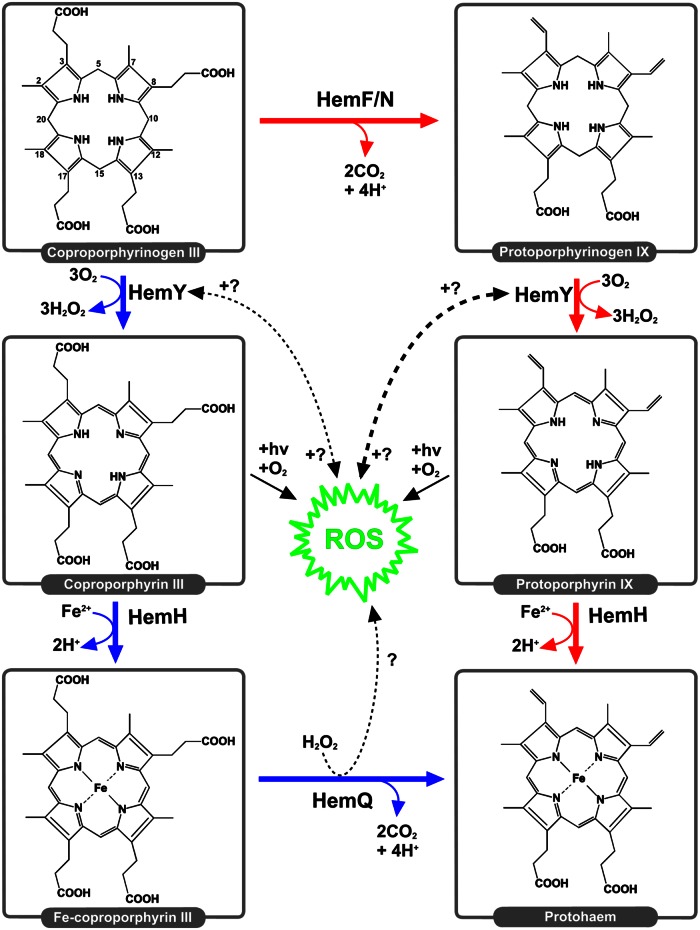
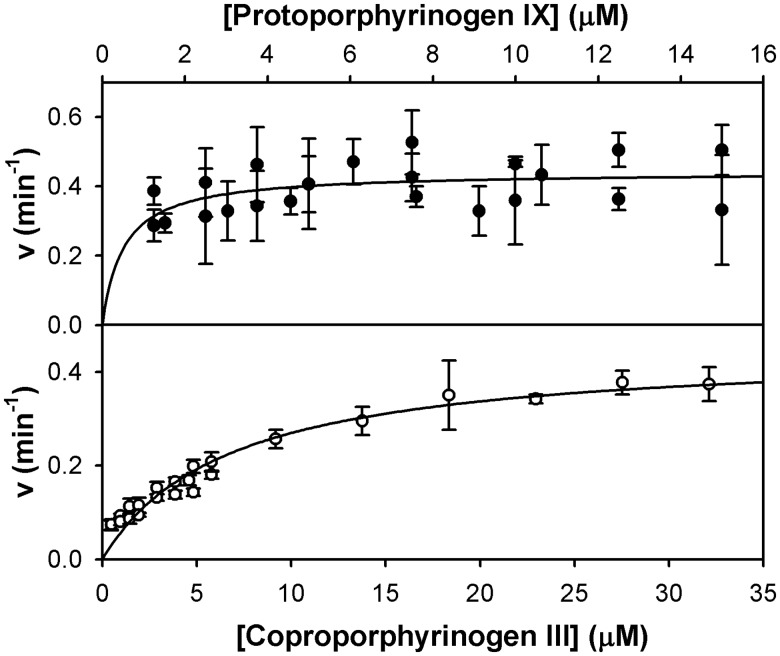
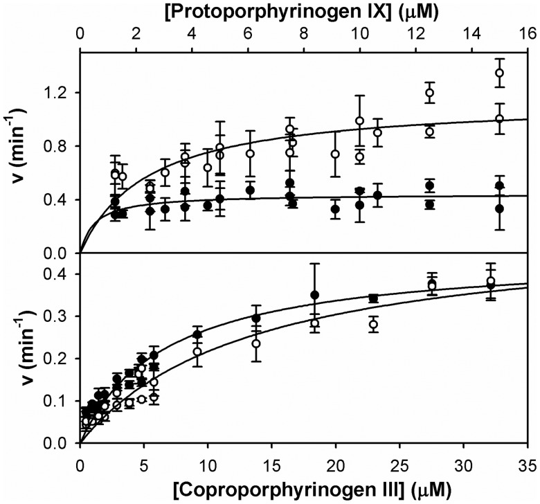
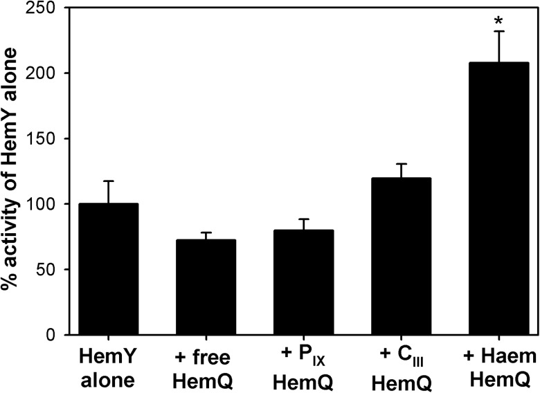
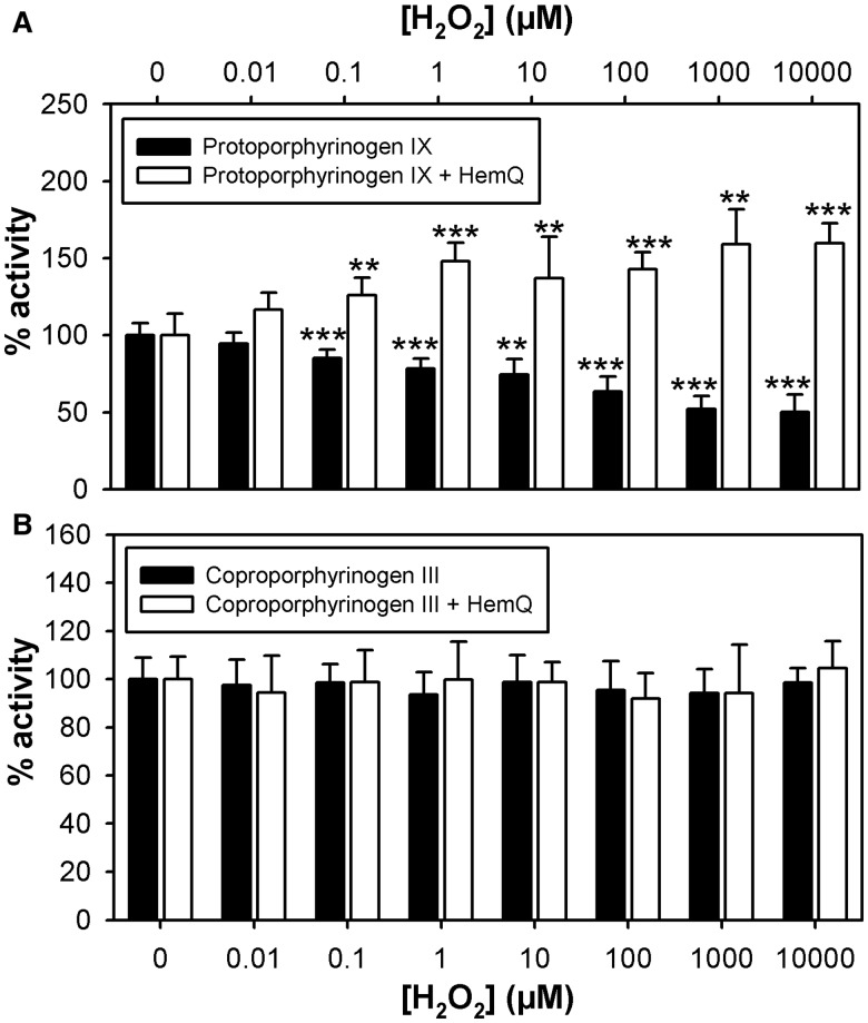
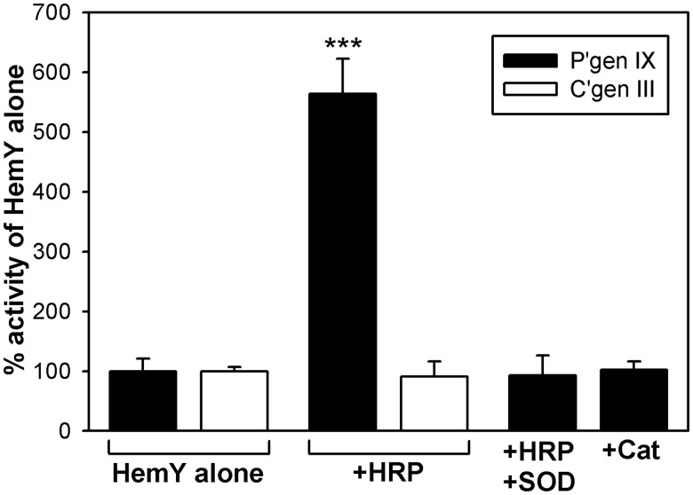
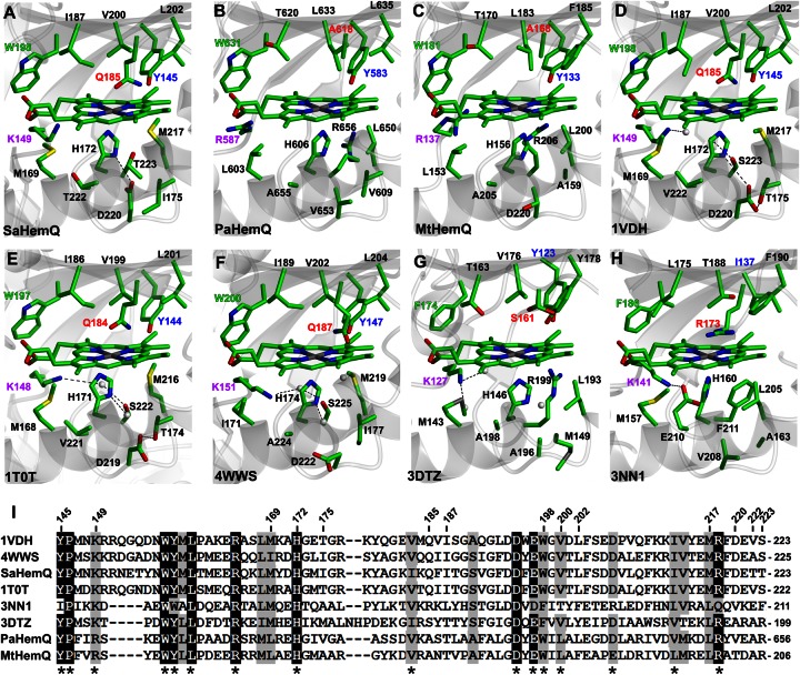
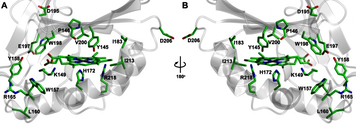
Bacteria require a haem biosynthetic pathway for the assembly of a variety of protein complexes, including cytochromes, peroxidases, globins, and catalase. Haem is synthesised via a series of tetrapyrrole intermediates, including non-metallated porphyrins, such as protoporphyrin IX, which is well known to generate reactive oxygen species in the presence of light and oxygen. Staphylococcus aureus has an ancient haem biosynthetic pathway that proceeds via the formation of coproporphyrin III, a less reactive porphyrin. Here, we demonstrate, for the first time, that HemY of S. aureus is able to generate both protoporphyrin IX and coproporphyrin III, and that the terminal enzyme of this pathway, HemQ, can stimulate the generation of protoporphyrin IX (but not coproporphyrin III). Assays with hydrogen peroxide, horseradish peroxidase, superoxide dismutase, and catalase confirm that this stimulatory effect is mediated by superoxide. Structural modelling reveals that HemQ enzymes do not possess the structural attributes that are common to peroxidases that form compound I [FeIV==O]+, which taken together with the superoxide data leaves Fenton chemistry as a likely route for the superoxide-mediated stimulation of protoporphyrinogen IX oxidase activity of HemY. This generation of toxic free radicals could explain why HemQ enzymes have not been identified in organisms that synthesise haem via the classical protoporphyrin IX pathway. This work has implications for the divergent evolution of haem biosynthesis in ancestral microorganisms, and provides new structural and mechanistic insights into a recently discovered oxidative decarboxylase reaction.

Keywords: coproporphyrinogen; protoporphyrinogen.


Figures

Figure 1.
Figure 1.. The classical and coproporphyrin-dependent pathways of haem synthesis.
This is a depiction of haem synthesis in a hypothetical ancestral organism in which the oxidation of coproporphyrinogen III to coproporphyrin III (blue) and the classical route, involving a protoporphyrin IX intermediate (red), takes place. HemQ is hypothesised to generate ROS, which, in turn, are hypothesised to have an impact on protoporphyrinogen IX oxidation to a greater extent than coproporphyrinogen III oxidation.
Figure 2.
Figure 2.. Kinetic analysis of the HemY protoporphyrinogen oxidase utilising protoporphyrinogen IX and coproporphyrinogen III substrates.
Non-linear regression analysis reveals kcat values of 0.44 ± 0.03 and 0.46 ± 0.02 min−1 for protoporphyrinogen IX (●) and coproporphyrinogen III (○), respectively. The Km for coproporphyrinogen III is 6.7 ± 0.8 µM.
Figure 3.
Figure 3.. Haem-loaded HemQ stimulates HemY-mediated oxidation of protoporphyrinogen but not coproporphyrinogen III.
Steady-state kinetics for HemY in the presence (○) and absence (●) of haem-loaded HemQ (1 µM), using protoporphyrinogen IX (top panel) and coproporphyrinogen III (bottom panel) substrates. Non-linear regression analysis reveals a HemQ-mediated 2.6-fold increase in kcat when protoporphyrinogen IX is used as a substrate, and a HemQ-mediated 1.1-fold increase in kcat when coproporphyrinogen III is used as a substrate.
Figure 4.
Figure 4.. HemQ must bind haem for the stimulation of protoporphyrinogen oxidase activity.
Protoporphyrinogen oxidase activity of HemY was assayed in the presence of HemQ (1 µM) and protoporphyrinogen IX (10 µM) with different tetrapyrroles bound. Error bars represent SD values. Asterisk indicates that rates measured are significantly different from those measured using HemY alone (Student's t-test, P < 0.05).
Figure 5.
Figure 5.. Hydrogen peroxide stimulates protoporphyrinogen oxidase activity in the presence of HemQ.
Protoporphyrinogen oxidase activity of HemY was assayed in the presence (1 µM) and absence of HemQ at various concentrations of hydrogen peroxide using 9.5 µM protoporphyrinogen IX (A) and 5.8 µM coproporphyrinogen III (B) as substrates. Error bars represent SD values. Asterisks indicate that rates measured are significantly different from those measured in the absence of peroxide (Student's t-test, *P < 0.05, **P < 0.01, ***P < 0.001).
Figure 6.
Figure 6.. Peroxidase-derived superoxide stimulates HemY-mediated oxidation of protoporphyrinogen IX.
Kinetic assays showing the effect of HRP (1 µM), superoxide dismutase (1 µM), and catalase (1 µM) upon the activity of HemY (0.5 µM) using protoporphyrinogen IX (8.6 µM) and coproporphyrinogen III (8.2 µM) substrates. Abbreviations: HRP, horseradish peroxidase; SOD, superoxide dismutase; Cat, catalase; P'gen IX, protoporphyrinogen IX; C'gen III, coproporphyrinogen III. Asterisks indicate that rates measured are significantly different from those measured in the absence of peroxide (Student's t-test, ***P < 0.001).
Figure 7.
Figure 7.. Structral analysis of the haem-binding cleft of HemQ family members.
Structural models of haem-binding clefts for HemQ enzymes from S. aureus (A, SaHemQ), P. acnes (B, PaHemQ), and M. tuberculosis (C, MtHemQ) were produced using the haem cofactor from the chlorite dismutase from Candidatus Nitrospira defluvii (H, 3NN1 [34]). (DG) Apoprotein crystal structures of HemQ enzymes from T. thermophilus (1VDH [33]), B. stearothermophilus (1T0T), L. monocytogenes (4WWS [18]), and T. acidophilum (3DTZ), all with the haem cofactor from 3NN1 superimposed. (I) Multiple sequence alignment of sections of the proteins described above with residue numbers for SaHemQ marked at the top and functionally conserved residues marked with an asterisk.
Figure 8.
Figure 8.. Conserved residues in the haem-binding cleft of HemQ family members.
Fully and functionally conserved residues in HemQ proteins from Figure 7I are represented on the structural model of S. aureus HemQ viewed from the entrance to the haem cleft (A) and from behind rotated 180° around the y-axis (B).

References

    1. Layer G., Reichelt J., Jahn D. and Heinz D.W. (2010) Structure and function of enzymes in heme biosynthesis. Prot. Sci. 19, 1137–1161 doi: 10.1002/pro.405 - DOI - PMC - PubMed
    1. Mobius K., Arias-Cartin R., Breckau D., Hannig A.-L., Riedmann K., Biedendieck R. et al. (2010) Heme biosynthesis is coupled to electron transport chains for energy generation. Proc. Natl Acad. Sci. USA 107, 10436–10441 doi: 10.1073/pnas.1000956107 - DOI - PMC - PubMed
    1. Jacobs N.J. and Jacobs J.M. (1976) Nitrate, fumarate, and oxygen as electron acceptors for a late step in microbial heme synthesis. Biochim. Biophys. Acta Bioenergetics 449, 1–9 doi: 10.1016/0005-2728(76)90002-5 - DOI - PubMed
    1. Kobayashi K., Masuda T., Tajima N., Wada H. and Sato N. (2014) Molecular phylogeny and intricate evolutionary history of the three isofunctional enzymes involved in the oxidation of protoporphyrinogen IX. Genome Biol. Evol. 6, 2141–2155 doi: 10.1093/gbe/evu170 - DOI - PMC - PubMed
    1. Buchenau B., Kahnt J., Heinemann I.U., Jahn D. and Thauer R.K. (2006) Heme biosynthesis in Methanosarcina barkeri via a pathway involving two methylation reactions. J. Bacteriol. 188, 8666–8668 doi: 10.1128/JB.01349-06 - DOI - PMC - PubMed

Publication types

MeSH terms

LinkOut - more resources