. 2016 Sep 5;4(1):22.
doi: 10.1186/s40349-016-0066-7. eCollection 2016.

Open-source, small-animal magnetic resonance-guided focused ultrasound system

Affiliations

Open-source, small-animal magnetic resonance-guided focused ultrasound system

Megan E Poorman et al. J Ther Ultrasound. .

Erratum in

Abstract

Background: MR-guided focused ultrasound or high-intensity focused ultrasound (MRgFUS/MRgHIFU) is a non-invasive therapeutic modality with many potential applications in areas such as cancer therapy, drug delivery, and blood-brain barrier opening. However, the large financial costs involved in developing preclinical MRgFUS systems represent a barrier to research groups interested in developing new techniques and applications. We aim to mitigate these challenges by detailing a validated, open-source preclinical MRgFUS system capable of delivering thermal and mechanical FUS in a quantifiable and repeatable manner under real-time MRI guidance.

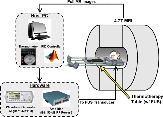
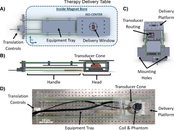
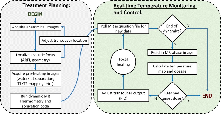
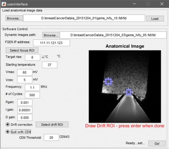
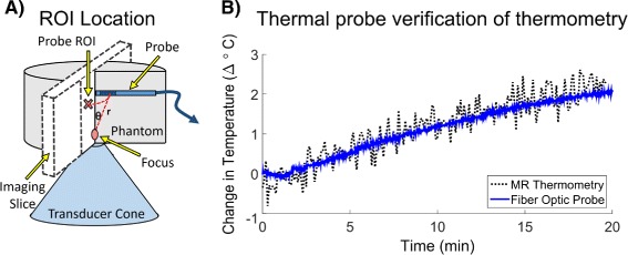
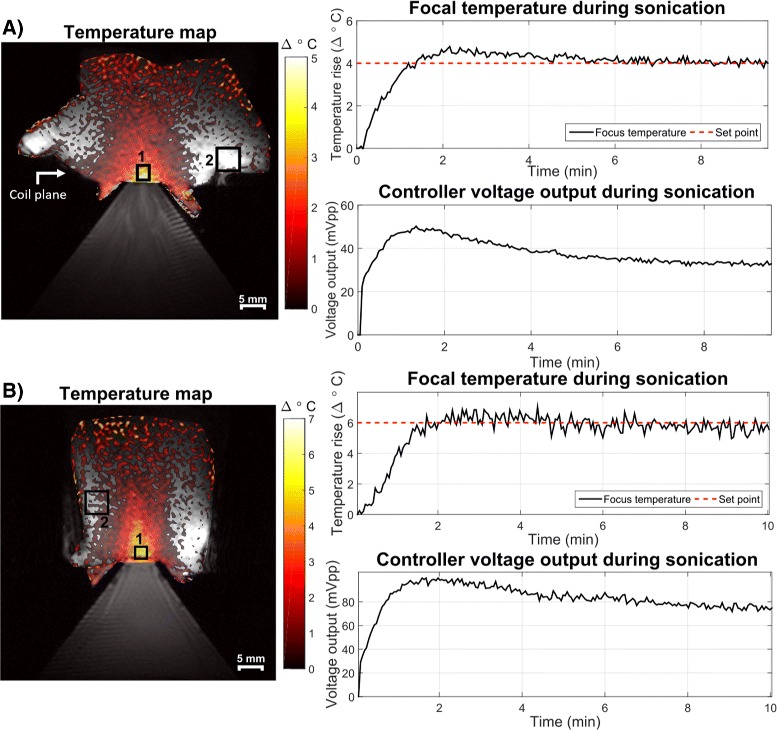
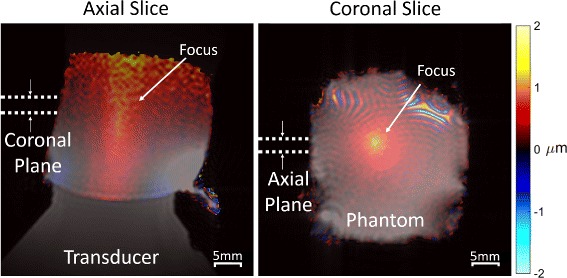
Methods: A hardware and software package was developed that includes closed-loop feedback controlled thermometry code and CAD drawings for a therapy table designed for a preclinical MRI scanner. For thermal treatments, the modular software uses a proportional integral derivative controller to maintain a precise focal temperature rise in the target given input from MR phase images obtained concurrently. The software computes the required voltage output and transmits it to a FUS transducer that is embedded in the delivery table within the magnet bore. The delivery table holds the FUS transducer, a small animal and its monitoring equipment, and a transmit/receive RF coil. The transducer is coupled to the animal via a water bath and is translatable in two dimensions from outside the magnet. The transducer is driven by a waveform generator and amplifier controlled by real-time software in Matlab. MR acoustic radiation force imaging is also implemented to confirm the position of the focus for mechanical and thermal treatments.

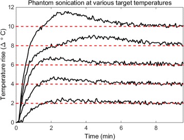
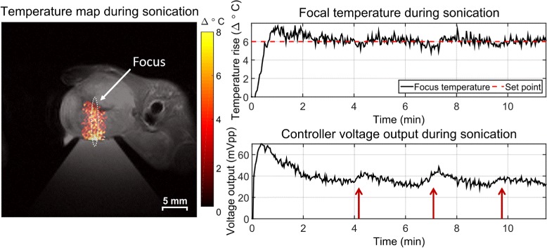
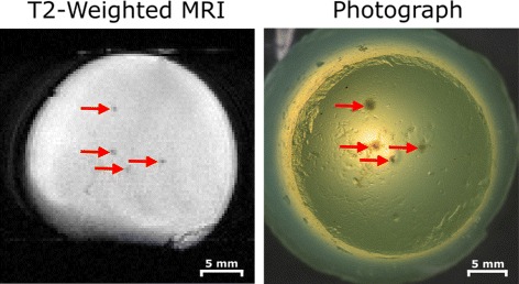
Results: The system was validated in tissue-mimicking phantoms and in vivo during murine tumor hyperthermia treatments. Sonications were successfully controlled over a range of temperatures and thermal doses for up to 20 min with minimal temperature overshoot. MR thermometry was validated with an optical temperature probe, and focus visualization was achieved with acoustic radiation force imaging.

Conclusions: We developed an MRgFUS platform for small-animal treatments that robustly delivers accurate, precise, and controllable sonications over extended time periods. This system is an open source and could increase the availability of low-cost small-animal systems to interdisciplinary researchers seeking to develop new MRgFUS applications and technology.

Keywords: High-intensity focused ultrasound (HIFU); MR-guided focused ultrasound (MRgFUS); Open source; Preclinical.


Figures

Fig. 1
Fig. 1
Open-source small-animal MRgFUS system overview. The delivery table holds the target and transducer at magnet isocenter while imaging is performed. Therapy control software for planning and closed-loop temperature control is implemented in Matlab on the MRI scanner’s host PC, which collects the real-time MR images, computes the focal temperature, and modulates the ultrasound output accordingly
Fig. 2
Fig. 2
Detailed view of the delivery table. a Top view showing placement inside magnet, positioning controls, and rectangular delivery window. b Side view showing the housing of the FUS transducer and coupling cone. c End view showing routing and mounting locations. d Photo of the table to illustrate arrangement of coil and sample
Fig. 3
Fig. 3
Software flow chart. The treatment protocol comprises a planning stage followed by real-time temperature monitoring and control. The software design allows anatomical and parametric imaging prior to sonication for treatment planning. The temperature monitoring control loop will adjust the FUS amplitude according to observed heating, automatically stopping treatment when a desired thermal dose is achieved
Fig. 4
Fig. 4
Optional GUI for the setup of the control software. The user can draw ROIs on an anatomical image for the acoustic focus and drift control, set the ultrasound parameters, tune the control parameters, and define a thermal dose target
Fig. 5
Fig. 5
Fiber optic probe thermometry validation. a Illustration of the experimental setup. To avoid artifacts and damage to the probe, it was placed above the focus. b Plots probe temperature compared to MR temperature measurements in a 5.7 mm2 ROI at a geometrically equivalent position within the slice
Fig. 6
Fig. 6
Sonications across temperature set points. After initial overshoots that did not exceed 1.5 °C of the set points (dashed red lines), focal temperature was maintained for 10 min with a mean standard deviation of the temperature error of 0.28 °C and a mean RMSE of 0.3 °C
Fig. 7
Fig. 7
Sonications at 1.1 (a) and 3.68 (b) MHz FUS frequencies, targeting temperature set points in ROI 1 for 10 min. Background phase drifts were corrected using an ROI outside of the area of heating (ROI 2). Controller voltage is also plotted for each case and also stabilizes after an initial rise and small overshoot. The white arrow indicates surface coil placement. Low-temperature SNR at the top of the phantoms (and far from the surface coil which sat at the level of the water-phantom interface) contributed to the apparent elevated temperatures there but did not interfere with the focus measurements. Stripe artifacts in the water cone are likely due to Moire fringes caused by poor field homogeneity in the water bath near the transducer causing aliasing
Fig. 8
Fig. 8
Sustained local hyperthermia in a murine mammary tumor for 12 min. Sustained, long-term sonication was achieved with minimal overshoot, a 0.49 °C standard deviation of the error, and 0.56 °C RMSE after the initial temperature rise. The PID controller responded to sudden changes in focal temperature as indicated by the red arrows. The 1.4 mm by 10 mm contour of the transducer focus is indicated by the white oval
Fig. 9
Fig. 9
Demonstration of transducer translation capabilities via multiple egg white phantom ablations. The transducer was moved using the controls outside of the magnet; no re-positioning of the phantom or removal of the platform was necessary. Lesions were visible on a T 2-weighted image (left) and on photographs taken outside the magnet (right) and matched geometrically
Fig. 10
Fig. 10
ARFI-measured displacement in the axial and coronal directions overlaid on T 1-weighted images of a tofu phantom. Localized displacements are apparent at the focus while smoother displacements appear throughout which may correspond to shear waves. The focal displacement location and size correspond well to the expected geometry of the transducer. Acoustic reflections with the air boundary and lower SNR near the top of the phantom likely contribute to the diffuse rise in displacement there. Moire fringes are visible at the top of the coronal image, which are likely due to field inhomogeneity near the surface of the transducer causing some water bath signals to be excited and alias into the image

Comment in

  • Ultrasound for the brain.
    Landhuis E. Landhuis E. Nature. 2017 Nov 9;551(7679):257-259. doi: 10.1038/d41586-017-05479-7. Nature. 2017. PMID: 29120442 No abstract available.

References

    1. Hokland SL, Pedersen M, Salomir R, Quesson B, Stødkilde-Jørgensen H, Moonen CTW. MRI-guided focused ultrasound: methodology and applications. IEEE Trans Med Imaging. 2006;25(6):723–31. doi: 10.1109/TMI.2006.873296. - DOI - PubMed
    1. Ran LF, Xie XP, Xia JZ, Xie FL, Fan YM, Wu F. Specific antitumour immunity of HIFU-activated cytotoxic T lymphocytes after adoptive transfusion in tumour-bearing mice. Int J Hyperth. 2015;1–7. doi:10.3109/02656736.2015.1112438. - DOI - PubMed
    1. Kheirolomoom A, Ingham ES, Mahakian LM, Tam SM, Silvestrini MT, Tumbale SK, Foiret J, Hubbard NE, Borowsky AD, Murphy WJ, Ferrara KW. CpG expedites regression of local and systemic tumors when combined with activatable nanodelivery. J Control Release. 2015;220:253–64. doi: 10.1016/j.jconrel.2015.10.016. - DOI - PMC - PubMed
    1. Elias WJ, Huss D, Voss T, Loomba J, Khaled M, Zadicario E, Frysinger RC, Sperling Sa, Wylie S, Monteith SJ, Druzgal J, Shah BB, Harrison M, Wintermark M. A pilot study of focused ultrasound thalamotomy for essential tremor. N Engl J Med. 2013;369:640–8. doi: 10.1056/NEJMoa1300962. - DOI - PubMed
    1. Yoo SS, Bystritsky A, Lee JH, Zhang Y, Fischer K, Min BK, McDannold NJ, Pascual-Leone A, Jolesz Fa. Focused ultrasound modulates region-specific brain activity. NeuroImage. 2011;56:1267–1275. doi: 10.1016/j.neuroimage.2011.02.058. - DOI - PMC - PubMed