

Bone Regeneration after Treatment with Covering Materials Composed of Flax Fibers and Biodegradable Plastics: A Histological Study in Rats
- PMID: 27597965
- PMCID: PMC4997065
- DOI: 10.1155/2016/5146285
Bone Regeneration after Treatment with Covering Materials Composed of Flax Fibers and Biodegradable Plastics: A Histological Study in Rats
Abstract
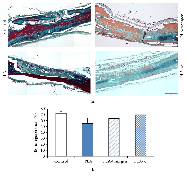
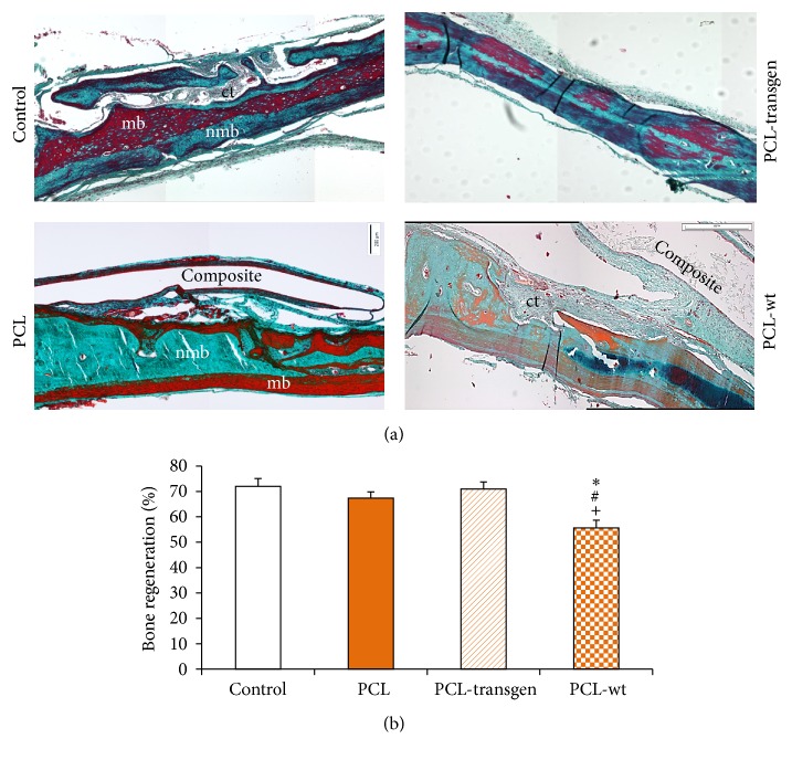
The aim of this study was to examine the osteogenic potential of new flax covering materials. Bone defects were created on the skull of forty rats. Materials of pure PLA and PCL and their composites with flax fibers, genetically modified producing PHB (PLA-transgen, PCL-transgen) and unmodified (PLA-wt, PCL-wt), were inserted. The skulls were harvested after four weeks and subjected to histological examination. The percentage of bone regeneration by using PLA was less pronounced than after usage of pure PCL in comparison with controls. After treatment with PCL-transgen, a large amount of new formed bone could be found. In contrast, PCL-wt decreased significantly the bone regeneration, compared to the other tested groups. The bone covers made of pure PLA had substantially less influence on bone regeneration and the bone healing proceeded with a lot of connective tissue, whereas PLA-transgen and PLA-wt showed nearly comparable amount of new formed bone. Regarding the histological data, the hypothesis could be proposed that PCL and its composites have contributed to a higher quantity of the regenerated bone, compared to PLA. The histological studies showed comparable bone regeneration processes after treatment with tested covering materials, as well as in the untreated bone lesions.
Figures
Similar articles
-
In vivo analysis of covering materials composed of biodegradable polymers enriched with flax fibers.Biomater Res. 2017 May 19;21:8. doi: 10.1186/s40824-017-0094-6. eCollection 2017. Biomater Res. 2017. PMID: 28529764 Free PMC article.
-
Osteogenic capacity of transgenic flax scaffolds.Biomed Tech (Berl). 2012 Jan 19;57(1):53-8. doi: 10.1515/bmt-2011-0035. Biomed Tech (Berl). 2012. PMID: 22718592
-
The influence of biocomposites containing genetically modified flax fibers on gene expression in rat skeletal muscle.Biomed Tech (Berl). 2010 Dec;55(6):323-9. doi: 10.1515/BMT.2010.048. Epub 2010 Oct 25. Biomed Tech (Berl). 2010. PMID: 20973615
-
The survival and proliferation of fibroblasts on biocomposites containing genetically modified flax fibers: an in vitro study.Ann Anat. 2012 Nov;194(6):513-7. doi: 10.1016/j.aanat.2011.12.006. Epub 2012 Feb 8. Ann Anat. 2012. PMID: 22377281
-
Bioabsorbable Composites Based on Polymeric Matrix (PLA and PCL) Reinforced with Magnesium (Mg) for Use in Bone Regeneration Therapy: Physicochemical Properties and Biological Evaluation.Polymers (Basel). 2023 Dec 11;15(24):4667. doi: 10.3390/polym15244667. Polymers (Basel). 2023. PMID: 38139919 Free PMC article. Review.
Cited by
-
Titanium or Biodegradable Osteosynthesis in Maxillofacial Surgery? In Vitro and In Vivo Performances.Polymers (Basel). 2022 Jul 7;14(14):2782. doi: 10.3390/polym14142782. Polymers (Basel). 2022. PMID: 35890557 Free PMC article. Review.
-
Revolutionizing bone regeneration: advanced biomaterials for healing compromised bone defects.Front Aging. 2023 Jul 14;4:1217054. doi: 10.3389/fragi.2023.1217054. eCollection 2023. Front Aging. 2023. PMID: 37520216 Free PMC article. Review.
-
Properties and Degradation of Novel Fully Biodegradable PLA/PHB Blends Filled with Keratin.Int J Mol Sci. 2020 Dec 18;21(24):9678. doi: 10.3390/ijms21249678. Int J Mol Sci. 2020. PMID: 33353232 Free PMC article.
-
Biomaterial design strategies to address obstacles in craniomaxillofacial bone repair.RSC Adv. 2021;11(29):17809-17827. doi: 10.1039/d1ra02557k. Epub 2021 May 17. RSC Adv. 2021. PMID: 34540206 Free PMC article.
-
In vivo analysis of covering materials composed of biodegradable polymers enriched with flax fibers.Biomater Res. 2017 May 19;21:8. doi: 10.1186/s40824-017-0094-6. eCollection 2017. Biomater Res. 2017. PMID: 28529764 Free PMC article.
References
-
- Albert A., Leemrijse T., Druez V., Delloye C., Cornu O. Are bone autografts still necessary in 2006? A three-year retrospective study of bone grafting. Acta Orthopaedica Belgica. 2006;72(6):734–740. - PubMed
-
- Berrey B. H., Jr., Lord C. F., Gebhardt M. C., Mankin H. J. Fractures of allografts. Frequency, treatment, and end-results. The Journal of Bone & Joint Surgery—American Volume. 1990;72(6):825–833. - PubMed
MeSH terms
Substances
LinkOut - more resources
Full Text Sources
Other Literature Sources
