. 2016 Sep 1;17(9):1448.
doi: 10.3390/ijms17091448.

Novel Anti-Melanogenesis Properties of Polydeoxyribonucleotide, a Popular Wound Healing Booster

Affiliations

Novel Anti-Melanogenesis Properties of Polydeoxyribonucleotide, a Popular Wound Healing Booster

Tai Kyung Noh et al. Int J Mol Sci. .

Abstract

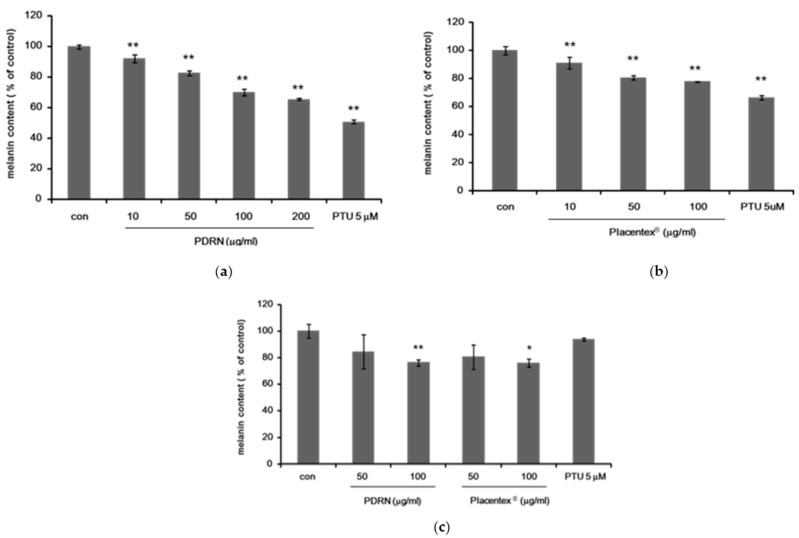
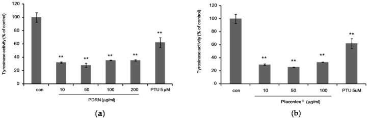
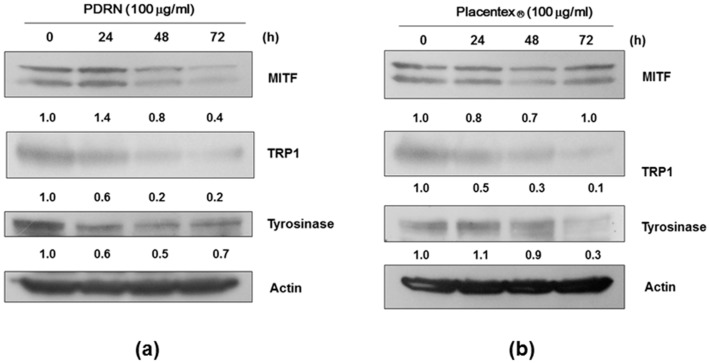
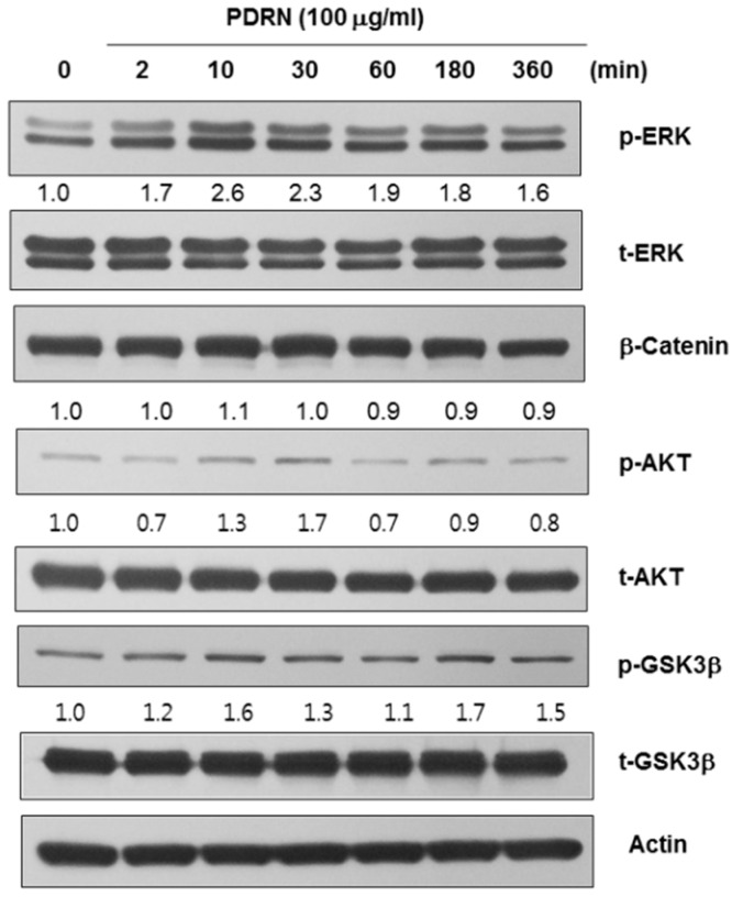
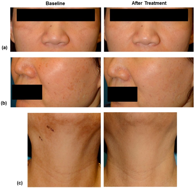
Polydeoxyribonucleotide (PDRN), a deoxyribonucleotide polymer, is popularly used for faster healing of cutaneous wounds and boosting of neocollagenesis of photoaged skin among current dermatologic practitioners. Some patients receiving PDRN injection treatment also reported improvement of photoaging-associated mottled pigmentation (PMP). To investigate the effect of PDRN on cutaneous melanogenesis, we examined the effect of PDRN and an available product (Placentex(®)) containing PDRN on melanogenesis using human melanocytes-keratinocytes cocultures and mouse melanocytes. Melanin content, tyrosinase activity, and levels of microphthalmia-associated transcription factor (MITF), tyrosinase, and tyrosinase-related protein (TRP-1) were determined. Intracellular signaling pathways were assessed by Western blotting. PDRN and Placentex(®) led to decreases in melanin content, tyrosinase activity, and MITF and TRP-1 expression with concomitant increases in phosphorylated forms of extracellular signal-regulated protein kinase (ERK) and AKT in mouse melanocytes. More importantly, both PDRN and Placentex(®) significantly suppressed the melanin content in human melanocyte-keratinocyte cocultures. Clinical evaluation of six female patients with facial hyperpigmentation after three sessions of intradermal PDRN injections using a 5-point scale revealed that PDRN led to more than noticeable improvements in hyperpigmented lesions. This is the first study to demonstrate that PDRN, which is known for its wound-healing properties, may have novel anti-melanogenesis and potential skin whitening properties.

Keywords: coculture; hyperpigmentation; melanogenesis; polydeoxyribonucleotide.


Conflict of interest statement

The authors declare no conflict of interest.

Figures

Figure 1
Figure 1
Polydeoxyribonucleotide and Placentex® inhibit melanogenesis in Mel-Ab cells and in human melanocyte–keratinocyte cocultures. (a) Polydeoxyribonucleotide (PDRN) inhibits melanogenesis in Mel-Ab cells. Melanin content of Mel-Ab cells treated with PDRN (10–200 μg/mL) for 4 days. N′-phenylthiourea (PTU, 5 μM) was used as a positive control. Data represent mean ± standard deviation (SD) of triplicate assays expressed as percentage of control. * p < 0.05, ** p < 0.01 compared to untreated control; (b) Placentex® inhibits melanogenesis in Mel-Ab cells. Melanin content of Mel-Ab cells treated with Placentex® (10–100 μg/mL) for 4 days. PTU (5 μM) was used as a positive control. Data represent mean ± SD of triplicate assays expressed as percentage of control. * p < 0.05, ** p < 0.01 compared to untreated control; (c) PDRN and Placentex® inhibit melanogenesis in normal human melanocytes cocultured with human keratinocytes. Melanin content of human melanocytes treated with PDRN (50–100 μg/mL) or Placentex® (50–100 μg/mL) for 5 days. PTU (5 μM) was used as a positive control. Data represent mean ± SD of triplicate assays expressed as percentage of control. * p < 0.05, ** p < 0.01 compared to untreated control.
Figure 2
Figure 2
Polydeoxyribonucleotide and Placentex® suppress intracellular tyrosinase activity in Mel-Ab cells. (a) Polydeoxyribonucleotide (PDRN) suppresses tyrosinase activity in Mel-Ab cells. Mel-Ab cells were incubated with 10–200 μg/mL PDRN for 4 days, and cellular tyrosinase activity was measured. N′-phenylthiourea (PTU, 5 μM) was used as a positive control. Data represent mean ± standard deviation (SD) of triplicate assays expressed as percentage of control. * p < 0.05, ** p < 0.01 compared to untreated control; (b) Placentex® represses tyrosinase activity in Mel-Ab cells. Mel-Ab cells were incubated with 10–100 μg/mL Placentex® for 4 days, and cellular tyrosinase activity was measured. PTU (5 μM) was used as a positive control. Data represent mean ± SD of triplicate assays expressed as percentage of control. ** p < 0.01 compared to untreated control.
Figure 3
Figure 3
Polydeoxyribonucleotide and Placentex® reduce the levels of microphthalmia-associated transcription factor and melanogenesis-related proteins in Mel-Ab cells. Whole-cell lysates were analyzed by western immunoblotting using antibodies against microphthalmia-associated transcription factor (MITF), tyrosinase-related protein (TRP)-1, and tyrosinase. Normalization was achieved by dividing the densitometric values for individual bands by the densitometric value for β-actin for the same sample. (a) Polydeoxyribonucleotide (PDRN) reduces the levels of melanogenesis-related proteins. Mel-Ab cells were incubated with 100 μg/mL PDRN for 24–72 h; (b) Placentex® reduces the levels of melanogenesis-related proteins. Mel-Ab cells were incubated with 100 μg/mL Placentex® for 24–48 h.
Figure 4
Figure 4
Polydeoxyribonucleotide affects the levels of melanogenesis-related signaling pathways. Polydeoxyribonucleotide (PDRN) induced the phosphorylation of extracellular signal-regulated protein kinase (ERK) and AKT within 10–30 min after treatment, whereas the levels of β-catenin or phosphorylated-GSK3β were not affected under identical conditions.
Figure 5
Figure 5
Standard digital photography images of patient number 1 (a), 5 (b) and 6 (c) at baseline and 4 weeks after 3 times of intradermal PDRN injection treatment. The clinical photographs of right side demonstrate significant improvement after 3 sessions of intralesional injection of Placentex® treatment.

References

    1. Lee S.H., Zheng Z., Kang J.S., Kim D.Y., Oh S.H., Cho S.B. Therapeutic efficacy of autologous platelet-rich plasma and polydeoxyribonucleotide on female pattern hair loss. Wound Repair Regen. 2015;23:30–36. doi: 10.1111/wrr.12250. - DOI - PubMed
    1. Squadrito F., Bitto A., Altavilla D., Arcoraci V., de Caridi G., de Feo M.E., Corrao S., Pallio G., Sterrantino C., Minutoli L., et al. The effect of PDRN, an adenosine receptor A2A agonist, on the healing of chronic diabetic foot ulcers: Results of a clinical trial. J. Clin. Endocrinol. Metab. 2014;99:E746–E753. doi: 10.1210/jc.2013-3569. - DOI - PubMed
    1. Galeano M., Bitto A., Altavilla D., Minutoli L., Polito F., Calo M., Lo Cascio P., Stagno d'Alcontres F., Squadrito F. Polydeoxyribonucleotide stimulates angiogenesis and wound healing in the genetically diabetic mouse. Wound Repair Regen. 2008;16:208–217. doi: 10.1111/j.1524-475X.2008.00361.x. - DOI - PubMed
    1. Bitto A., Polito F., Irrera N., D'Ascola A., Avenoso A., Nastasi G., Campo G.M., Micali A., Bagnato G., Minutoli L., et al. Polydeoxyribonucleotide reduces cytokine production and the severity of collagen-induced arthritis by stimulation of adenosine A2A receptor. Arthritis Rheum. 2011;63:3364–3371. doi: 10.1002/art.30538. - DOI - PubMed
    1. Koroskenyi K., Kiss B., Szondy Z. Adenosine A2A receptor signaling attenuates LPS-induced pro-inflammatory cytokine formation of mouse macrophages by inducing the expression of DUSP1. Biochim. Biophys. Acta. 2016;1863:1461–1471. doi: 10.1016/j.bbamcr.2016.04.003. - DOI - PubMed

MeSH terms