Case Reports
. 2016 Sep 6;11(9):e0161789.
doi: 10.1371/journal.pone.0161789. eCollection 2016.

SCN10A Mutation in a Patient with Erythromelalgia Enhances C-Fiber Activity Dependent Slowing

Affiliations
Case Reports

SCN10A Mutation in a Patient with Erythromelalgia Enhances C-Fiber Activity Dependent Slowing

Andreas M Kist et al. PLoS One. .

Abstract

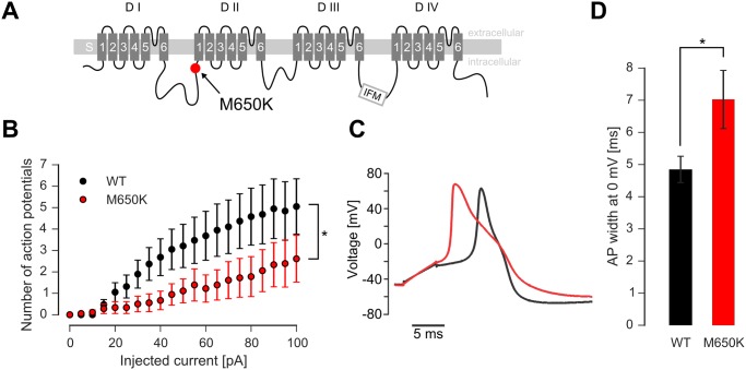
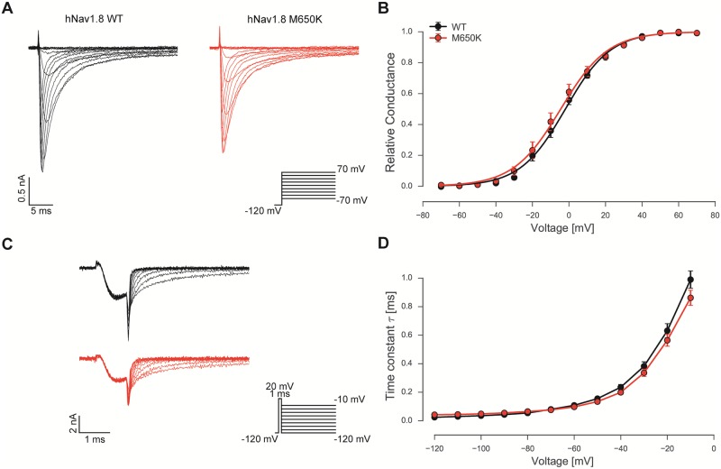
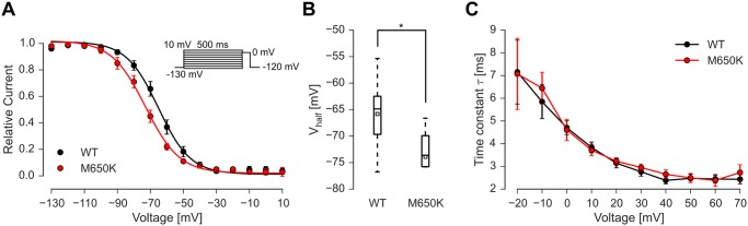
Gain-of-function mutations in the tetrodotoxin (TTX) sensitive voltage-gated sodium channel (Nav) Nav1.7 have been identified as a key mechanism underlying chronic pain in inherited erythromelalgia. Mutations in TTX resistant channels, such as Nav1.8 or Nav1.9, were recently connected with inherited chronic pain syndromes. Here, we investigated the effects of the p.M650K mutation in Nav1.8 in a 53 year old patient with erythromelalgia by microneurography and patch-clamp techniques. Recordings of the patient's peripheral nerve fibers showed increased activity dependent slowing (ADS) in CMi and less spontaneous firing compared to a control group of erythromelalgia patients without Nav mutations. To evaluate the impact of the p.M650K mutation on neuronal firing and channel gating, we performed current and voltage-clamp recordings on transfected sensory neurons (DRGs) and neuroblastoma cells. The p.M650K mutation shifted steady-state fast inactivation of Nav1.8 to more hyperpolarized potentials and did not significantly alter any other tested gating behaviors. The AP half-width was significantly broader and the stimulated action potential firing rate was reduced for M650K transfected DRGs compared to WT. We discuss the potential link between enhanced steady state fast inactivation, broader action potential width and the potential physiological consequences.


Conflict of interest statement

AMR, ZZ, HS, DM, PW, JK were employees of AstraZeneca (Sweden) at the time of the study. THC is a current employee of AstraZeneca (UK) and owns shares in AstraZeneca. This did not alter the authors' adherence to PLOS ONE policies on sharing data and materials.

Figures

Fig 1
Fig 1
A: Microneurography setup. Action potentials are evoked electrically in the receptive field of the pierced fascicle, travel proximally along the peroneal nerve and are recorded at the fibular head. B: The interval between the first electrical stimulus and the arrival time of the evoked AP at the recording needle t0 is set as reference conduction latency. Conduction latencies of subsequent pulses (Δt) are calculated relative to the first latency (Δt/t0). C: Plot of the relative conduction latency (Δt/t0) versus pulse number. The increase in Δt/t0 indicates the slowing of the AP conduction upon repetitive stimulation.
Fig 2
Fig 2. ADS during 2 Hz stimulation protocol of CMi-nociceptors in the M650K patient (A) and EM controls without Nav mutations (B) and age-matched healthy controls (C).
The lower panel shows the first 15 pulses of the upper panel (subset indicated with gray shadow in the upper panel). Note the prominent abnormal progressive increase in ADS in the last part of the stimulation in two of the CMi-nociceptors from the M650K patient (red), none in EM patients without mutation and only one less pronounced in age-matched healthy controls showed this behavior.
Fig 3
Fig 3. The M650K mutation decreases the firing rate of DRG neurons.
A: Schematic overview of a eukaryotic Nav showing the four domains DI to DIV, each consisting of the transmembrane segments S1 to S6. The rare genetic variant M650K in Nav1.8 (red dot, [8]) is located at the adjacent intracellular side of DII/S1. B: Number of APs evoked by 500 ms square step current injections to WT (black markers) or M650K (red markers) transfected cells (n = 18 and 19). * < 0.05 in a 2-sided t-test C: Example of an AP recorded from a WT (black trace) and a M650K (red trace) transfected DRG neuron evoked by a 5 ms depolarizing pulse. Injected current was twice threshold current. D: The AP half width was significantly broader in M650K than in WT transfected neurons (n = 13 and 19). *< 0.05 in a 2-sided t-test.
Fig 4
Fig 4. Voltage-dependence of activation and deactivation of Nav1.8 is unaltered by the M650K mutation.
A: Pulse protocol and representative voltage-clamp recordings of hNav1.8 WT and hNav1.8 M650K expressed heterologously in ND7/23 cells. Traces were digitally low pass filtered for display.B: Conductance-voltage relations for WT (black circles, n = 10) and the Nav1.8 M650K mutation (red circles, n = 12) are shown. Solid lines represent Boltzmann-fits to the mean data points. C: Pulse protocol for deactivation measurements and representative recordings of hNav1.8 WT and hNav1.8 M650K mutant expressed heterologously in ND7/23 cells. D: Deactivation time constants as derived from single exponential fits of current decay during repolarization for WT (black circles, n = 13) and M650K (red circles, n = 15) at various voltages.
Fig 5
Fig 5. The M650K mutation shifts steady-state fast inactivation to more hyperpolarized potentials.
A: Voltage dependence of steady-state fast inactivation as determined with the indicated pulse protocol. Black circles indicate WT (n = 7–8) and red circles the M650K mutation (n = 7–8). B: Distribution of the Vhalf determined by Boltzmann fits to each cell. Mean is indicated as an open square, the median is depicted as a line inside the box, the boxes indicate 50% confidence interval and whiskers indicate 95% confidence interval. C: Time constants of single exponential fits of currents evoked with the pulse protocol shown in Fig 4A did not differ between WT and M650K mutation at any investigated voltage.

References

    1. Lampert A, O’Reilly AO, Reeh P, Leffler A. Sodium channelopathies and pain. Pflüg Arch—Eur J Physiol. 2010;460: 249–263. 10.1007/s00424-009-0779-3 - DOI - PubMed
    1. Yang Y, Wang Y, Li S, Xu Z, Li H, Ma L, et al. Mutations in SCN9A, encoding a sodium channel alpha subunit, in patients with primary erythermalgia. J Med Genet. 2004;41: 171–174. 10.1136/jmg.2003.012153 - DOI - PMC - PubMed
    1. Magerl W, Krumova EK, Baron R, Tölle T, Treede R-D, Maier C. Reference data for quantitative sensory testing (QST): Refined stratification for age and a novel method for statistical comparison of group data. PAIN®. 2010;151: 598–605. 10.1016/j.pain.2010.07.026 - DOI - PubMed
    1. Rolke R, Baron R, Maier C, Tölle TR, Treede R-D, Beyer A, et al. Quantitative sensory testing in the German Research Network on Neuropathic Pain (DFNS): standardized protocol and reference values. Pain. 2006;123: 231–243. 10.1016/j.pain.2006.01.041 - DOI - PubMed
    1. Namer B, Ørstavik K, Schmidt R, Kleggetveit IP, Weidner C, Mørk C, et al. Specific changes in conduction velocity recovery cycles of single nociceptors in an erythromelalgia patient with the I848T gain-of-function mutation of NaV1.7. Pain. 2015; 10.1097/j.pain.0000000000000229 - DOI - PubMed

Publication types

Substances