. 2016 Sep 6;11(9):e0161596.
doi: 10.1371/journal.pone.0161596. eCollection 2016.

The Envelope Cytoplasmic Tail of HIV-1 Subtype C Contributes to Poor Replication Capacity through Low Viral Infectivity and Cell-to-Cell Transmission

Affiliations

The Envelope Cytoplasmic Tail of HIV-1 Subtype C Contributes to Poor Replication Capacity through Low Viral Infectivity and Cell-to-Cell Transmission

Eveline Santos da Silva et al. PLoS One. .

Abstract

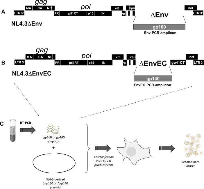
The cytoplasmic tail (gp41CT) of the HIV-1 envelope (Env) mediates Env incorporation into virions and regulates Env intracellular trafficking. Little is known about the functional impact of variability in this domain. To address this issue, we compared the replication of recombinant virus pairs carrying the full Env (Env viruses) or the Env ectodomain fused to the gp41CT of NL4.3 (EnvEC viruses) (12 subtype C and 10 subtype B pairs) in primary CD4+ T-cells and monocyte-derived-macrophages (MDMs). In CD4+ T-cells, replication was as follows: B-EnvEC = B-Env>C-EnvEC>C-Env, indicating that the gp41CT of subtype C contributes to the low replicative capacity of this subtype. In MDMs, in contrast, replication capacity was comparable for all viruses regardless of subtype and of gp41CT. In CD4+ T-cells, viral entry, viral release and viral gene expression were similar. However, infectivity of free virions and cell-to-cell transmission of C-Env viruses released by CD4+ T-cells was lower, suggestive of lower Env incorporation into virions. Subtype C matrix only minimally rescued viral replication and failed to restore infectivity of free viruses and cell-to-cell transmission. Taken together, these results show that polymorphisms in the gp41CT contribute to viral replication capacity and suggest that the number of Env spikes per virion may vary across subtypes. These findings should be taken into consideration in the design of vaccines.


Conflict of interest statement

The authors have declared that no competing interests exist.

Figures

Fig 1
Fig 1. Generation of recombinant viruses and molecular constructs pNL4.3ΔEnv and pNL4.3ΔEC.
A and B: The molecular constructs were based on the pNL4.3 infectious clone deleted of the entire Env (gp160, ΔEnv) (A) or of the Env gp120+gp41 ectodomain (gp140, ΔEnvEC) (B) by inverse PCR. The deleted portion is replaced by the AfeI restriction site for linearization prior to transfection. C: Infectious viral particles expressing patient-derived Env or EnvEC sequences were generated by co-transfecting HEK 293T cells with the linearized pNL4.3ΔEnv or pNL4.3ΔEC backbones and a PCR amplicon spanning the target region (gp160 (Env) or gp140 (EnvEC) respectively).
Fig 2
Fig 2. Viruses with subtype C gp41CT have lower replication than viruses with subtype B gp41CT in CD4+ T-cells but not in MDMs.
A and B. Replication of subtype B and subtype C recombinants in CD4+ T-cells from two different donors. 105 CD4+ T-cells were infected with equivalent amounts (1 ng/ml) of Env or EnvEC recombinant viruses. Infection was monitored by measuring p24 in culture supernatants at days 2 or 3, 4 or 5, 7 and 9 or 10. Examples of replication in CD4+ T cells of two different donors are shown after infection with subtype B (left panels) and subtype C (right panels) Env and EnvEC recombinant viruses. All infections were performed in triplicate wells. (C and D) P24 in supernatants of 105 CD4+ T-cells 5 days post-infection. The same results at day 5 post-infection are presented and statistically significant means (paired t-test for viral pairs, unpaired t-test for inter-subtype comparisons) are indicated (C). In panel (D), the results are shown per pair. The ratio of p24 released in supernatants of cells exposed to Env recombinant viruses divided by p24 in the supernatants of cells exposed to the corresponding EnvEC recombinant virus is reported. A positive ratio indicates that the full Env recombinant replicates better than its EnvEC counterpart containing the gp41CT of NL4.3. A negative ratio implies the EnvEC recombinant virus replicates better than the Env recombinant. Because of inter-donor variability, results are presented as individual experiments performed with CD4+ T-cells from independent donors. E. Replication of subtype B and subtype C recombinants in MDMs. 105 MDMs were infected with equivalent amounts (3 ng/ml) of subtype B (left panel) and subtype C (right panel) Env and EnvEC recombinant viruses. Infection was monitored by measuring p24 in culture supernatants at days 3, 7, 10 and 14. All infections were performed in triplicate or quadruplicate wells. (F and G) p24 in supernatants of 3x10⁵ MDMs 7 days post-infection. All infections were performed in triplicate or quadruplicate wells. Standard deviation is indicated. In panel (G), the same results as in panel (F) are represented for each individual strain. The ratio of p24 released in supernatants of cells exposed to Env recombinant viruses divided by p24 in the supernatants of cells exposed to the corresponding EnvEC recombinant virus is reported. Tropism is indicated as follows: R5: blue circle; X4: red circle; R5X4: purple circle.
Fig 3
Fig 3. Lower replication of C-Env recombinants in CD4+ T-cells is not associated with lower viral gene expression.
A and B. Western Blot analyses of intracellular Env and Gag in CD4+ T-cells exposed to subtype B and subtype C recombinant virus pairs at day 5 post infection (A) and 40 hours post infection (B). CD4+ T-cells (105 cells/well) infected in triplicate wells for 5 days (A) or for 40 hours (B) were washed, pooled and lysed in 75μl of reducing Lämmli buffer. Polybrene was added at the time of infection of C-EnvEC and C-Env virions for WB detection in 40-hour lysates. Proteins were resolved by SDS-PAGE and immunoblotted with a rabbit polyclonal antibody recognizing the immature p55Gag immature precursor and the mature p17 and p24 proteins, and a goat polyclonal antibody against gp120. β-actin was immunoblotted to control for loading. The corresponding supernatant (30 μl) were immunoblotted using a mouse monoclonal antibody against p24. One representative experiment of three is shown. Samples are arranged by subtype to ease visual comparison. C and B Env samples were also run on the same gel, and similar subtype and gp41CT-related differences were observed (not shown).
Fig 4
Fig 4. The gp41CT of subtype C Envs does not tether virus to CD4+ T-cells.
CD4+ T-cells (105 cells/well) were infected with Env or EnvEC recombinant virus pairs for 5 days, treated with subtilisin or with buffer alone, and virus in each fraction was measured by p24 ELISA. The amount of p24 that released upon subtilisin-treatment was reported to total p24 (i.e. released in the supernatant+virus attached to the PM). Three experiments with CD4+ T-cells from different donors are shown (average). Error bars represent standard deviation.
Fig 5
Fig 5. Infectivity of C-Env virions, Env incorporation in virions produced by CD4+ T-cells and cell-to-cell transmission are lessened.
(A and B) Infectivity of virions produced by HEK 293T cells (A) and CD4+ T-cells (B) in a single round TZM-bl assay. TZM-bl cells (2x10⁴ cells/well) were infected with 2 ng/ml equivalent p24 of Env or EnvEC recombinant viruses produced from HEK 293T cells (A) or from CD4+ T-cells 5 days post infection (B). Viral entry was monitored in cell lysates 48h post-infection. Infections were performed in triplicated wells. The means of three independent experiments with HEK293T cells and of 4 independent infections with CD4+ T cells from 4 different donors are shown. Error bars report standard deviation C. Env incorporated in virions produced by CD4+ T-cells. P24 and gp120 in day 5 supernatants of CD4+ T-cells were quantified by Western Blot. One representative experiment of two is shown. D and E. Cell-to-cell transmission. CD4+ T-cells were infected as above with 1 ng/ml equivalent p24. After 48 hours, cell were cocultured with TZM-bl cells in the presence of IDV to avoid any contribution of free virus. The next morning, MVC and AMD3100 were added to ensure single-round infection. Luciferase in TZM-bl cells was measured after 48 hours. (D) The mean and standard deviation of four independent experiments performed with CD4+ T-cells from four different donors are reported. (E) The same data is shown for each individual virus pair.
Fig 6
Fig 6. Subtype C MA decreases viral production, only partially rescues viral replication in CD4+ T-cells but does not restore infectivity.
A and B. Production of viruses containing subtype C MA. The matrix sequences of two subtype C strains with intermediate replication capacity (MA1671) and with poor replication capacity (MA0266) were cloned into the EnvEC and Env backbones. Recombinant viruses with subtype C MAs were generated by transfecting HEK 293T cells with these backbones and the corresponding subtype C EnvEC and Env amplicons from patient samples. p24 produced was measured by ELISA 48 hours post transfection. At least three independent productions with MANL4.3 and two with MA1671 and MA0266 are averaged and error bars show standard deviation. Results are shown for all viruses as groups (A) and per pairs (B). C and D. Replication of C-EnvEC and C-Env recombinant viruses containing subtype C MA1671 in CD4+ T-cells. CD4+ T-cells were infected with C-EnvEC and C-Env recombinants containing the MA of NL4.3 or of strain 1671, and p24 was measured in the supernatants 5 days post-infection. The average and standard deviation of four independent experiments performed with CD4+ T-cells from four different donors are shown. Results are shown for all viruses as groups (C) and per pairs (D) In (D), relative infectivity with respect to the corresponding C-EnvEC value is reported. E and F. Infectivity of virions produced by CD4+ T-cells in a TZM-bl single round assay. TZM-bl cells were infected with supernatants from infected CD4+ T-cells collected 5 days after infection and adjusted to equivalent p24 contents (2 ng/well). Infection was monitored 48h post-infection as in Fig 3. The mean of two independent experiments performed with CD4+ T-cell supernatants from two independent donors are shown and error bars report standard deviation. G and H. Cell-to-cell transmission of viruses with subtype C MA1671. CD4+ T-cells were infected with C-EnvEC, C-Env or C-EnvMA1671 viral particles for 40 hours, extensively washed, then co-cultured with TZM-bl cells for a further 48 hours in the presence of IDV. MVC and AMD3100 were added the next morning. The mean of two independent experiments is shown. Error bars represent standard deviation. Panel G shows the same data as panel F detailed for each individual pair.

References

    1. Blanco J, Bosch B, Fernandez-Figueras MT, Barretina J, Clotet B, Este JA. High level of coreceptor-independent HIV transfer induced by contacts between primary CD4 T cells. The Journal of biological chemistry. 2004;279(49):51305–14. Epub 2004/09/17. 10.1074/jbc.M408547200 M408547200 [pii]. . - DOI - PubMed
    1. Chen P, Hubner W, Spinelli MA, Chen BK. Predominant mode of human immunodeficiency virus transfer between T cells is mediated by sustained Env-dependent neutralization-resistant virological synapses. Journal of virology. 2007;81(22):12582–95. Epub 2007/08/31. JVI.00381-07 [pii] 10.1128/JVI.00381-07 - DOI - PMC - PubMed
    1. Hubner W, McNerney GP, Chen P, Dale BM, Gordon RE, Chuang FY, et al. Quantitative 3D video microscopy of HIV transfer across T cell virological synapses. Science. 2009;323(5922):1743–7. Epub 2009/03/28. 323/5922/1743 [pii] 10.1126/science.1167525 - DOI - PMC - PubMed
    1. Ruggiero E, Bona R, Muratori C, Federico M. Virological consequences of early events following cell-cell contact between human immunodeficiency virus type 1-infected and uninfected CD4+ cells. Journal of virology. 2008;82(16):7773–89. Epub 2008/05/30. JVI.00695-08 [pii] 10.1128/JVI.00695-08 - DOI - PMC - PubMed
    1. Sourisseau M, Sol-Foulon N, Porrot F, Blanchet F, Schwartz O. Inefficient human immunodeficiency virus replication in mobile lymphocytes. Journal of virology. 2007;81(2):1000–12. Epub 2006/11/03. JVI.01629-06 [pii] 10.1128/JVI.01629-06 - DOI - PMC - PubMed

Substances