. 2016 Oct 3;126(10):3799-3813.
doi: 10.1172/JCI87041. Epub 2016 Sep 6.

TGF-β receptor maintains CD4 T helper cell identity during chronic viral infections

TGF-β receptor maintains CD4 T helper cell identity during chronic viral infections

Gavin M Lewis et al. J Clin Invest. .

Abstract

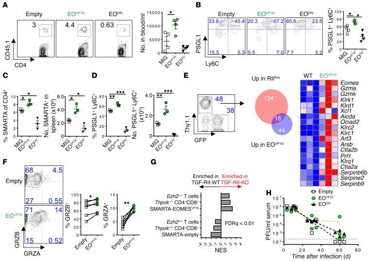
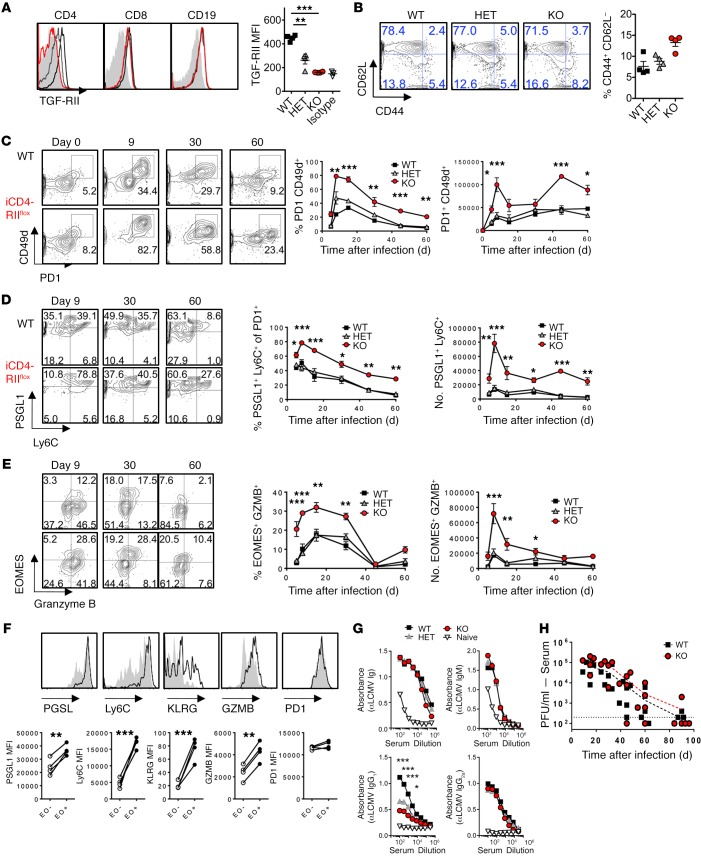
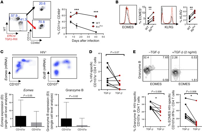
Suppression of CD8 and CD4 T cells is a hallmark in chronic viral infections, including hepatitis C and HIV. While multiple pathways are known to inhibit CD8 T cells, the host molecules that restrict CD4 T cell responses are less understood. Here, we used inducible and CD4 T cell-specific deletion of the gene encoding the TGF-β receptor during chronic lymphocytic choriomeningitis virus infection in mice, and determined that TGF-β signaling restricted proliferation and terminal differentiation of antiviral CD4 T cells. TGF-β signaling also inhibited a cytotoxic program that includes granzymes and perforin expression at both early and late stages of infection in vivo and repressed the transcription factor eomesodermin. Overexpression of eomesodermin was sufficient to recapitulate in great part the phenotype of TGF-β receptor-deficient CD4 T cells, while SMAD4 was necessary for CD4 T cell accumulation and differentiation. TGF-β signaling also restricted accumulation and differentiation of CD4 T cells and reduced the expression of cytotoxic molecules in mice and humans infected with other persistent viruses. These data uncovered an eomesodermin-driven CD4 T cell program that is continuously suppressed by TGF-β signaling. During chronic viral infection, this program limits CD4 T cell responses while maintaining CD4 T helper cell identity.


Figures

Figure 1
Figure 1. Cell-intrinsic TGFβ-RII signaling limits CD4 T cell proliferation but not prototypical T helper subset differentiation early after chronic LCMV infection.
Reconstituted 1:1 mix of WT (CD45.1, black) and ERCre+ Tgfbr2fl/fl (RIIflox-CD45.2, red) bone marrow–chimeric mice were tamoxifen treated, rested, and infected with 2 × 10⁶ PFU of LCMV Cl13. Blood was analyzed prior to infection (A and B) and spleens, livers, and lungs were analyzed on postinfection day 9 for the presence of LCMV-specific CD4 T cells by flow cytometry (CJ). (A) Surface TGFβ-RII on circulating leukocytes after tamoxifen treatment over isotype staining (gray). (B) CD44 and CD62L activation markers on total CD4 and CD8 T cells. (C) Percentage of PD1+ CD4 T cells after gating on CD4 T cells from each donor compartment in the indicated tissue. (D) Incorporation of 7-aminoactinomycin D (7AAD) and BrdU (left) after a 16-hour pulse in splenic CD4 PD1+ T cells or annexin V staining (right) from either WT or RIIflox compartments. (E) Percentages of virus-specific I-Ab:GP67–77+ cells of CD4 T cells. (F) Coproduction of intracellular IFN-γ and TNF-α, or TNF-α and IL-2 after a 5-hour stimulation of splenocytes with GP67–77 cognate peptide, graphed as percentage of I-Ab:GP67–77+ cells from C. (G) Representative overlays and mean fluorescence intensity (MFI) plotted for TBET expression in CD4 I-Ab:GP61–80 T cells in WT and RIIflox compared with naive CD4 T cells (gray). (H and I) CXCR5 vs. BCL6 (H) and SLAM vs. CXCR5 (I) staining on CD4 I-Ab:GP67–77 T cells. (J) FOXP3 expression in PD1+ CD4 T cells. Representative of 3 independent experiments, with n = 4 or 5 mice/experiment. Paired t test, *P < 0.05, **P < 0.005.
Figure 2
Figure 2. TGFβ-RII signaling in CD4 T cells suppressed terminal differentiation and the cytotoxic gene program early after chronic LCMV infection.
Reconstituted 1:1 mix of WT (CD45.1, black) and ERCre+ Tgfbr2fl/fl (RIIflox-CD45.2, red) bone marrow–chimeric mice were tamoxifen treated, rested, and infected with 2 × 10⁶ PFU of LCMV Cl13 and spleens, livers, and lungs were analyzed after 9 days. (A) FACS-sorted CD4+CD8PD1+CD49d+ cells from each compartment were analyzed by microarray. Representative genes shown as a heat map of relative expression values (blue = min; red = max) from differentially regulated genes (P < 10–7). (B) GSEA and normalized enrichment score (NES) of TGFβ-RII CD4 T cell array signature for virus-specific CD4 and CD8 T cell microarrays from acute (effector and memory) and chronic (exhausted) LCMV infection (FDR q < 0.01). (C) PSGL1 vs. Ly6C gated on CD4 I-Ab:GP67–77 in the indicated tissue. (D) EOMES expression gated on PSGL1+Ly6C+ cells from C. (E) EOMES and PD1 expression on virus-specific CD4 T cells. (F) PSGL1 and Ly6C on cells from E. (G) Granzyme B expression in IFN-γ+ cells stimulated with cognate GP67–77 peptide from (Figure 1F). Representative of 3 independent experiments, with n = 4 or 5 mice/experiment. Paired t test, *P < 0.05, **P < 0.005, ***P < 0.0005.
Figure 3
Figure 3. Cell-intrinsic TGFβ-RII signaling in CD4 T cells continuously suppressed terminal differentiation and the cytotoxic gene program late after chronic LCMV infection.
Eight weeks after bone marrow reconstitution with 1:1 mix of WT (CD45.1, black) and ERCre+ Tgfbr2fl/fl (RIIflox-CD45.2, red) bone marrow, mice were first infected with 2 × 10⁶ PFU of LCMV Cl13, tamoxifen treated on postinfection days 12–17, and spleens analyzed on postinfection day 30 for the presence of LCMV-specific T cells by flow cytometry. (A) PD1+ activated CD4 T cells from each compartment in the indicated tissue. (B) BrdU and 7-aminoactinomycin D (7AAD) incorporation after 16 hours in CD4 PD1+ T cells. (C) Accumulation of CD4 I-Ab:GP67–77 T cells by tetramer staining in the indicated tissue. (D) Coproduction of intracellular IFN-γ, TNF-α, and IL-2 after a 5-hour splenocyte stimulation with GP67–77 peptide, graphed as percentage of I-Ab:GP67–77+ cells from C. (EG) Gating on CD4 I-Ab:GP67–77 T cells from C, overlays and mean fluorescence intensity (MFI) plotted for TBET (E) and BCL6 (F) expression over naive CD4 T cells (gray), or SLAM and CXCR5 staining (G). (H) FOXP3 expression on CD4 PD1+ cells. (I and J) PSGL1 and Ly6C (I) or EOMES (J) gated on CD4 I-Ab:GP67–77. (K) Granzyme B expression gated on IFN-γ+ cells from D. Representative of 3 independent experiments, with n = 4 or 5 mice/experiment. Paired t test, *P < 0.05, **P < 0.005.
Figure 4
Figure 4. SMAD4 is required for accumulation and differentiation of CD4 and CD8 T cells during chronic LCMV infection.
Reconstituted 1:1 mix of WT (CD45.1, black) and ERCre+ Smad4fl/fl (iSM4flox, red) bone marrow–chimeric mice were tamoxifen treated for 5 days and 1 week later infected with 2 × 10⁶ PFU of LCMV Cl13. T cells were analyzed in blood by flow cytometry. (A) Percentage of PD1+ CD4 T cells after gating on CD4 T cells from each donor. (B) Expression of indicated markers in PD1+ CD4 T cells. (C) Percentage of PD1+ CD8 T cells after gating on CD8 T cells from each donor. (D) Expression of indicated markers in PD1+ CD8 T cells. WT (black) and iSM4flox (red). Representative of 3 independent experiments, with n = 4 or 5 mice/experiment. Paired t test, *P < 0.05, **P < 0.005, ***P < 0.0005.
Figure 5
Figure 5. EOMES regulates accumulation and differentiation of CD4 T cells during chronic LCMV infection.
LCMV-specific SMARTA+ CD4 T cells (congenic CD45.1) were retrovirally transduced with constitutively active EOMES (EOVP16), dominant-negative EOMES (EODN), or empty vector MSCV-GFP (MIG) and transferred into C57BL/6 mice 24 hours before infection and analyzed 9 days later in the indicated tissue. (A) Number of SMARTA T cells in the blood. (B) Expression of PSGL1 and Ly6C in cells from A. (C and D) Number and percentage of transduced SMARTA cells (C) expressing PSGL1 and Ly6C (D) in the spleen. (E) MIG-EOVP16 and MSCV-Thy1.1+ empty vector–transduced SMARTA+ T cells were cotransferred into the same host and splenic SMARTA cells analyzed by microarray. Overlap of significantly regulated genes by TGFβ-RII KO (from Figure 3) and EOMES overexpression. (F) Protein expression of granzyme A and B on cotransferred SMARTA T cells in spleen. (G) GSEA of TGFβ-RII–regulated genes against CD4+ CD8+ double-positive cells from Thpok–/–, CD4 T cells from Ezh2–/– mice, or EOMES overexpression from E. (H) Kinetics of viremia (PFU/ml) in mice receiving EOMES-modified cells from A. Representative of 2 to 3 independent experiments, with n = 3–6 mice per group. ANOVA (AD and H), paired t test (F), *P < 0.05, **P < 0.005, ***P < 0.0005.
Figure 6
Figure 6. Exclusive ablation of TGFβ-RII in CD4 T cells enhanced their numbers and terminal differentiation but limited LCMV-specific IgG1 during chronic infection.
(A) Cd4-ERCre+ Tgfbr2fl/fl (iCD4-RIIflox, red) mice, Cd4-ERCre+ Tgfbr2fl/+ heterozygotes (HET, gray) and Cre littermate controls (WT, black) were infected with 2 × 10⁶ PFU of LCMV Cl13. Blood was monitored by flow cytometry prior to infection (A and B) and at the indicated time points after infection (CG). (A) TGFβ-RII expression over isotype (filled histogram) on B cells and CD4 and CD8 T cells prior to infection. Mean fluorescence intensity (MFI) for TGFβ-RII on CD4 T cells is graphed. (B) Percentage of activated CD44+CD62L CD4 T cells prior to infection. (C) Percentage and number of virus-specific PD1+CD49d+ cells over time after infection. (D) Percentage and number of PSGL1+Ly6C+ cells within activated CD4 T cells from C. (E) Percentage and number of EOMES- and granzyme B–expressing cells within activated CD4 T cells from C. (F) Expression overlay of indicated marker in EOMES+ (black line) vs. EOMES (gray fill) activated TGFβ-RII–deficient CD4 T cells from C. (G) Anti-LCMV (αLCMV) Ig levels in serum at postinfection day 30. (H) Viremia by plaque assay, as pooled results from 2 experiments. Representative of 3 independent experiments, with n = 4 or 5 mice/group. Two-way ANOVA (AH), paired t test (F), *P < 0.05, **P < 0.005, ***P < 0.0005.
Figure 7
Figure 7. TGF-β suppression of EOMES-driven responses was common to CD4 T cells from mice infected with MCMV and HIV-infected patients.
(A and B) Mixed chimeras with 1:1 ratio of WT (CD45.1, black) and ERCre+ Tgfbr2flox (CD45.2, red) bone marrow reconstituted prior to TGFβ-RII deletion, were infected with 2 × 10⁴ PFU MCMV i.p. (A) Proportion of CD4 T cells expressing activation markers CD11a and CD49d over time after gating on congenic marker, is shown for postinfection day 14. (B) Overlays of EOMES and KLRG expression in activated CD4 T cells. (C) Upper panels show density plots of mRNA expression level vs. CD107 protein and quantification of Eomes and GrzB transcripts in CD107+ vs. CD107 responding CD4 T cells. Lower panels show single-cell analysis of Eomes and granzyme B mRNA expression levels in HIV-specific IFN-γ+CD107a+ CD4 T cells after a 5-hour Gag peptide stimulation using Biomark Fluidigm analysis. The transcript expression threshold (Et) was defined as the qPCR cycle number above background at which the transcript was detected. (D and E) Gag-responsive HIV-specific CD4 T cells were isolated and cultured for 5 days in the presence or absence of TGF-β and restimulated with GAG peptides in the presence of CD28/CD49d, and monensin for percentage of CD107+ CD4 T cells (D) and eomesodermin and granzyme B expression in HIV-specific CD107+ CD4 T cells (E). (A and B) Representative data from 2 independent experiments, with n = 4 or 5 mice/group. (CE) Representative data from n = 10 HIV+ treatment-naive subjects. Two-way ANOVA (A), paired t test (BE), *P < 0.05, **P < 0.005, ***P < 0.0005.

Similar articles

Cited by

References

    1. Stelekati E, Wherry EJ. Chronic bystander infections and immunity to unrelated antigens. Cell Host Microbe. 2012;12(4):458–469. doi: 10.1016/j.chom.2012.10.001. - DOI - PMC - PubMed
    1. Zuniga EI, Macal M, Lewis GM, Harker JA. Innate and adaptive immune regulation during chronic viral infections. Annu Rev Virol. 2015;2(1):573–597. doi: 10.1146/annurev-virology-100114-055226. - DOI - PMC - PubMed
    1. Wherry EJ. T cell exhaustion. Nat Immunol. 2011;12(6):492–499. - PubMed
    1. Nelson DR, et al. Transforming growth factor-β1 in chronic hepatitis C. J Viral Hepat. 1997;4(1):29–35. doi: 10.1046/j.1365-2893.1997.00124.x. - DOI - PubMed
    1. Tinoco R, Alcalde V, Yang Y, Sauer K, Zuniga EI. Cell-intrinsic transforming growth factor-β signaling mediates virus-specific CD8+ T cell deletion and viral persistence in vivo. Immunity. 2009;31(1):145–157. doi: 10.1016/j.immuni.2009.06.015. - DOI - PMC - PubMed

MeSH terms

Substances