

Hydroxysafflor yellow A exerts antioxidant effects in a rat model of traumatic brain injury
- PMID: 27599591
- PMCID: PMC5042747
- DOI: 10.3892/mmr.2016.5720
Hydroxysafflor yellow A exerts antioxidant effects in a rat model of traumatic brain injury
Abstract
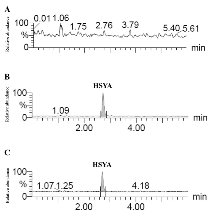
Free radical-induced oxidative damage occurs rapidly and is of primary importance during the secondary pathophysiological cascades of traumatic brain injury (TBI). Hydroxysafflor yellow A (HSYA) is a constituent of the flower petals of Carthamus tinctorius (safflower) and may represent a potential therapeutic strategy to improve outcomes following TBI. The present study aimed to identify HSYA in the brain tissues of rats exposed to TBI to determine its absorption and to investigate the underlying effects of HSYA on antioxidant enzymes in the brain tissues of TBI rats. To determine the absorption of HSYA for the investigation of the underlying antioxidant effects of HSYA in TBI, the presence of HSYA in the brain tissues of the TBI rats was identified using an ultra performance liquid chromatography‑tandem mass spectrometry method. Subsequently, the state of oxidative stress in the TBI rat model following the administration of HSYA was investigated by determining the levels of antioxidant enzymes, including superoxide dismutase (SOD), malondialdehyde (MDA) and catalase (CAT), and the ratio of glutathione (GSH)/glutathione disulfide (GSSG). The data obtained demonstrated that HSYA was absorbed in the brain tissues of the TBI rats. HSYA increased the activities of SOD and CAT, the level of GSH and the GSH/GSSG ratio. However, HSYA concomitantly decreased the levels of MDA and GSSG. These preliminary data suggest that HSYA has the potential to be utilized as a neuroprotective drug in cases of TBI.
Figures


Similar articles
-
Hydroxysafflor yellow A protects rat brains against ischemia-reperfusion injury by antioxidant action.Neurosci Lett. 2005 Sep 23;386(1):58-62. doi: 10.1016/j.neulet.2005.05.069. Neurosci Lett. 2005. PMID: 16002219
-
Restorative effects of hydroxysafflor yellow A on hepatic function in an experimental regression model of hepatic fibrosis induced by carbon tetrachloride.Mol Med Rep. 2017 Jan;15(1):47-56. doi: 10.3892/mmr.2016.5965. Epub 2016 Nov 24. Mol Med Rep. 2017. PMID: 27909717 Free PMC article.
-
Effects of hydroxysafflor yellow A on the experimental traumatic brain injury in rats.J Asian Nat Prod Res. 2010 Mar;12(3):239-47. doi: 10.1080/10286020903510636. J Asian Nat Prod Res. 2010. PMID: 20390772
-
Pharmacological Actions, Molecular Mechanisms, Pharmacokinetic Progressions, and Clinical Applications of Hydroxysafflor Yellow A in Antidiabetic Research.J Immunol Res. 2021 Dec 13;2021:4560012. doi: 10.1155/2021/4560012. eCollection 2021. J Immunol Res. 2021. PMID: 34938814 Free PMC article. Review.
-
Oxidative stress in traumatic brain injury.Curr Med Chem. 2014 Apr;21(10):1201-11. doi: 10.2174/0929867321666131217153310. Curr Med Chem. 2014. PMID: 24350853 Review.
Cited by
-
Three Ingredients of Safflower Alleviate Acute Lung Injury and Inhibit NET Release Induced by Lipopolysaccharide.Mediators Inflamm. 2020 Feb 29;2020:2720369. doi: 10.1155/2020/2720369. eCollection 2020. Mediators Inflamm. 2020. PMID: 32189992 Free PMC article.
-
Nucleolin mediated pro-angiogenic role of Hydroxysafflor Yellow A in ischaemic cardiac dysfunction: Post-transcriptional regulation of VEGF-A and MMP-9.J Cell Mol Med. 2018 May;22(5):2692-2705. doi: 10.1111/jcmm.13552. Epub 2018 Mar 7. J Cell Mol Med. 2018. PMID: 29512890 Free PMC article.
-
Safflower Yellow and Its Main Component HSYA Alleviate Diet-Induced Obesity in Mice: Possible Involvement of the Increased Antioxidant Enzymes in Liver and Adipose Tissue.Front Pharmacol. 2020 Apr 21;11:482. doi: 10.3389/fphar.2020.00482. eCollection 2020. Front Pharmacol. 2020. PMID: 32372961 Free PMC article.
-
Nanoemulsions of Hydroxysafflor Yellow A for Enhancing Physicochemical and In Vivo Performance.Int J Mol Sci. 2023 May 12;24(10):8658. doi: 10.3390/ijms24108658. Int J Mol Sci. 2023. PMID: 37240000 Free PMC article.
-
Metabolomics reveals the effects of hydroxysafflor yellow A on neurogenesis and axon regeneration after experimental traumatic brain injury.Pharm Biol. 2023 Dec;61(1):1054-1064. doi: 10.1080/13880209.2023.2229379. Pharm Biol. 2023. PMID: 37416997 Free PMC article.
References
MeSH terms
Substances
LinkOut - more resources
Full Text Sources
Other Literature Sources
Medical
Miscellaneous
