

Molecular subtypes and imaging phenotypes of breast cancer
- PMID: 27599892
- PMCID: PMC5040136
- DOI: 10.14366/usg.16030
Molecular subtypes and imaging phenotypes of breast cancer
Abstract
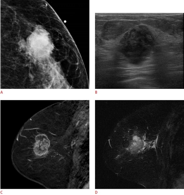
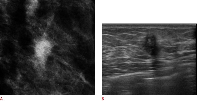
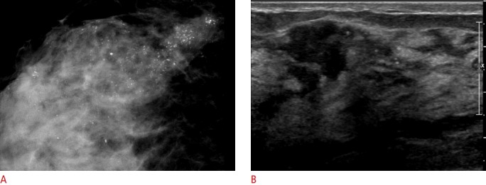
During the last 15 years, traditional breast cancer classifications based on histopathology have been reorganized into the luminal A, luminal B, human epidermal growth factor receptor 2 (HER2), and basal-like subtypes based on gene expression profiling. Each molecular subtype has shown varying risk for progression, response to treatment, and survival outcomes. Research linking the imaging phenotype with the molecular subtype has revealed that non-calcified, relatively circumscribed masses with posterior acoustic enhancement are common in the basal-like subtype, spiculated masses with a poorly circumscribed margin and posterior acoustic shadowing in the luminal subtype, and pleomorphic calcifications in the HER2-enriched subtype. Understanding the clinical implications of the molecular subtypes and imaging phenotypes could help radiologists guide precision medicine, tailoring medical treatment to patients and their tumor characteristics.
Keywords: Breast neoplasms; Diagnosis; Gene expression profiling; Ultrasonography.
Conflict of interest statement
No potential conflict of interest relevant to this article was reported.
Figures
References
-
- Ng CK, Schultheis AM, Bidard FC, Weigelt B, Reis-Filho JS. Breast cancer genomics from microarrays to massively parallel sequencing: paradigms and new insights. J Natl Cancer Inst. 2015;107:djv015. - PubMed
-
- Goldhirsch A, Winer EP, Coates AS, Gelber RD, Piccart-Gebhart M, Thurlimann B, et al. Personalizing the treatment of women with early breast cancer: highlights of the St Gallen International Expert Consensus on the Primary Therapy of Early Breast Cancer 2013. Ann Oncol. 2013;24:2206–2223. - PMC - PubMed
Publication types
LinkOut - more resources
Full Text Sources
Other Literature Sources
Research Materials
Miscellaneous
