Review
. 2015 Mar 24;4(2):133-61.
doi: 10.3390/microarrays4020133.

3D Cell Culture in Alginate Hydrogels

Affiliations
Review

3D Cell Culture in Alginate Hydrogels

Therese Andersen et al. Microarrays (Basel). .

Abstract

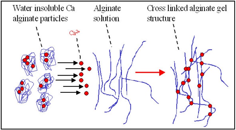
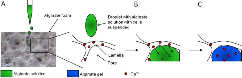
This review compiles information regarding the use of alginate, and in particular alginate hydrogels, in culturing cells in 3D. Knowledge of alginate chemical structure and functionality are shown to be important parameters in design of alginate-based matrices for cell culture. Gel elasticity as well as hydrogel stability can be impacted by the type of alginate used, its concentration, the choice of gelation technique (ionic or covalent), and divalent cation chosen as the gel inducing ion. The use of peptide-coupled alginate can control cell-matrix interactions. Gelation of alginate with concomitant immobilization of cells can take various forms. Droplets or beads have been utilized since the 1980s for immobilizing cells. Newer matrices such as macroporous scaffolds are now entering the 3D cell culture product market. Finally, delayed gelling, injectable, alginate systems show utility in the translation of in vitro cell culture to in vivo tissue engineering applications. Alginate has a history and a future in 3D cell culture. Historically, cells were encapsulated in alginate droplets cross-linked with calcium for the development of artificial organs. Now, several commercial products based on alginate are being used as 3D cell culture systems that also demonstrate the possibility of replacing or regenerating tissue.

Keywords: 3D; AlgiMatrix®; NovaMatrix®-3D; alginate; beads; bioprinting; drug development; drug discovery; hydrogel; tissue regeneration.


Figures

Figure 1
Figure 1
The structure of alginate shown as the segment of ..MMGG.. residues [18]. Epimerisation of the M residues changes the conformation of the sugar from 4C1 to 1C4 [19,20].
Figure 2
Figure 2
Four consecutive G-residues from two chain segments of one or two alginate molecules creating a binding site for divalent cations (A). Formation of an intermolecular network of alginate molecules formed in presence of gelling ions such as Ca2+ (B). Gelling ions organized in alternative junction zones (C) [18].
Figure 3
Figure 3
Chemical structure of RGD-alginate (arginine-glycine-aspartic acid conjugated to sodium alginate) (prepared using ISIS Draw).
Figure 4
Figure 4
Cellular organization within the alginate gel obtained by confocal microscopy of vital stained (Calcein AM) cells. Panels A–D and E–F show cells cultured without and with RGD-alginate, respectively. C2C12: murine myoblasts, HT-29: human colorectal adenocarcinoma cells, V79-379A: Chinese hamster lung fibroblasts, L1210: murine leukemia suspension cells, NIH:OVCAR-3: human ovarian adenocarcinoma cells, HCT 116: human colorectal carcinoma cells. Magnification ×100, scale bar 100 µm. Adapted from [60].
Figure 5
Figure 5
Elasticity scale of soft tissues. Adapted from reference [74]. Reprinted with permission from AAAS.
Figure 6
Figure 6
Encapsulation of cells with alginate. (From FMC internal archive.)
Figure 7
Figure 7
Principle of electrostatic (left) and coaxial air flow (right) bead generators [80].
Figure 8
Figure 8
Examples of encapsulated cells in alginate. (From FMC internal archive.)
Figure 9
Figure 9
Principle of delayed gelling of a hydrogel made from alginate only [99]. By mixing a sodium alginate solution with a dispersion of insoluble calcium alginate, using for example connected syringes, a homogeneous hydrogel can be made. Upon mixing, gel forming ions rearrange between insoluble and soluble alginate molecules resulting in gel formation.
Figure 10
Figure 10
Schematic presentation of the steps for in situ gelation in macroporous alginate scaffolds. (A) Am alginate solution with cells is applied on top of a dry scaffold containing calcium ions, (B) rehydration of the scaffold by the alginate solution filling its pores and diffusion of calcium ions from the foam lamellas to the absorbed alginate, and (C) formation of a calcium cross-linked alginate hydrogel inside the pores of the foam. From reference [77].

References

    1. Lee J., Cuddihy M.J., Kotov N.A. Three-Dimensional Cell Culture Matrices: State of the Art. Tissue Eng. 2008;14:61–86. doi: 10.1089/teb.2007.0150. - DOI - PubMed
    1. Gevaert M. Engineering 3D Tissue Systems to Better Mimic Human Biology. Bridge. 2012;42:48–55.
    1. Weigelt B., Lo A.T., Park C.C., Gray J.W., Bissell M.J. HER2 Signaling Pathway Activation and Response of Breast Cancer Cells to HER2-Targeting Agents Is Dependent Strongly on the 3D Microenvironment. Breast. Cancer Res. Treat. 2010;122:35–43. doi: 10.1007/s10549-009-0502-2. - DOI - PMC - PubMed
    1. Kenny P.A., Lee G.Y., Myers C.A., Neve R.M., Semeiks J.R., Spellman P.T., Lorenz K., Lee E.H., Barcellos-Hoff M.H., Petersen O.W., et al. The Morphologies of Breast Cancer Cell Lines in Three-Dimensional Assays Correlate With Their Profiles of Gene Expression. Mol. Oncol. 2007;1:84–96. doi: 10.1016/j.molonc.2007.02.004. - DOI - PMC - PubMed
    1. Paul S.M., Mytelka D.S., Dunwiddie C.T., Persinger C.C., Munos B.H., Lindborg S.R., Schacht A.L. How to Improve R&D Productivity: The Pharmaceutical Industry's Grand Challenge. Nat. Rev. Drug Discov. 2010;9:203–214. - PubMed