. 2016 Sep 7:7:12630.
doi: 10.1038/ncomms12630.

ATRA mechanically reprograms pancreatic stellate cells to suppress matrix remodelling and inhibit cancer cell invasion

Affiliations

ATRA mechanically reprograms pancreatic stellate cells to suppress matrix remodelling and inhibit cancer cell invasion

Antonios Chronopoulos et al. Nat Commun. .

Abstract

Pancreatic ductal adenocarcinoma (PDAC) is a highly aggressive malignancy with a dismal survival rate. Persistent activation of pancreatic stellate cells (PSCs) can perturb the biomechanical homoeostasis of the tumour microenvironment to favour cancer cell invasion. Here we report that ATRA, an active metabolite of vitamin A, restores mechanical quiescence in PSCs via a mechanism involving a retinoic acid receptor beta (RAR-β)-dependent downregulation of actomyosin (MLC-2) contractility. We show that ATRA reduces the ability of PSCs to generate high traction forces and adapt to extracellular mechanical cues (mechanosensing), as well as suppresses force-mediated extracellular matrix remodelling to inhibit local cancer cell invasion in 3D organotypic models. Our findings implicate a RAR-β/MLC-2 pathway in peritumoural stromal remodelling and mechanosensory-driven activation of PSCs, and further suggest that mechanical reprogramming of PSCs with retinoic acid derivatives might be a viable alternative to stromal ablation strategies for the treatment of PDAC.


Figures

Figure 1
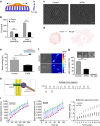
Figure 1. ATRA increases focal adhesion size and cell–ECM adhesion strength of PSCs.
(a) Immunostaining images of talin and paxillin. Scale bar, 20 μm. Zoomed areas, 10 μm. (b,c) Quantification of normalized focal adhesion area and intensity. (d) Schematic representation of the magnetic tweezers device used in this study to apply forces on the cell surface. (e) Quantification of cell-bound beads that detached after force application. (f) Quantification of cell spreading area over time. (g) Time-elapsed sequential images of PSCs spreading until PSCs attained maximum area. Scale bars, 10 μm. In all panels, error bars represent s.e.m. *P<0.05; **P<0.01; ***P<0.001; (t-test); three experimental replicates.
Figure 2
Figure 2. ATRA reduces traction forces and impairs mechanosensing capacity in PSCs.
(a) Schematic representation of the elastic pillars microsensors (F, force; x, distance). (b) Average force applied on each pillar for early and late spreading. n>10 in all cases. (c) Bright-field images of representative PSCs on top of the micropillars—early spreading phase. Scale bar, 10 μm. (d) The respective force vector maps indicating the magnitude and direction of applied forces calculated from the maximum pillar displacement through a custom-built tracking algorithm. (e) Strain energy (mean±s.e.m., n>300 cells per condition) imparted by stationary PSCs. (f) Bright-field images of representative PSCs on FN-coated, polyacrylamide gels and corresponding traction maps. (g) Images show gel contraction by PSCs. Histogram shows quantification of n>10 gels per condition assessed over multiple experiments. (h) Left, diagram of the cell–matrix interface showing application of mechanical tension onto integrin receptors to mimic the transmission of mechanical force from the extracellular matrix to the cytoskeleton via the mechanical bridge formed by integrins and focal adhesions. Right, pulsatile force regimen applied with magnetic tweezers. (i) Left and middle, representative traces tracking the displacement of the beads in response to the force (mechanosensing). The amplitude of the bead displacement was progressively reduced in control PSCs, showing that cells detected force application and responded by stiffening their cytoskeleton. In ATRA-treated PSCs, the reduction in bead displacement was severely suppressed indicating impaired ability to detect external mechanical stimuli. Black and red arrows indicate initial and final amplitude of the bead oscillation, respectively. Right, cell stiffening response to force, (P<0.001, n=26 traces for control, n=34 for ATRA). When not specified, results are expressed as mean±s.e.m. In all panels, error bars represent s.e.m. *P<0.05; **P<0.01; ***P<0.001; (t-test); three or more experimental replicates.
Figure 3
Figure 3. ATRA impairs PSCs capacity to remodel the ECM to promote cancer invasion.
(a) Elastic modulus for collagen Matrigel matrices previously remodelled by PSCs expressed as mean±s.e.m.; control n=113 and ATRA n=114 measurements obtained in three independent experiments. (b) Collagen second-harmonic signals (green) and cells (red). Scale bar, 20 μm. (c) Representative images of haematoxylin and eosin immunofluorescence staining showing AsPC1 cancer cell invasions. Scale bar, 50 μm. (d) AsPC1 invasion expressed as number of invading particles and area, respectively. Each point represents a different section. In all panels, ***P<0.001; ****P<0.0001; (t-test).
Figure 4
Figure 4. ATRA downregulates MLC-2 actomyosin contractility.
(a) Immunofluorescent images of total myosin light-chain 2 (MLC-2) and phosphorylated levels (pMLC-2). (b) Quantification of staining intensity (n>20 cells in all cases and three experimental replicates). Scale bar, 50 μm. (c,d) MLC-2 level expression at the protein (western blotting) and mRNA (qPCR) levels. All results are expressed as mean±s.e.m. and *P<0.05; **P<0.01; ***P<0.001; (t-test), three independent experiments for western blotting and qPCR.
Figure 5
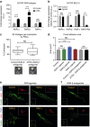
Figure 5. ATRA biomechanically reprograms PSCs in a RAR-β-dependent manner.
(a) qPCR mRNA levels of the different retinoic acid receptor (RAR) subtypes, RAR-α, RAR-β and RAR-γ in untreated control and ATRA-treated PSCs. Bars represent mean±s.e.m. (b) qPCR mRNA levels of MLC-2 in control PSCs and in PSCs treated with agonists for RAR-α, RAR-β and RAR-γ. Bars represent mean±s.e.m. (c) 3D gel contraction assay using control and ATRA-treated PSCs in the presence of RAR-β antagonist. In the box-and-whisker plot, the central box represents values from the lower to the upper quartile. The middle line represents the mean. The vertical line extends from the minimum to the maximum value. Control and ATRA 12 and 15 gels, respectively, assessed in three independent experiments. NS, no significant differences. Dotted yellow lines represent the gels contours of representative images of gel contraction. (d) Quantification of focal adhesions size of the images presented in e,f. Bars represent mean±s.e.m. (e) Representative images of focal adhesions size in PSCs treated with agonists for RAR-α, RAR-β and RAR-γ. (f) Representative images of focal adhesion sizes in PSCs untreated control and ATRA treated in the presence of RAR-β antagonist. Scale bars for e,f: main image 20 μm; focal adhesion inset 5 μm; and blue/red channels inset 20 μm. In all panels, data were collected during three or more independent experiments and *P<0.05; **P<0.01; ***P<0.001; (t-test).
Figure 6
Figure 6. Model for the biomechanical reprogramming of PSCs.
ATRA downregulates myosin II-dependent force generation and mechanosensing in PSCs. PSCs are reprogrammed to a more quiescent phenotype, cannot migrate, remodel the ECM or promote cancer invasion.

References

    1. Winter J. M. et al. Survival after resection of pancreatic adenocarcinoma: results from a single institution over three decades. Ann. Surg. Oncol. 19, 169–175 (2012). - PubMed
    1. Apte M. V. et al. Desmoplastic reaction in pancreatic cancer: role of pancreatic stellate cells. Pancreas 29, 179–187 (2004). - PubMed
    1. Vonlaufen A. et al. Pancreatic stellate cells: partners in crime with pancreatic cancer cells. Cancer Res. 68, 2085–2093 (2008). - PubMed
    1. Wilson J. S., Pirola R. C. & Apte M. V. Stars and stripes in pancreatic cancer: role of stellate cells and stroma in cancer progression. Front. Physiol. 5, 52 (2014). - PMC - PubMed
    1. Erkan M. et al. The role of stroma in pancreatic cancer: diagnostic and therapeutic implications. Nat. Rev. Gastroenterol. Hepatol. 9, 454–467 (2012). - PubMed

Publication types

MeSH terms