Review
. 2016 Aug 26;14(1):129.
doi: 10.1186/s12916-016-0676-5.

Post-mortem assessment in vascular dementia: advances and aspirations

Affiliations
Review

Post-mortem assessment in vascular dementia: advances and aspirations

Kirsty E McAleese et al. BMC Med. .

Abstract

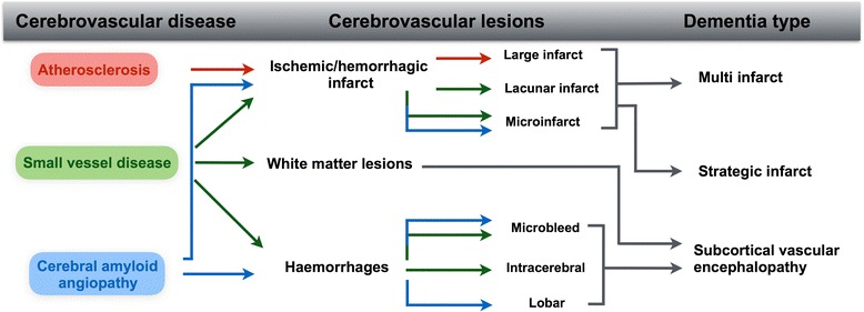
Background: Cerebrovascular lesions are a frequent finding in the elderly population. However, the impact of these lesions on cognitive performance, the prevalence of vascular dementia, and the pathophysiology behind characteristic in vivo imaging findings are subject to controversy. Moreover, there are no standardised criteria for the neuropathological assessment of cerebrovascular disease or its related lesions in human post-mortem brains, and conventional histological techniques may indeed be insufficient to fully reflect the consequences of cerebrovascular disease.

Discussion: Here, we review and discuss both the neuropathological and in vivo imaging characteristics of cerebrovascular disease, prevalence rates of vascular dementia, and clinico-pathological correlations. We also discuss the frequent comorbidity of cerebrovascular pathology and Alzheimer's disease pathology, as well as the difficult and controversial issue of clinically differentiating between Alzheimer's disease, vascular dementia and mixed Alzheimer's disease/vascular dementia. Finally, we consider additional novel approaches to complement and enhance current post-mortem assessment of cerebral human tissue.

Conclusion: Elucidation of the pathophysiology of cerebrovascular disease, clarification of characteristic findings of in vivo imaging and knowledge about the impact of combined pathologies are needed to improve the diagnostic accuracy of clinical diagnoses.

Keywords: Cerebrovascular disease; Cerebrovascular lesions; Magnetic resonance imaging; Mixed dementia; Neuropathology; Post-mortem MRI; Vascular cognitive impairment; Vascular dementia.


Figures

Fig. 1
Fig. 1
Schematic diagram illustrating the three most commonly observed cerebrovascular diseases and their resulting cerebrovascular lesions that may lead to specific types of vascular dementia
Fig. 2
Fig. 2
A series of images for three separate cases indicating normal-appearing white matter and the similarity of white matter changes with differing pathogenesis in the deep white matter of the parietal lobe (Brodman area 39/40), as seen on both T2-weighted magnetic resonance imaging (MRI) and on histology. (AAiv) Normal-aged control brain with no obvious white matter changes or small vessel disease (SVD), and no Alzheimer’s disease (AD)-related pathology: (A) post-mortem T2-weighted MRI scan of normal-appearing white matter; (Ai, Aii) corresponding histological magnified image of normal-appearing white matter and a normal white matter artery (Aii); (Aiv) overlying cortex with no hyperphosphorylated tau (HPτ) pathology. (BBiv) Normal-aged case that exhibited severe white matter hyperintensities (WMHs)/lesions with SVD but no AD pathology: (B) post-mortem T2-weighted MRI scan indicating confluent WMH; (Bi) corresponding histological magnified image of white matter lesion indicated by widespread pallor of the central white matter with typical sparing of the subcortical U-fibres (arrow); (Bii) higher magnification of white matter lesion exhibiting severe rarefaction, that is, myelin and axonal loss; (Biii) white matter arterioles from white matter lesion area exhibiting arteriolosclerosis with hyalinisation (arrows) of vessel walls; (Biv) overlying cortex with no HPτ pathology. In this case, one may speculate SVD-related hypoperfusion was the primary cause of white matter changes. (CCiv) AD brain exhibiting severe WMHs/lesions and no obvious SVD: (C) post-mortem T2-weighted MRI scan indicating confluent white WMH; (Ci) white matter lesion with severe white matter pallor; (Cii) magnified image of severe white matter rarefaction; (Ciii) white matter arteriole with enlarged perivascular space but no SVD-related fibrosis or hyalinisation; (Civ, overlying parietal cortex exhibiting severe HPτ pathology. In this case, one may speculate white matter changes were the result of degenerative myelin and axonal loss as a result of grey matter atrophy in the overlying cortex or via protease-mediated degradation, activated by AD pathology-related axonal transport dysfunction. MRI scans captured in sagittal plane. Microphotoimages captured from serial sections. Histological stain Luxol fast blue was used for images Ai–ii, Bi–ii and Ci–ii; hematoxylin and eosin stain was used for Aiii, Biii and Ciii. Immunohistochemistry with the AT8 antibody was performed in Aiv, Biv and Civ. Scale bars represent 1000 μm in images A, B and C and 20 μm in images Ai–iii, Bi–iii and Ci–iii
Fig. 3
Fig. 3
Magnetic resonance imaging (MRI) and histological sections of cerebral tissue exhibiting microhaemorrhages. (A) Radiological characteristics of microhaemorrhages inclusive of small, well-demarcated hypointense ovoid lesions (arrow). (BCi) Images from an 81-year-old man with dementia and severe cerebral amyloid angiopathy on pathology: (B) post-mortem 7 T MRI scan of hypointense ovoid lesion (arrow); (C) magnified image of cortical microhaemorrhage; (Ci) increased magnified image of cortical microhaemorrhage – brown deposits are haemosiderin (arrow) and yellow deposit is haematoidin (arrow head), indicating the microhaemorrhage is subacute. Histological stain hematoxylin and eosin used on images C and Ci. Scale bars represent 1000 μm in image C, and 100 μm in image Ci. Images prepared by Dr S. van Veluw
Fig. 4
Fig. 4
Schematic illustration of the distribution of myelin-associated glycoprotein (MAG; pink dots) and proteolipid protein 1 (PLP1; green dots) in the myelin sheath. When the supply of oxygen and glucose is insufficient to meet the metabolic needs of the oligodendrocyte, as occurs in hypoperfusion, the first part of the cell to degenerate is the adaxonal loop of myelin – the part of the oligodendrocyte that is furthest away from the cell body (so-called dying back oligodendrogliopathy). Because MAG is restricted to the adaxonal loop of myelin whereas PLP1 is widely distributed throughout the myelin sheath, hypoperfusion leads to greater loss of MAG than PLP1. In contrast, degeneration of nerve fibres causes loss of both MAG and PLP1. The severity of ante mortem hypoperfusion can be assessed by measuring the ratio of MAG to PLP1. Illustration from [175] with permission from Prof. S. Love
Fig. 5
Fig. 5
Neuroinflammatory markers in donated human brain tissue from older people. a Immunohistochemical labelling for the pan-selective microglial marker Iba-1. b Activated microglia in a phagocytic state, with amoeboid morphology, immunoreactive for lysosomal marker CD68 (clone PGM1). c Immunoreactivity for endothelial marker thrombomodulin (TM) in a small penetrating artery of the anterior putamen. d Immunoreactivity for the large plasma protein fibrinogen (FGEN) in deep subcortical white matter. Perivascular cells with astrocytic morphology show cellular labelling (arrows). e A localised cluster of activated microglia (CD68+ (PGM1)), indicating a focal white matter lesion within deep subcortical white matter. f Magnified image of E exhibiting a small arterial vessel. Haematoxylin counterstain was used in a–f. Scale bars represent 20 μm in images a, b and c; 100 μm in image e, and 50 μm in images d and f

References

    1. Beach TG, Wilson JR, Sue LI, Newell A, Poston M, Cisneros R, Pandya Y, Esh C, Connor DJ, Sabbagh M, et al. Circle of Willis atherosclerosis: association with Alzheimer's disease, neuritic plaques and neurofibrillary tangles. Acta Neuropathol. 2007;113(1):13–21. doi: 10.1007/s00401-006-0136-y. - DOI - PubMed
    1. Stary HC. Natural history and histological classification of atherosclerotic lesions: an update. Arterioscler Thromb Vasc Biol. 2000;20(5):1177–1178. doi: 10.1161/01.ATV.20.5.1177. - DOI - PubMed
    1. Grinberg LT, Thal DR. Vascular pathology in the aged human brain. Acta Neuropathol. 2010;119(3):277–290. doi: 10.1007/s00401-010-0652-7. - DOI - PMC - PubMed
    1. Thal DR, Ghebremedhin E, Orantes M, Wiestler OD. Vascular pathology in Alzheimer disease: correlation of cerebral amyloid angiopathy and arteriosclerosis/lipohyalinosis with cognitive decline. J Neuropathol Exp Neurol. 2003;62(12):1287–1301. doi: 10.1093/jnen/62.12.1287. - DOI - PubMed
    1. Vinters HV. Cerebral amyloid angiopathy. A critical review. Stroke. 1987;18(2):311–324. doi: 10.1161/01.STR.18.2.311. - DOI - PubMed