. 2016 Sep 6;11(1):64.
doi: 10.1186/s13024-016-0128-x.

Deimmunization for gene therapy: host matching of synthetic zinc finger constructs enables long-term mutant Huntingtin repression in mice

Affiliations

Deimmunization for gene therapy: host matching of synthetic zinc finger constructs enables long-term mutant Huntingtin repression in mice

Carmen Agustín-Pavón et al. Mol Neurodegener. .

Abstract

Background: Synthetic zinc finger (ZF) proteins can be targeted to desired DNA sequences and are useful tools for gene therapy. We recently developed a ZF transcription repressor (ZF-KOX1) able to bind to expanded DNA CAG-repeats in the huntingtin (HTT) gene, which are found in Huntington's disease (HD). This ZF acutely repressed mutant HTT expression in a mouse model of HD and delayed neurological symptoms (clasping) for up to 3 weeks. In the present work, we sought to develop a long-term single-injection gene therapy approach in the brain.

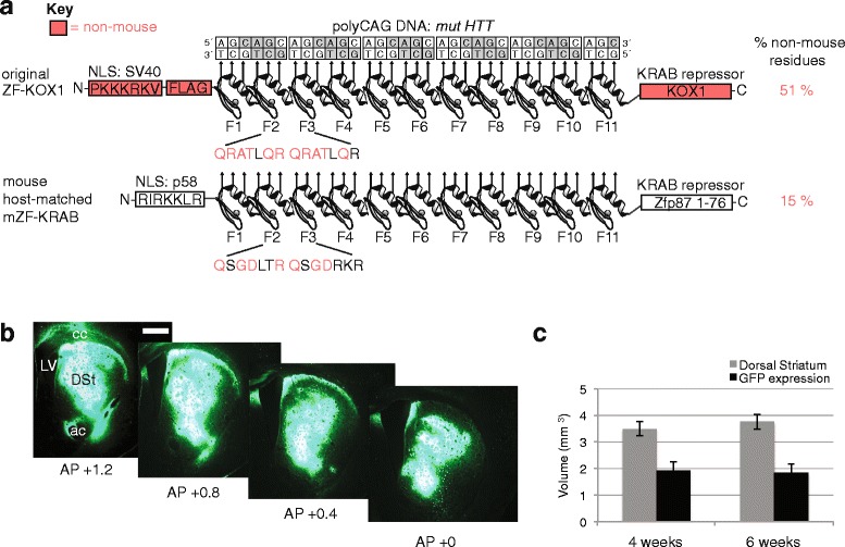
Method: Since non-self proteins can elicit immune and inflammatory responses, we designed a host-matched analogue of ZF-KOX1 (called mZF-KRAB), to treat mice more safely in combination with rAAV vector delivery. We also tested a neuron-specific enolase promoter (pNSE), which has been reported as enabling long-term transgene expression, to see whether HTT repression could be observed for up to 6 months after AAV injection in the brain.

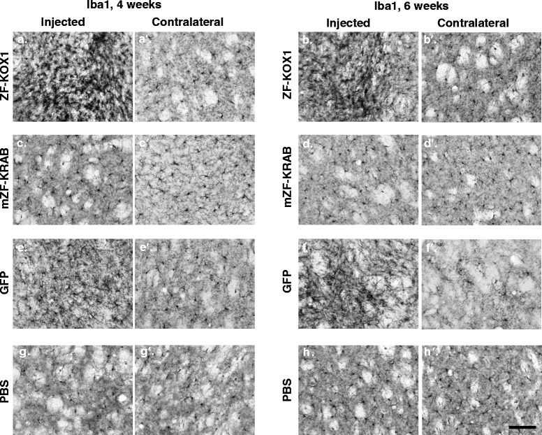
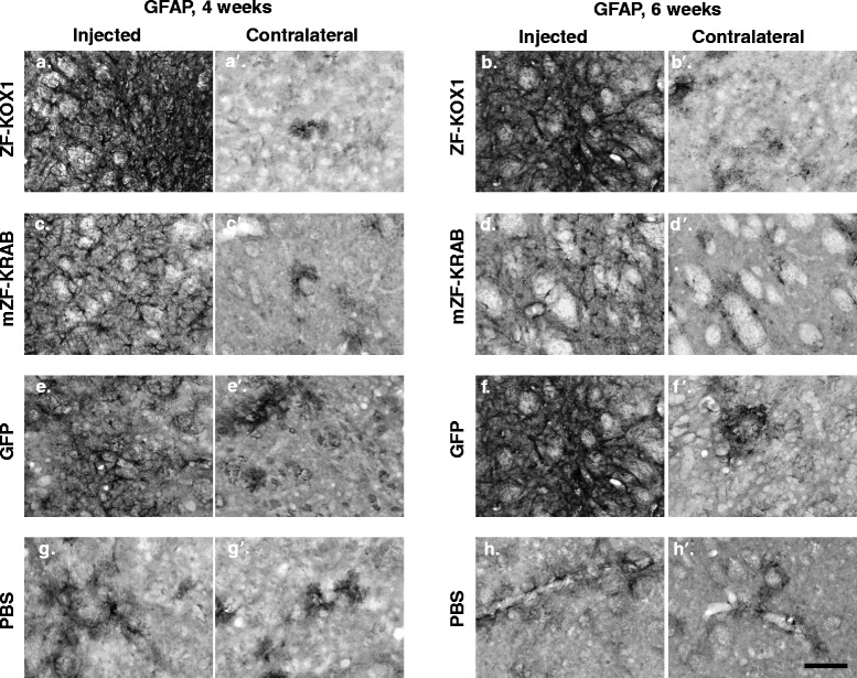
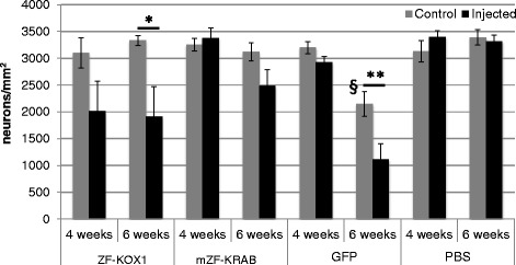
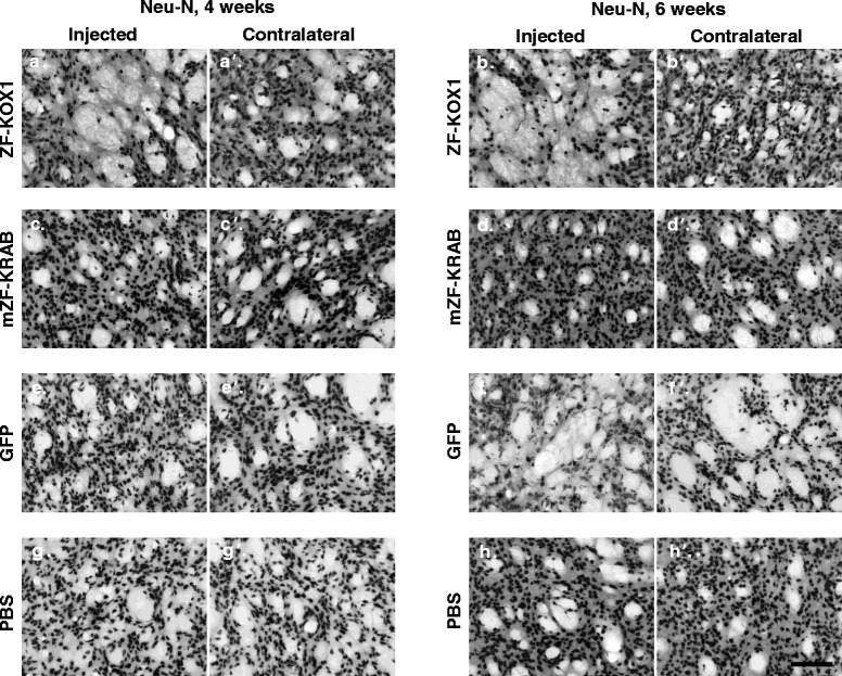
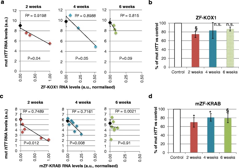
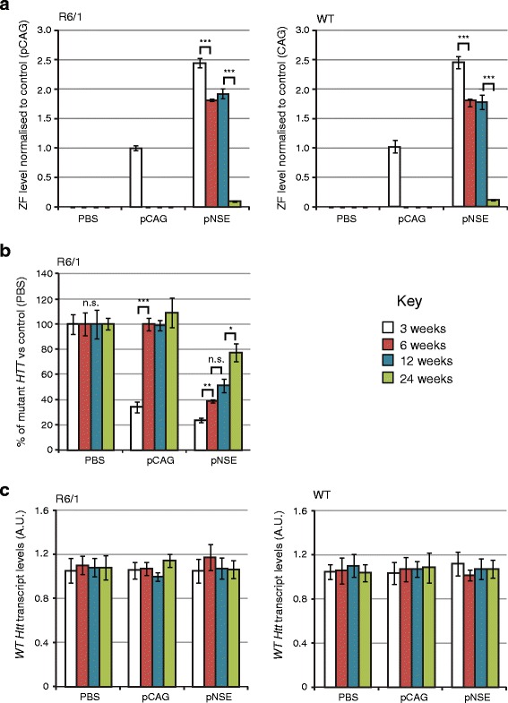
Results: After rAAV vector delivery, we found that non-self proteins induce significant inflammatory responses in the brain, in agreement with previous studies. Specifically, microglial cells were activated at 4 and 6 weeks after treatment with non-host-matched ZF-KOX1 or GFP, respectively, and this was accompanied by a moderate neuronal loss. In contrast, the host-matched mZF-KRAB did not provoke these effects. Nonetheless, we found that using a pCAG promoter (CMV early enhancer element and the chicken β-actin promoter) led to a strong reduction in ZF expression by 6 weeks after injection. We therefore tested a new non-viral promoter to see whether the host-adapted ZF expression could be sustained for a longer time. Vectorising mZF-KRAB with a promoter-enhancer from neuron-specific enolase (Eno2, rat) resulted in up to 77 % repression of mutant HTT in whole brain, 3 weeks after bilateral intraventricular injection of 10(10) virions. Importantly, repressions of 48 % and 23 % were still detected after 12 and 24 weeks, respectively, indicating that longer term effects are possible.

Conclusion: Host-adapted ZF-AAV constructs displayed a reduced toxicity and a non-viral pNSE promoter improved long-term ZF protein expression and target gene repression. The optimized constructs presented here have potential for treating HD.

Keywords: Gene therapy; Host optimization; Huntington’s disease; Immune response; Monogenetic disease; Neurodegenerative disorder; Synthetic transcription factors; rAAV.


Figures

Fig. 1
Fig. 1
Zinc finger (ZF) mouse host-adaptation design and rAAV2/1 transfer into the striatum. a Comparison of the ZF-KOX1 and mZF-KRAB zinc finger repressor designs, showing the 11-finger constructs aligned to their target poly(CAG) DNA sequence (mut HTT). Protein domains containing non-mouse peptide sequences (containing potential foreign epitopes) are shaded in red. The sequences of representative DNA recognition helices from fingers 2 and 3 (F2, F3) are displayed below the ZF arrays, with foreign sequences in red font. The percentage totals of non-mouse residues within the full length peptide sequences are given to show that the mouse-adapted design reduces overall non-host sequences. Full annotated sequences are provided in Additional file 1. b Representative specimen showing GFP expression in mouse coronal slices of hemi-brains, from anterior to posterior view. The anteroposterior (AP) location of each slice within the brain of the mouse is shown as AP distance from Bregma, following [39]. c Bar chart showing the average volume (±S.E.M) of the whole dorsal striatum and the volume covered by GFP fluorescence. Abbreviations: NLS, nuclear localization signal; ac, anterior commissure; cc, corpus callosum; DSt, dorsal striatum; LV, lateral ventricle. Scale bar: 1 mm
Fig. 2
Fig. 2
Relative O.D. values of the striatal samples immunostained for glial markers. Relative O.D values, representing inflammatory responses to various treatments, were calculated for the microglial marker IBA1 (a) and the reactive astroglial marker GFAP (b), at 4 and 6 weeks after injection. Unoptimized ZF-KOX1 treatment was compared to expression of a host optimized mZF-KRAB, GFP or a control PBS injection. Relative O.D. is calculated as the mean O.D. of four coronal slices, separated by 240 μm in the injected hemisphere, minus the average O.D. in the contralateral control hemisphere. Data are displayed as Relative O.D. ± S.E.M, *** P < 0.001, **P < 0.01, *P < 0.05
Fig. 3
Fig. 3
Microglial activation in the striatum after various treatments. Representative micrographs of IBA1 immunostained striatal coronal slices, for the control and injected hemispheres, for each treatment at 4 or 6 weeks. ZF-KOX1 samples displayed an apparent increase in Iba1 immunoreactivity in the injected hemispheres, at 4 and 6 weeks after treatment (a, b). This was not observed in the contralateral hemispheres (a’, b’). Hemispheres treated with mZF-KRAB showed similar levels of Iba1+ cells compared with their contralateral non-injected hemispheres (c, c’, d, d’). Certain GFP-treated samples showed a slight increase in Iba1 immunoreactivity 4 weeks after treatment (e). Iba1 immunoreactivity was significantly increased 6 weeks after GFP injections, compared with the contralateral hemispheres (f, f’). PBS-injected samples show similar Iba1 immunoreactivity between hemispheres at both time points (g, g’, h, h’). Scale bar: 100 μm
Fig. 4
Fig. 4
Astroglial activation in the striatum and cortex after various treatments. Representative micrographs of GFAP immunostained striatal coronal slices for the control and injected hemispheres, for each treatment at each time point. ZF-KOX1 samples displayed a strong and sustained increase in GFAP immunoreactivity in the injected hemispheres, 4 and 6 weeks after treatment (a, b). In contrast, mZF-KRAB treatment provoked a transient upregulation of GFAP immunoreactivity, which started to decline at 6 weeks post-injection (c, d). GFAP immunoreactivity after GFP injections followed the pattern of Iba1 staining, with a slight increase 4 weeks post-treatment and significant signal increase at 6 weeks post-treatment (e, f). Isolated, non-reactive astrocytes can be observed at PBS-injected samples and their contralateral hemispheres (a’, b’, c’, d’, e’, d’, g, g’, h, h’). Scale bar: 100 μm
Fig. 5
Fig. 5
Quantification of striatal neuronal density after various treatments. Bar chart representing the estimated neuronal density in the striata of mice after the different treatments. Data are expressed as mean ± S.E.M. *p < 0.05; **p < 0.01; § p < 0.01 (§compares cell counts in the contralateral hemispheres of the 6-week GFP and PBS samples. GFP is the only treatment in this study where cell numbers are reduced in the contralateral, non-injected hemisphere)
Fig. 6
Fig. 6
Visualising striatal neuronal density after various treatments. Representative micrographs of Neu-N immunostained striatal coronal slices for the control and injected hemisphere of each treatment at each time point. ZF-KOX1 toxicity is observed in areas of the injected striata that are devoid of marked neurons, 4 and 6 weeks after treatment (a, b), whereas the contralateral hemispheres (a’, b’) show neuronal densities similar to PBS injected (g, h) and untreated hemispheres (g’, h’). Conversely, mZF-KRAB treatment did not significantly affect neuronal density (c, c’, d, d’). Strikingly, GFP injections did not affect neuronal density at 4 weeks after treatment (e, e’), but caused a delayed strong toxic response that reduced neuronal density both in the injected (f) and the contralateral hemisphere (f’), 6 weeks post-injection. Scale bar: 100 μm
Fig. 7
Fig. 7
Mutant huntingtin gene expression analysis after treatment with ZFs. a Linear regression showing negative correlations of mut HTT RNA levels and ZF-KOX1 expression 2, 4, and 6 weeks after treatment, suggesting an effective repression of mut HTT by the treatment. Black diamonds show the mean mut HTT expression values (±1 S.E.M.) of the control hemispheres of each group. ZF-KOX1 expression levels are in arbitrary units (a.u), normalised to the maximum ZF-KOX1 qRT-PCR signal across all samples. b Percentage of mut HTT with respect to the average value in the control hemispheres, over the same period. The data show an average of ~25 % reduction of mut HTT, 2 weeks post-treatment (previously reported in [14]), with an individual mouse showing up to ~40 % reduction. The average percentage increases with time, but later values should be interpreted cautiously because of ZF-KOX1 expression leakage to the contralateral hemisphere, and because of the significant neuronal loss. c Linear regression analysis testing for negative correlations between mut HTT RNA levels and mZF-KRAB expression, at 2, 4 and 6 weeks after treatment. mZF-KRAB expression levels are in arbitrary units (a.u), normalised to the maximum mZF-KRAB qRT-PCR signal across all samples. d Percentage of mut HTT with respect to the average value in the control hemispheres over the same period. The columns show mean RNA expression levels; error bars: ±1 S.E.M. *p < 0.05; § p < =0.06
Fig. 8
Fig. 8
Long-term effects of bilateral intraventricular injection of AAV expressing mZF-KRAB under pCAG or pNSE promoters. a Zinc finger expression over time. mZF-KRAB transcript levels from whole brains were assayed by qRT-PCR at 3, 6, 12 and 24 weeks after viral (or PBS control) injections, in WT or R6/1 neonates. b Zinc finger repression of mutant Huntingtin in R6/1 mice. mut HTT (exon 1) expression levels in the whole brain samples from the various treatments were compared to transcript levels in PBS controls, by qRT-PCR. c Verification of lack of cross-reactivity of mZF-KRAB with short WT Htt alleles. WT Htt (exon 1) expression levels were quantified in the same treatment samples as above. Housekeeping genes and other control data are shown in Additional file 5. Error bars are S.E.M (n = 3). ** p < 0.01, *** p < 0.001, n.s. = not significant

References

    1. Basner-Tschakarjan E, Bijjiga E, Martino AT. Pre-clinical assessment of immune responses to adeno-associated virus (AAV) vectors. Front. Immunol. 2014;5:28. - PMC - PubMed
    1. Papayannakos C, Daniel R. Understanding lentiviral vector chromatin targeting: working to reduce insertional mutagenic potential for gene therapy. Gene Ther. [Internet] 2013;20:581–8. doi: 10.1038/gt.2012.88. - DOI - PubMed
    1. Sillay K, Hinchman A, Kumbier L, Schomberg D, Ross C, Kubota K, et al. Strategies for the delivery of multiple collinear infusion clouds in convection-enhanced delivery in the treatment of Parkinson’s disease. Stereotact Funct Neurosurg. 2013;91:153–61. doi: 10.1159/000345270. - DOI - PubMed
    1. Yin D, Zhai Y, Gruber HE, Ibanez CE, Robbins JM, Kells a P, et al. Convection-enhanced delivery improves distribution and efficacy of tumor-selective retroviral replicating vectors in a rodent brain tumor model. Cancer Gene Ther [Internet] 2013;20:336–41. doi: 10.1038/cgt.2013.25. - DOI - PMC - PubMed
    1. Biffi A, Montini E, Lorioli L, Cesani M, Fumagalli F, Plati T, et al. Lentiviral hematopoietic stem cell gene therapy benefits metachromatic leukodystrophy. Science [Internet] 2013;341:1233158. - PubMed

Publication types

Substances