. 2016 Sep 7:6:32670.
doi: 10.1038/srep32670.

Transparent crosslinked ultrashort peptide hydrogel dressing with high shape-fidelity accelerates healing of full-thickness excision wounds

Affiliations

Transparent crosslinked ultrashort peptide hydrogel dressing with high shape-fidelity accelerates healing of full-thickness excision wounds

Wei Yang Seow et al. Sci Rep. .

Abstract

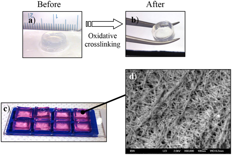
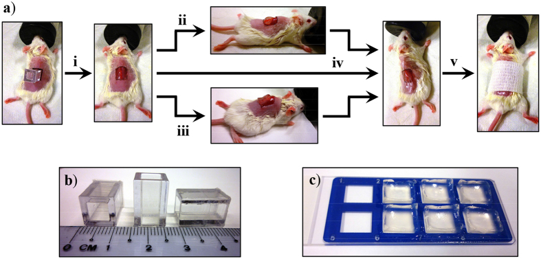
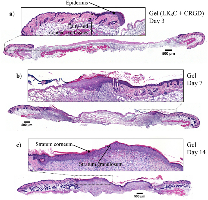
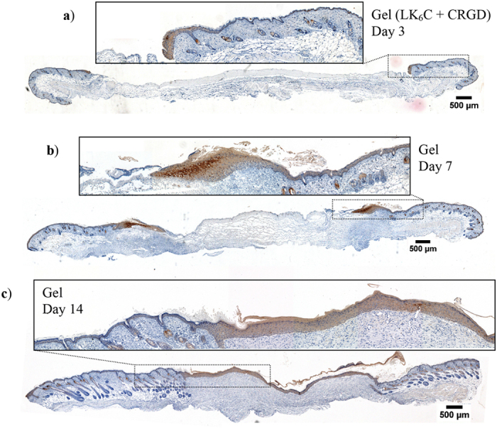
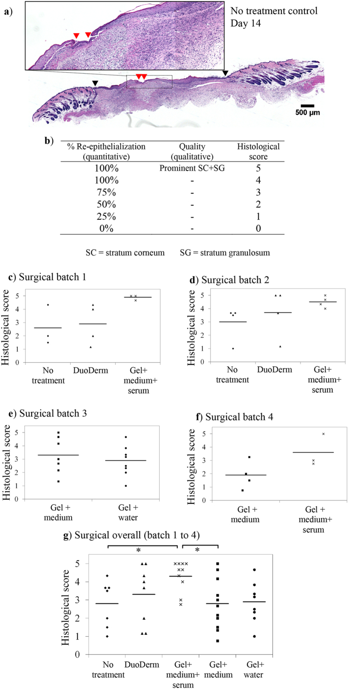
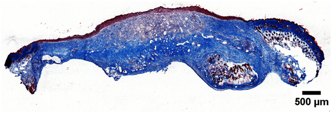
Wound healing is a major burden of healthcare systems worldwide and hydrogel dressings offer a moist environment conducive to healing. We describe cysteine-containing ultrashort peptides that self-assemble spontaneously into hydrogels. After disulfide crosslinking, the optically-transparent hydrogels became significantly stiffer and exhibited high shape fidelity. The peptide sequence (LIVAGKC or LK6C) was then chosen for evaluation on mice with full-thickness excision wounds. Crosslinked LK6C hydrogels are handled easily with forceps during surgical procedures and offer an improvement over our earlier study of a non-crosslinked peptide hydrogel for burn wounds. LK6C showed low allergenic potential and failed to provoke any sensitivity when administered to guinea pigs in the Magnusson-Kligman maximization test. When applied topically as a dressing, the medium-infused LK6C hydrogel accelerated re-epithelialization compared to controls. The peptide hydrogel is thus safe for topical application and promotes a superior rate and quality of wound healing.


Figures

Figure 1
Figure 1
(a,b) The gel stiffness increased after crosslinking and can be easily picked up with a pair of forceps, while remaining optically transparent. (c) LK6C + CRGD gels (99% water) were clear, transparent and easily handled. Phenol red was added to aid visualisation. d) FESEM analysis revealed a dense mesh of fibrous network. The thickness of the fibers was on the order of tens of nm, mimicking the fibrous microenvironment found in the ECM.
Figure 2
Figure 2
CD studies performed with LK6C either in (a) water or (b) 0.06% H2O2. (a) The secondary structure of LK6C transited from being predominantly β-turn to α-helical, followed by random coil as it was serially diluted in water. (b) In H2O2, LK6C made the transition from being predominantly β-turn to α-helical at a lower concentration of 0.2–0.4 mg/mL, compared to 0.6–0.8 mg/mL in water. This suggests that the disulfide bridges restrict the peptide fibers to a greater extent, thereby reducing their propensity to change secondary conformations.
Figure 3
Figure 3. Rate of water lost from wounds covered with Tegaderm.
(a) Small containers were filled with 200 μL of either water or LK6C + CRGD hydrogels and sealed with Tegaderm. The containers were then left on a heated plate (37 °C) exposed to ambient conditions to simulate the wound/air interface. (b) The weight of the containers was then recorded regularly to quantify the amount of moisture remaining and reported as average ± s.d. of quadruplicates. The rate of water lost was linearly constant and there was no significant difference between the samples.
Figure 4
Figure 4
(a) Schematic of the full-thickness excision mouse model used to evaluate the wound healing property of LK6C + CRGD hydrogel dressings. (i) The wound boundaries were marked out on dorsally shaved mice using a template (as shown in b). Both the epidermis and dermis were then surgically removed to create 1 × 1 cm full-thickness wounds. Either the (ii) positive control (DuoDerm hydroactive gel) or (iii) peptide hydrogel (cast as ready-to-use sterile gel blocks, as shown in c) was applied to the wounds. (iv) All wounds were then covered up with Tegaderm, including the negative control group which received no gel application. (v) The wound was lightly bandaged and left to heal. Animals were sacrificed on day 3, 7 and 14 for histological examination.
Figure 5
Figure 5. Representative cross-sectional histological sections of wounds treated with peptide hydrogel infused with completed medium.
Skin samples were obtained on day (a) 3, (b) 7, (c) 14 for staining with H&E. Over time, the leading front of the epidermal “tongue” was observed to migrate inwards to close up the wound and restore the critical barrier function of skin. Re-epithelialization was completed by day 14, along with the appearance of a visible layer of stratum corneum and stratum granulosum. All insets show magnified images of the respective boxed regions.
Figure 6
Figure 6. Representative cross-sections of wounds stained for K14 protein (using monoclonal antibody LL001).
Wounds were treated with the completed medium-infused peptide hydrogel and animals were sacrificed on (a–c) day 3, 7 and 14 for immunohistochemistry. K14 is a marker for basal keratinocytes, as evidenced by its basal localization in unwounded epidermis. In these healing wounds, K14 was detected in both the basal and suprabasal layers of the newly regenerated epidermis. This suggests that keratinocytes in the newly regenerated epidermis have yet to fully differentiate as the neo-epidermis gradually matures to a more homeostatic morphology (2–4 cell layers thick).
Figure 7
Figure 7. Comparison of different treatment strategies on day 14.
(a) Representative H&E cross-section of a wound from the no-treatment group. The black arrow heads mark out the edges of the original excision sites, upon which the wound gap remaining (red arrow heads) was normalized against to derive the % re-epithelialization achieved. (b) The scoring criteria used in this semi-quantitative scale for grading skin samples. (c–f) Surgery was performed over four batches of animals. Skin sections were obtained on day 14 and scored based on the quality of re-epithelialization. (g) The overall scores of all treatment groups (n = 7–11). Wounds treated with peptide hydrogel infused with completed medium (average group score: 4.3) received higher and more consistent histological scores than wounds treated with the DuoDerm gel positive control (average: 3.3), no-treatment negative control (average: 2.8), or peptide gels infused with medium alone (average: 2.8) or water (average: 2.9).
Figure 8
Figure 8. Representative frozen cross-section of a wound treated with the completed medium-infused peptide hydrogel after Masson’s trichrome staining on day 14.
Collagen is stained blue. Collagen deposition was denser near the wound edge and in the deeper tissue layers, indicative of the inwards and upwards progression of wound healing. There was no obvious difference between the treatment groups in terms of density and location of collagen deposition.

References

    1. Sonnemann K. J. & Bement W. M. Wound repair: toward understanding and integration of single-cell and multicellular wound responses. Annual Review of Cell and Developmental Biology 27, 237–263 (2011). - PMC - PubMed
    1. Guo S. & DiPietro L. A. Factors affecting wound healing. Journal of Dental Research 89, 219–229 (2010). - PMC - PubMed
    1. Martin P. Wound healing - aiming for perfect skin regeneration. Science 276, 75–81 (1997). - PubMed
    1. Gurtner G. C., Werner S., Barrandon Y. & Longaker M. T. Wound repair and regeneration. Nature 453, 314–321 (2008). - PubMed
    1. Porter S. The role of the fibroblast in wound contraction and healing. Wounds UK 3, 33–40 (2007).

Publication types