. 2016 Sep 7:6:31613.
doi: 10.1038/srep31613.

Functional Magnetic Resonance Imaging of Electrical and Optogenetic Deep Brain Stimulation at the Rat Nucleus Accumbens

Affiliations

Functional Magnetic Resonance Imaging of Electrical and Optogenetic Deep Brain Stimulation at the Rat Nucleus Accumbens

Daniel L Albaugh et al. Sci Rep. .

Abstract

Deep brain stimulation of the nucleus accumbens (NAc-DBS) is an emerging therapy for diverse, refractory neuropsychiatric diseases. Although DBS therapy is broadly hypothesized to work through large-scale neural modulation, little is known regarding the neural circuits and networks affected by NAc-DBS. Using a healthy, sedated rat model of NAc-DBS, we employed both evoked- and functional connectivity (fc) MRI to examine the functional circuit and network changes achieved by electrical NAc stimulation. Optogenetic-fMRI experiments were also undertaken to evaluate the circuit modulation profile achieved by selective stimulation of NAc neurons. NAc-DBS directly modulated neural activity within prefrontal cortex and a large number of subcortical limbic areas (e.g., amygdala, lateral hypothalamus), and influenced functional connectivity among sensorimotor, executive, and limbic networks. The pattern and extent of circuit modulation measured by evoked-fMRI was relatively insensitive to DBS frequency. Optogenetic stimulation of NAc cell bodies induced a positive fMRI signal in the NAc, but no other detectable downstream responses, indicating that therapeutic NAc-DBS might exert its effect through antidromic stimulation. Our study provides a comprehensive mapping of circuit and network-level neuromodulation by NAc-DBS, which should facilitate our developing understanding of its therapeutic mechanisms of action.


Figures

Figure 1
Figure 1
(A) Schematic of experimental imaging setup with a custom single loop surface coil and a tungsten microwire electrode. (B) Electrode tip mapping to the NAc for all electrical DBS subjects (n = 9). Tip placements were estimated using T2-weighted anatomical scans, which we deemed satisfactory given the relatively large size of the NAc (including anteroposterior distance), as well as the reduced electrode artifact.
Figure 2
Figure 2. Functional CBV activation maps by 130 Hz NAc-DBS (300 μA; n = 5).
CBV modulation was largely ipsilateral to the stimulated hemisphere, was predominantly positive in direction, and included both cortical and subcortical CBV modulation. Notable regions demonstrating CBV increases included the prefrontal cortex (including prelimbic, infralimbic, and orbitofrontal), NAc, lateral hypothalamus, amygdala, ventral hippocampus and others. CBV decreases were also detected within a small region of the ipsilateral dorsal striatum. 12 slices were acquired in each scan, with numbers below slices denoting relative distance from bregma (in mm). Color bar denotes t score values obtained by GLM analyses, with a significance threshold of p < 0.05 (corrected). Functional activation maps for all additional tested frequencies are located in Supplemental Figures S4.
Figure 3
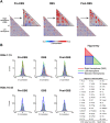
Figure 3. fcMRI modulation by 130 Hz NAc-DBS.
(A) Mean correlation matrices (n = 7) for each stimulus condition (Pre-DBS, DBS, Post-DBS) using 45 regions-of-interest (ROIs, see Figure Key). ROIs were chosen a priori, with reference to anatomical regions described in a standard rat brain atlas. Note the presence of between-hemispheric regional connectivity (displayed as a red diagonal line) in all matrices. (B) Histograms and t-distribution fits for each stimulus condition, network (NAc-DBS [ROIs #s 1–15] and Other [ROIs #’s 16–45]) and connectivity grouping (within and between hemispheres) using correlation measures from the average correlation matrices. Abbreviations: PLC: Prelimbic Cortex; ILC: Infralimbic Cortex; OFC: Orbitofrontal Cortex; CC: Cingulate Cortex; Insula: Insular Cortex; NAc: Nucleus Accumbens; AS; Anterior Striatum; vPAll: Ventral Pallidum; Sept: Septum; lHyp: Lateral Hypothalamus; Amyg: Amygdala; BNST: Bed Nucleus of the Stria Terminalis; MDT: Mediodorsal Thalamus; vHipp: Ventral Hippocampus; VTA: Ventral Tegmental Area; AC: Auditory Cortex; AOB: Accessory Olfactory Bulb; DLS: Dorsolateral Striatum; DMS: Dorsomedial Striatum; ENT: Entorhinal Cortex; GPe: External Globus Pallidus; Motor: Motor Cortex (Primary and Secondary); OT: Olfactory Tubercle; PAG: Periaqueductal Grey; PPTg: Pedunculopontine Tegmental Nucleus; PC: Parietal Cortex; Piriform: Piriform Cortex; pHyp: Posterior Hypothalamus; pThal: Posterior Thalamus; S2: Secondary Somatosensory Cortex; SN: Substantia Nigra; Somato: Primary Somatosensory Cortex; STN: Subthalamic Nucleus; TeA: Temporal Association Cortex; VL: Ventrolateral Thalamus; VPL: Ventral Posterolateral Thalamus; Visual: Visual Cortex (Primary and Secondary); ZI: Zona Incerta; dHipp: Dorsal Hippocampus; dRaphe: Dorsal Raphe Nucleus; lHab: Lateral Habenula; mPOA: Medial Preoptic Area; SC: Superior Colliculus; vHyp: Ventral Hypothalamus.
Figure 4
Figure 4. Network-level visualization of pair-wise fcMRI modulations during 130 Hz NAc-DBS.
Significant (rANOVA, p ≤ 0.05 uncorrected, ΔZ-Corr > 0.10) enhanced (red) or suppressed (blue) individual pair-wise connections grouped by functionally-defined networks (Sensorimotor, Executive, Limbic, and Between Network Connections). Thick red lines represent ΔZ-Corr > 0.20 at R-NAc ↔ R-Sept, R-PLC and R-ILC ↔ R-PLC.
Figure 5
Figure 5
Temporal dynamics (A) and amplitudes (B) of CBV responses to NAc-DBS across five stimulation frequencies (10, 40, 70, 130, 200 Hz; n = 8 per frequency), demonstrating that CBV responses to NAc-DBS were largely stimulation frequency-insensitive. All subjects were scanned with 500 μA DBS, except one subject with 600 μA. CBV responses are expressed as a percent change from pre-stimulation baseline values. Amplitudes were calculated as mean percent CBV changes during stimulation epochs (10 seconds; scan frames 21–30). Anatomically-defined ROIs are highlighted as figure inserts in 5A (Single slice shown; note that many ROIs encompassed multiple slices). *p ≤ 0.05 for 10 Hz compared to all other frequencies (no other statistically significant comparisons). Datapoints are presented as mean ± SEM.
Figure 6
Figure 6. Optogenetic stimulation at NAc evokes local CBV increases.
(A) Schematic of optogenetic stimulation at NAc (top), representative confocal image confirming ChR2 expression (green) in the NAc (bottom). Counterstain is DAPI (blue). ChR2 expression appeared strongest in the core subregion of the NAc. (B) Functional CBV responses induced by 40 Hz optogenetic stimulation at NAc in animals expressing ChR2 or EYFP (AAV5 using the CaMKIIα promoter; n = 4 and 2, respectively). Note that optogenetic stimulation of the NAc resulted in CBV increases locally within the stimulated region, with no detected downstream responses. No responses were observed in EYFP subjects. Anteroposterior slice coordinates are as described for Fig. 2. Temporal CBV dynamics (C) and amplitudes (D) within the NAc during local optogenetic stimulation. Stimulation-evoked CBV amplitude changes were calculated as described in Methods.

References

    1. Wichmann T. & Delong M. R. Deep brain stimulation for neurologic and neuropsychiatric disorders. Neuron 52, 197–204 (2006). - PubMed
    1. Goodman W. K. & Alterman R. L. Deep brain stimulation for intractable psychiatric disorders. Annu Rev Med 63, 511–524 (2012). - PubMed
    1. Floresco S. B. The nucleus accumbens: an interface between cognition, emotion, and action. Annu Rev Psychol 66, 25–52 (2015). - PubMed
    1. de Koning P. P., Figee M., van den Munckhof P., Schuurman P. R. & Denys D. Current status of deep brain stimulation for obsessive-compulsive disorder: a clinical review of different targets. Curr Psychiatry Rep 13, 274–282 (2011). - PubMed
    1. Bewernick B. H., Kayser S., Sturm V. & Schlaepfer T. E. Long-term effects of nucleus accumbens deep brain stimulation in treatment-resistant depression: evidence for sustained efficacy. Neuropsychopharmacology 37, 1975–1985 (2012). - PMC - PubMed

Publication types