. 2016 Jan;25(1):100-110.
doi: 10.1038/ejhg.2016.108. Epub 2016 Sep 7.

Hereditary spastic paraplegias: identification of a novel SPG57 variant affecting TFG oligomerization and description of HSP subtypes in Sudan

Affiliations

Hereditary spastic paraplegias: identification of a novel SPG57 variant affecting TFG oligomerization and description of HSP subtypes in Sudan

Liena E O Elsayed et al. Eur J Hum Genet. 2016 Jan.

Abstract

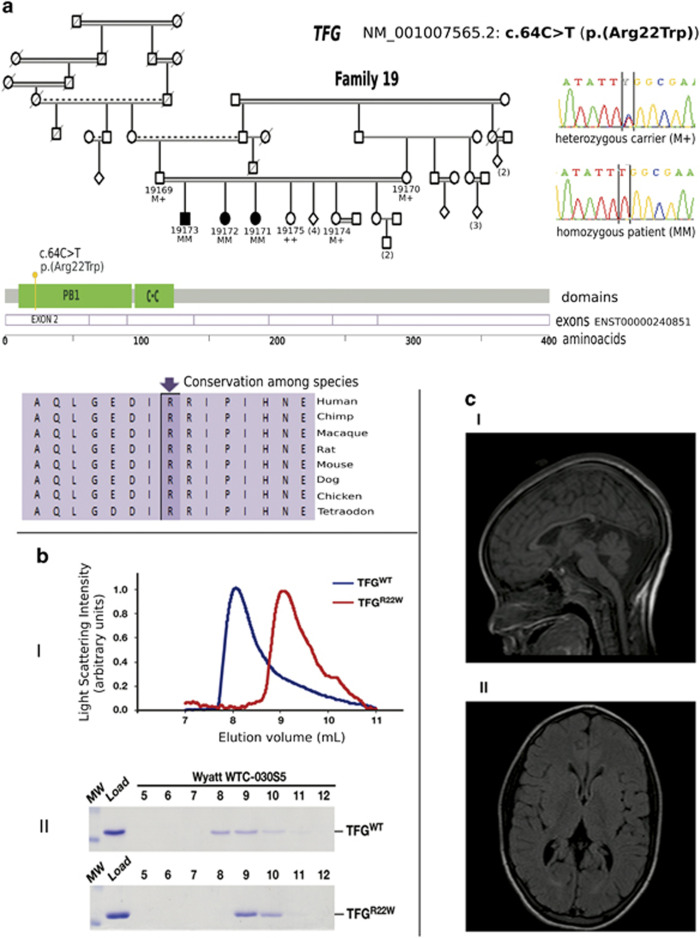
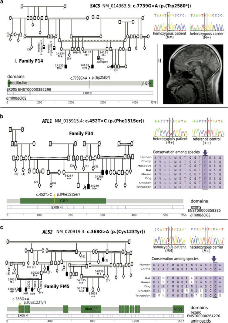
Hereditary spastic paraplegias (HSP) are the second most common type of motor neuron disease recognized worldwide. We investigated a total of 25 consanguineous families from Sudan. We used next-generation sequencing to screen 74 HSP-related genes in 23 families. Linkage analysis and candidate gene sequencing was performed in two other families. We established a genetic diagnosis in six families with autosomal recessive HSP (SPG11 in three families and TFG/SPG57, SACS and ALS2 in one family each). A heterozygous mutation in a gene involved in an autosomal dominant HSP (ATL1/SPG3A) was also identified in one additional family. Six out of seven identified variants were novel. The c.64C>T (p.(Arg22Trp)) TFG/SPG57 variant (PB1 domain) is the second identified that underlies HSP, and we demonstrated its impact on TFG oligomerization in vitro. Patients did not present with visual impairment as observed in a previously reported SPG57 family (c.316C>T (p.(Arg106Cys)) in coiled-coil domain), suggesting unique contributions of the PB1 and coiled-coil domains in TFG complex formation/function and a possible phenotype correlation to variant location. Some families manifested marked phenotypic variations implying the possibility of modifier factors complicated by high inbreeding. Finally, additional genetic heterogeneity is expected in HSP Sudanese families. The remaining families might unravel new genes or uncommon modes of inheritance.


Conflict of interest statement

The authors declare no conflict of interest.

Figures

Figure 1
Figure 1
Families F1, F6 and F16 with autosomal recessive hereditary spastic paraplegia with thin corpus callosum (TCC) caused by SPG11 variants. (a (I)) Pedigree of family F1 in which a nonsense variant segregates with the disease in subfamily I. Electrophoregrams show the variant in a homozygous patient and a heterozygous carrier. Subfamily II showed dissimilarities in HSP phenotypes. (a (II)) Brain MRI (sagittal T1W section) of an SPG11 patient showing severe TCC. (b (I)) Pedigree of family F6 showing the segregation of a frameshift variant. Electropherograms show the variant in a patient (homozygous) and in a heterozygous carrier. (b (II)) Brain MRI (sagittal T1W section) of the index patient showing severe TCC and cortical atrophy. (c (I)) Pedigree of family F16 showing the segregation of a nonsense variant. Electropherograms show the variant in a patient (homozygous mutant) and in a heterozygous carrier. (c (II)) Brain MRI (axial T2 FLAIR) of the index patient showing TCC and periventricular hyperintensities. (d) SPG11 gene model showing the position of detected variants as red lollipops. Pedigree symbols: full shading indicates affected individuals, half horizontal shading indicates other diseases, numbers between brackets are multiple siblings, diamonds indicate multiple siblings of different sex. Individual ID numbers and genotypes are indicated. Genotypes: ++ homozygous reference; M+ heterozygous; MM homozygous mutant. Others are standard medical pedigree symbols.
Figure 2
Figure 2
Family F19 with autosomal recessive hereditary spastic paraplegia caused by TFG/SPG57. (a) Pedigree of family F19 in which a missense variant segregates with the disease. Electropherograms show homozygous and heterozygous genotypes. Conservation of the amino-acid sequence is shown as well as a gene model showing the position of detected variant. (b) Purified, untagged forms of TFG (amino acids 1–96; wild-type or mutated to include the c.64C>T (p.(Arg22Trp)) substitution), were separated over a gel filtration column (Wyatt TWC-030S5) that was coupled to a multi-angle light-scattering device. Representative light-scattering profiles (wild-type TFG, blue; TFG c.64C>T (p.(Arg22Trp)), red) are plotted (b (I)). Eluted fractions were separated by SDS-PAGE and stained using Coomassie to highlight the elution profiles of the both forms of TFG (b (II)). Based on three independent experiments for each protein, wild-type TFG (amino acids 1–96) exhibited a molecular mass of 92.4 kD ± 3.7 kD, and c.64C>T (p.(Arg22Trp)) TFG (amino acids 1–96) exhibited a molecular mass of 24.4 kD ± 1.0 kD. (c) Brain MRIs of individuals 19172 (sagittal T1W section) and 19171 (T2 FLAIR axial section) showing thinning of the body of the corpus callosum (c (I)) and TCC with mild periventricular signal hyperintensity lesions at the occipital pole (c (II)). Pedigree symbols: full shading indicates affected individuals, half horizontal shading indicates other diseases, numbers between brackets are multiple siblings, diamonds indicate multiple siblings of different sex. Individual ID numbers and genotypes are indicated. Genotypes: ++ homozygous reference; M+ heterozygous; MM homozygous mutant. Others are standard medical pedigree symbols.
Figure 3
Figure 3
Families F14, F34 and FM5 with spastic ataxia and hereditary spastic paraplegia caused by SACS, SPG3A and ALS2 variants. (a (I)) Pedigree of family F14 with recessive spastic ataxia of Charlevoix Saguenay (ARSACS) caused by a nonsense variant in SACS. Other diseases are seen in the family. Electropherograms show the variant in a patient (homozygous mutant) and a heterozygous carrier. (a (II)) Brain MRI (Sagittal section T1W) of individual 14133 showing thinning of the posterior half of the body of corpus callosum and moderate thinning of cerebellum. (b) Pedigree of Family F34 with autosomal dominant HSP caused by a missense variant in ATL1. Electropherograms show the variant in a patient (heterozygous) and a control (reference alleles). (c) Pedigree of family FM5 with autosomal recessive HSP caused by missense variant in ALS2. Electropherograms are shown for a patient (homozygous mutant) and a heterozygous carrier. Pedigree symbols: shading indicates affected individuals, numbers between brackets are multiple siblings, diamonds indicate multiple siblings of different sex. Individual ID numbers and genotypes are indicated. Gene models show the position of detected variant in each gene. For missense variants (b and c) conservation of the (predicted) affected amino-acid is shown.

References

    1. Tesson C, Koht J, Stevanin G: Delving into the complexity of hereditary spastic paraplegias: how unexpected phenotypes and inheritance modes are revolutionizing their nosology. Hum Genet 2015; 134: 511–538. - PMC - PubMed
    1. Fink J: Hereditary spastic paraplegia overview. In: Pagon R, Adam M, Ardinger H et al: (eds): GeneReviews. University of Washington: Seattle (WA), USA, 1993. Available at: http://www.ncbi.nlm.nih.gov/books/NBK1509/. - PubMed
    1. Schmitz-Hübsch T, Klockgether T: An update on inherited ataxias. Curr Neurol Neurosci Rep 2008; 8: 310–319. - PubMed
    1. Finsterer J: Ataxias with autosomal, X-chromosomal or maternal inheritance. Can J Neurol Sci 2009; 36: 409–428. - PubMed
    1. Coutelier M, Stevanin G, Brice A: Genetic landscape remodelling in spinocerebellar ataxias: the influence of next-generation sequencing. J Neurol 2015; 262: 2382–2395. - PubMed

Publication types

MeSH terms

Supplementary concepts

LinkOut - more resources