. 2016 Oct 21;291(43):22671-22685.
doi: 10.1074/jbc.M116.739573. Epub 2016 Sep 6.

Arginine Demethylation of G3BP1 Promotes Stress Granule Assembly

Affiliations

Arginine Demethylation of G3BP1 Promotes Stress Granule Assembly

Wei-Chih Tsai et al. J Biol Chem. .

Abstract

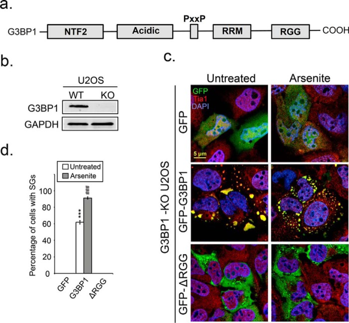
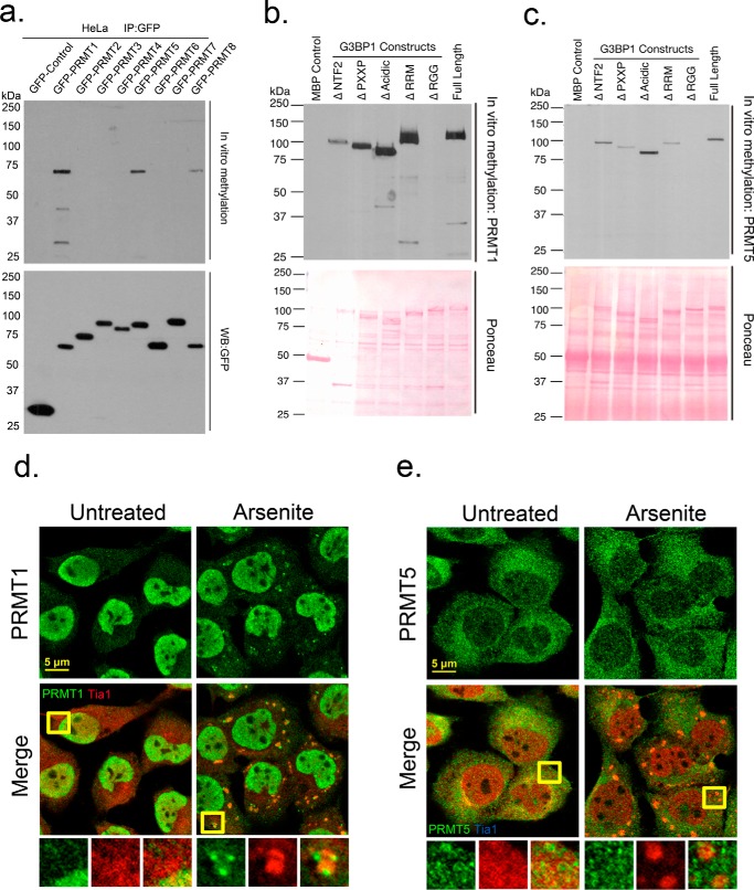
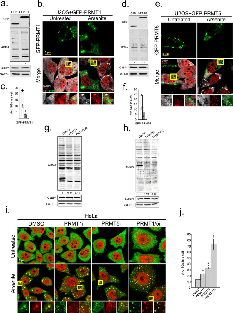
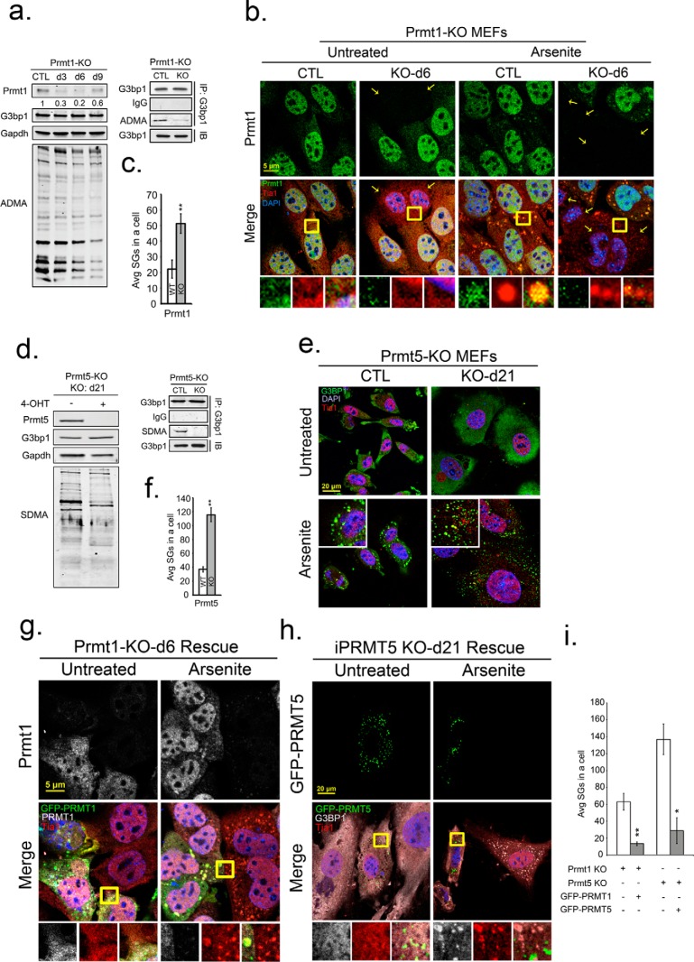
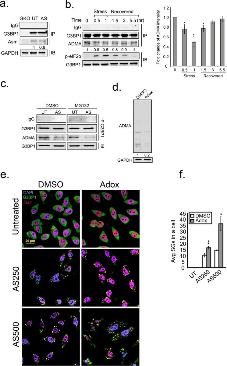
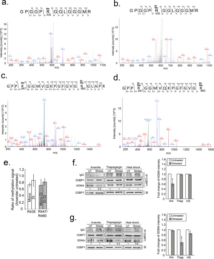
Stress granules (SGs) are cytoplasmic condensates of stalled messenger ribonucleoprotein complexes (mRNPs) that form when eukaryotic cells encounter environmental stress. RNA-binding proteins are enriched for arginine methylation and facilitate SG assembly through interactions involving regions of low amino acid complexity. How methylation of specific RNA-binding proteins regulates RNA granule assembly has not been characterized. Here, we examined the potent SG-nucleating protein Ras-GAP SH3-binding protein 1 (G3BP1), and found that G3BP1 is differentially methylated on specific arginine residues by protein arginine methyltransferase (PRMT) 1 and PRMT5 in its RGG domain. Several genetic and biochemical interventions that increased methylation repressed SG assembly, whereas interventions that decreased methylation promoted SG assembly. Arsenite stress quickly and reversibly decreased asymmetric arginine methylation on G3BP1. These data indicate that arginine methylation in the RGG domain prevents large SG assembly and rapid demethylation is a novel signal that regulates SG formation.

Keywords: G3BP1; RNA binding protein; mRNA; post-translational modification (PTM); protein arginine N-methyltransferase (PRMT); protein arginine N-methyltransferase 5 (PRMT5); stress granule.


Figures

FIGURE 1.
FIGURE 1.
Deletion of the RGG domain of G3BP1 impairs SG assembly. a, diagram depicting the domain structure of G3BP1. b, endogenous G3BP1 in U2OS cells was depleted by CRISPR/Cas9 knock-out. A single G3BP1 KO clone was selected and verified by Western blot analysis. c, G3BP1 KO U2OS cells were transfected with GFP, GFP-G3BP1, or GFP-G3BP1-ΔRGG constructs and SG responses following arsenite stress (0.2 mm for 30 min) monitored by IFA. Cells were counterstained with the SG marker Tia1 in red, and DAPI for nuclei in blue. d, bar graph shows the percentage of cells that produced SGs in each condition. White bars indicate untreated cells, and gray bars refer to arsenite-stressed cells. The results shown are representative of three independent experiments counting >100 cells. ***, p < 0.001 versus untreated GFP control; ###, p < 0.001 against arsenite-treated GFP control. Original magnification: 63×.
FIGURE 2.
FIGURE 2.
G3BP1 is methylated by PRMT1 and PRMT5 in vitro. Recombinant MBP-G3BP1 was incubated with a panel of purified GFP-tagged PRMTs in in vitro methylation reactions as described under “Experimental Procedures.” b and c, recombinant proteins bearing deletions of the indicated domains were incubated with GFP-PRMT1 or GFP-PRMT5, respectively. Loading controls of PRMTs are shown as Western blots against GFP (a, lower panel) or for G3BP constructs via ponceau staining (b, c, lower panel). U2OS cells were arsenite-treated and processed for IFA. Cells were stained for endogenous PRMT1 (d) or PRMT5 (e) in green, and counterstained with Tia1 in red. Yellow squares indicate regions depicted in vignettes. Original magnification: 63×.
FIGURE 3.
FIGURE 3.
Enzymatic activity of PRMT1 and PRMT5 suppresses SG formation. U2OS cells were transfected with GFP, GFP-PRMT1, or GFP-PRMT5. Antibodies specific for GFP (top panels in a and d), asymmetric (ADMA) methyl modifications (a and g), and symmetric methyl modifications (SDMA) (b and h) were used in Western blot analysis. b and e, to measure effects of PRMT overexpression on SGs, cells expressing PRMT1 or PRMT5 were stressed with arsenite and processed for IFA with antibodies against G3BP1 in gray and Tia1 in red. c and f, bar graphs represent average number of SGs/cell in cells that did express (white bars) or did not express transgenes (gray bars), **, p < 0.01. i, enzymatic activities of PRMTs in HeLa cells were inhibited by PRMT1 inhibitor (PRMT1i), PRMT5 inhibitor (PRMT5i) or both together (PRMT1/5i). HeLa cells were fixed after arsenite treatment and stained for SG markers G3BP1 in green and Tia1 in red. Yellow squares indicate regions depicted in vignettes, and the merged channel vignette is on the right of each series of three panels. j, quantification of average SGs/cell after inhibitor treatments. *, p < 0.05; **, p < 0.01; and ***, p < 0.001 versus DMSO-treated cells. The results shown are representative of three independent experiments in which 100 cells were counted in each. Original magnification: 63×.
FIGURE 4.
FIGURE 4.
Knockdown of PRMT1 or PRMT5 promotes SG assembly. Inducible PRMT1 (PRMT1-KO) and PRMT5 (PRMT5-KO) knock-out MEFs were treated with 4-OHT for 6 and 21 days, respectively, to deplete PRMT1 and PRMT5. a and d, endogenous PRMT1, PRMT5, G3BP1 protein levels, and methylation status of total proteins was determined by Western blot with antibodies against PRMT1, PRMT5, G3BP1, asymmetric (ADMA), or symmetric (SDMA) dimethylarginine as indicated. GAPDH protein levels serve as loading controls (a, d). b, PRMT1-KO MEFs were assessed for knock-out efficiency and effect on methylation (small panel shown in a), arsenite treated and processed for IFA with antibodies against PRMT1 in green, Tia1 in red, and DAPI in blue. Average SGs/cell were quantified before or after PRMT1 KO (c; white versus gray bars). **, p < 0.01. e, similarly, PRMT5-null MEFs (shown by small panel in d, were arsenite treated and processed for IFA. Cells were counterstained for G3BP1, Tia1, and DAPI, and the average SGs/cell were quantified with or without PRMT5 KO (f), **, p < 0.01 versus untreated PRMT5-KO MEFs. (g) and (h), PRMT1 and PRMT5 expression was rescued in PRMT-KO MEFs by transfection of GFP-tagged PRMT expression constructs. Cells were arsenite treated, fixed, and stained in IFA for PRMT1 in gray (g, top panels), G3BP1 in gray (h), Tia1 in red, and DAPI. Yellow squares indicate regions depicted in vignettes. Prmt1 KO was always incomplete, thus staining for both endogenous and transgene Prmt1 is shown (i). Quantification of average SGs/cell in rescue experiments with PRMT KO MEFs (white bar), and PRMT KO rescued MEFs (gray bar). *, p < 0.05; **, p < 0.01 versus PRMT-KO MEFs. The results shown are representative of three independent experiments counting >100 cells in each. Original magnification: 63×.
FIGURE 5.
FIGURE 5.
Mutation of putative methylation sites in the G3BP1 RGG domain modulates SG nucleation. a, a schematic showing potential arginine methylation sites in the RGG domain of G3BP1. Two types of GFP-tagged G3BP1 mutants were created at each site, charge-neutral mutants substituted Arg to Phe and methyl-deficient mutants replaced Arg to Lys. c and d, G3BP1 KO U2OS cells were transfected with GFP-tagged G3BP1 methylation mutants and left untreated or stressed, processed for IFA with staining for SGs with Tia1 (red). b, percentages of cells with SGs were quantified in charge-neutral (white bars) transfected cells and methyl-deficient (dark gray bars) transfected cells by analyzing 100 cells. *, p < 0.05; **, p < 0.01; and ***, p < 0.001 versus GFP-G3BP1-transfected cells (light gray bars). The results shown are representative of three independent experiments. e, in vitro methylation reactions (top panels) using recombinant MBP-G3BP1 variants bearing individual Arg point mutations were incubated with purified GFP-tagged PRMT 1 or PRMT5 as described under “Experimental Procedures.” Protein levels are indicated by Ponceau stain of the blots (in the lower panels).
FIGURE 6.
FIGURE 6.
Demethylation of G3BP1 promotes SG formation. a, pulldown of endogenous G3BP1 in G3BP1 KO (GKO), unstressed (UT), and arsenite-treated (AS) HeLa cells. The methylation status of G3BP1 was detected with the asymmetric dimethylation (ADMA) antibody in Western blot analysis. b, asymmetric dimethylation was detected on G3BP1 (ADMA) through stress and recovery periods. b, right panel, quantification of ADMA intensity is shown. The results are representative of three independent experiments. *, p < 0.05; **, p < 0.01 against the 0 h time point. Arsenite stress was applied for 60 min, removed, then cells were washed and incubated for the indicated recovery period. The level of phospho-eIF2α (p-eIF2α) was monitored by immunoblot analysis as a proxy for translation inhibition during the recovery period. G3BP1 asymmetric dimethyl (ADMA) status was determined by immunoblot analysis of immunoprecipitated G3BP1. c, HeLa cells were treated with either DMSO or MG132 simultaneously with either vehicle or arsenite for 1.5 h. The methylation status (ADMA) of G3PB1 was detected as described above. d, HeLa cells were treated with Adox for 48 h, and endogenous ADMA levels were determined by immunoblot. e, Adox-treated cells were stressed with 250 mm (AS250) or 500 mm (AS500) arsenite, and processed for IFA with G3BP1 in green, Tia1 in red, and DAPI. f, quantification of average SGs/cell were calculated in >100 cells for each condition. White bar indicates DMSO control, and gray bars indicates Adox treatment. *, p < 0.05; **, p < 0.01 against DMSO control. Original magnification: 63×.
FIGURE 7.
FIGURE 7.
Demethylation of G3PB1 confirmed by MS analysis and detected in ER stress and heat shock. a–d, fragment ion (MS/MS) spectrums isolated from immunoprecipitated G3BP1 analysis. The first arginine residue (R435) within the peptide GPGGPRGGLGGGMR was identified as either monomethylated (a) or dimethylated (b). Two methylated arginine residues (Arg-447 and Arg-460) were identified in the peptide GPPRGGMVQKPGFGVGR in two different forms; either (c) monomethylated Arg-447 and dimethylated Arg-460, or (d) dimethylation of Arg-447 and dimethylation of Arg-460. e, bar graph indicates triplicate MS data of methylation status of G3BP1 under oxidative stress comparing the ratio of methylated over non-methylated peptides. White bars represent two methylated forms of Arg-435. Gray bars represent either monomethylated (mono) Arg-447 and dimethylated (di) Arg-460 or dimethylated Arg-447 and Arg-460 (di-di). Immunoblot for ADMA (f) or SDMA (g) of G3BP1 that was immunoprecipitated from either arsenite-treated, thapsigargin-treated or heat shocked cells. UT indicates untreated, and Stress indicates the different stressors. The quantification is the intensity of ADMA (f, right panel) and SDMA (g, right panel) shown as bar graphs and normalized against untreated conditions. The results shown are representative of three independent experiments. White bars indicate untreated, and gray bars indicate different stressors. *, p < 0.05; **, p < 0.01; and ***, p < 0.01 against untreated controls.

References

    1. Kedersha N. L., Gupta M., Li W., Miller I., and Anderson P. (1999) RNA-binding proteins TIA-1 and TIAR link the phosphorylation of eIF-2α to the assembly of mammalian stress granules. J. Cell Biol. 147, 1431–1442 - PMC - PubMed
    1. Kedersha N., Chen S., Gilks N., Li W., Miller I. J., Stahl J., and Anderson P. (2002) Evidence that ternary complex (eIF2-GTP-tRNA(i)(Met))-deficient preinitiation complexes are core constituents of mammalian stress granules. Mol. Biol. Cell. 13, 195–210 - PMC - PubMed
    1. Kedersha N., Stoecklin G., Ayodele M., Yacono P., Lykke-Andersen J., Fritzler M. J., Scheuner D., Kaufman R. J., Golan D. E., and Anderson P. (2005) Stress granules and processing bodies are dynamically linked sites of mRNP remodeling. J. Cell Biol. 169, 871–884 - PMC - PubMed
    1. Reineke L. C., Dougherty J. D., Pierre P., and Lloyd R. E. (2012) Large G3BP-induced granules trigger eIF2α phosphorylation. Mol. Biol. Cell. 23, 3499–3510 - PMC - PubMed
    1. Tourrière H., Chebli K., Zekri L., Courselaud B., Blanchard J. M., Bertrand E., and Tazi J. (2003) The RasGAP-associated endoribonuclease G3BP assembles stress granules. J. Cell Biol. 160, 823–831 - PMC - PubMed

MeSH terms

LinkOut - more resources