. 2016 Sep 6;7(5):e00908-16.
doi: 10.1128/mBio.00908-16.

FtsZ-Dependent Elongation of a Coccoid Bacterium

Affiliations

FtsZ-Dependent Elongation of a Coccoid Bacterium

Ana R Pereira et al. mBio. .

Abstract

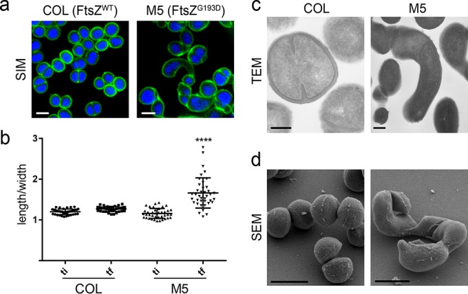
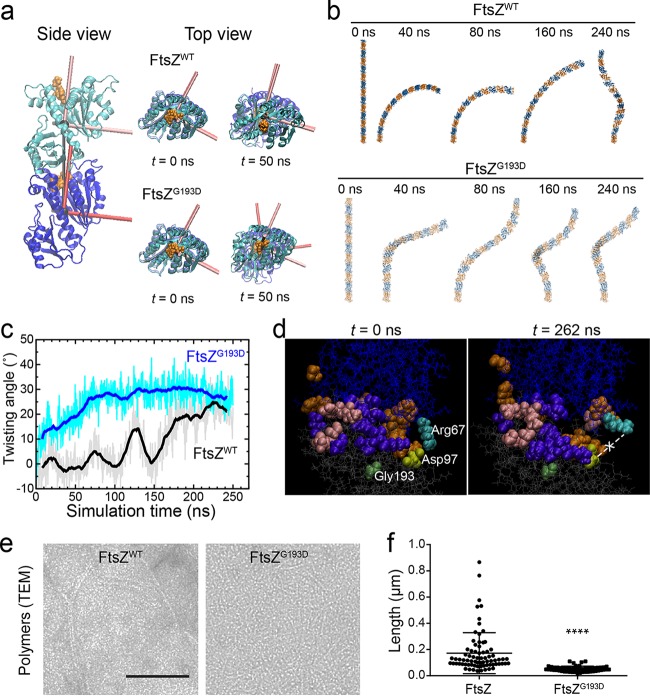
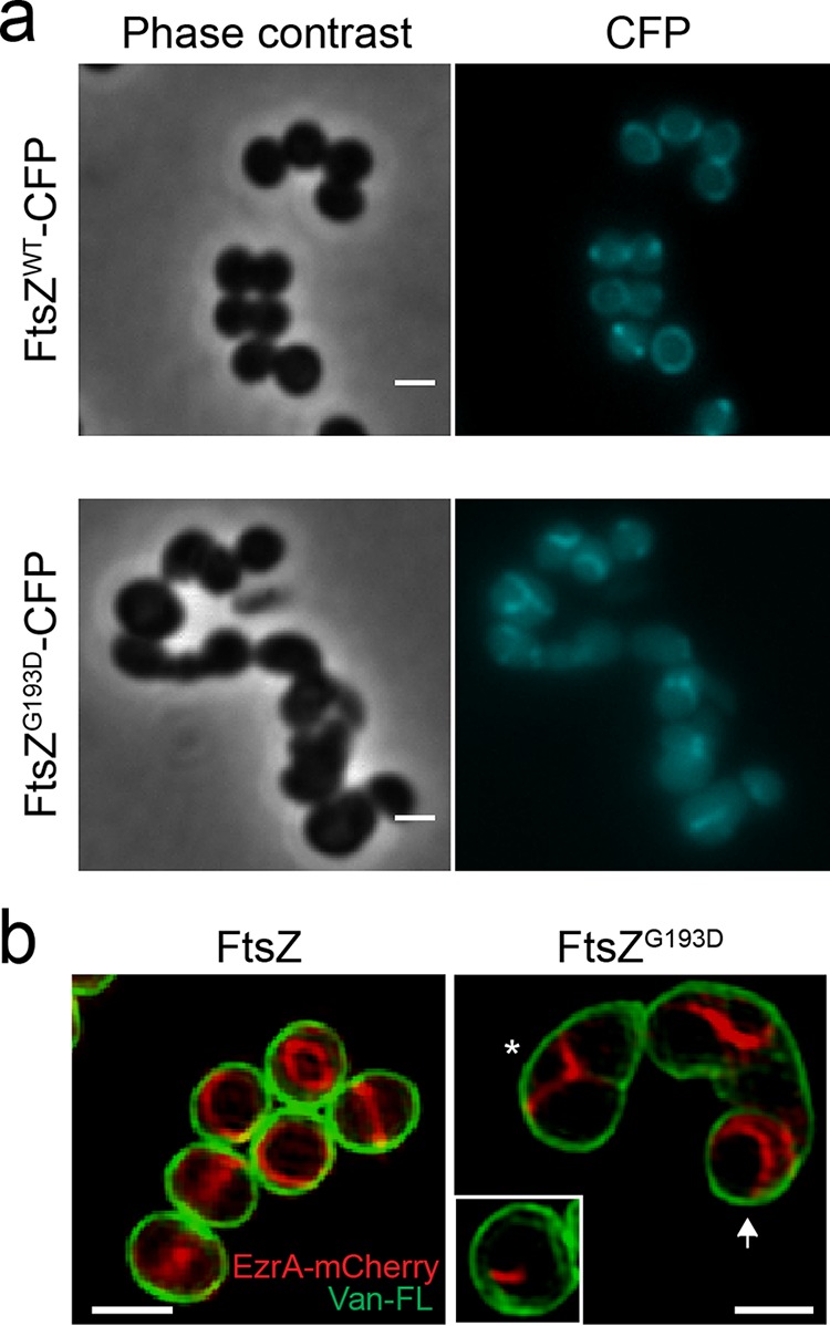
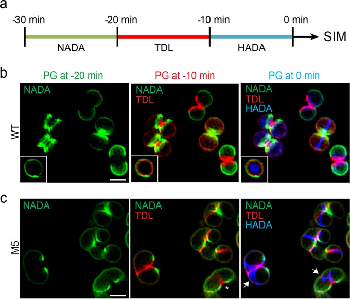
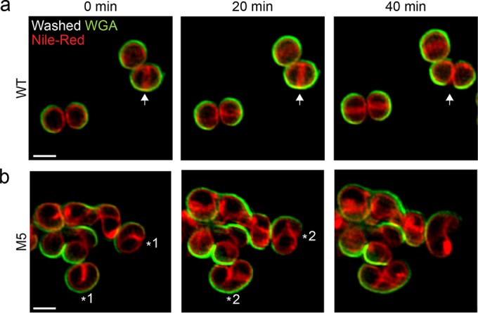
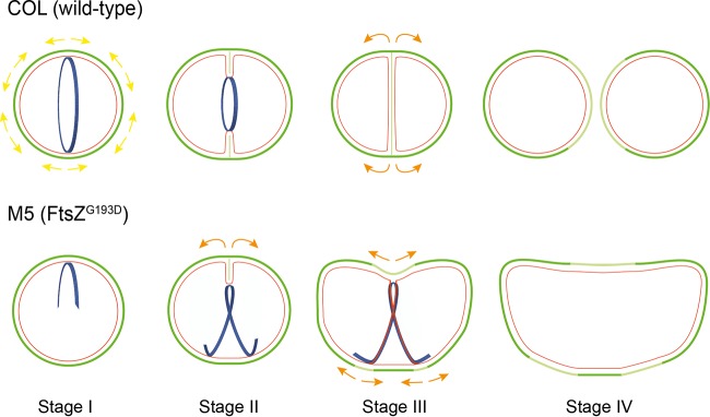
A mechanistic understanding of the determination and maintenance of the simplest bacterial cell shape, a sphere, remains elusive compared with that of more complex shapes. Cocci seem to lack a dedicated elongation machinery, and a spherical shape has been considered an evolutionary dead-end morphology, as a transition from a spherical to a rod-like shape has never been observed in bacteria. Here we show that a Staphylococcus aureus mutant (M5) expressing the ftsZ(G193D) allele exhibits elongated cells. Molecular dynamics simulations and in vitro studies indicate that FtsZ(G193D) filaments are more twisted and shorter than wild-type filaments. In vivo, M5 cell wall deposition is initiated asymmetrically, only on one side of the cell, and progresses into a helical pattern rather than into a constricting ring as in wild-type cells. This helical pattern of wall insertion leads to elongation, as in rod-shaped cells. Thus, structural flexibility of FtsZ filaments can result in an FtsZ-dependent mechanism for generating elongated cells from cocci.

Importance: The mechanisms by which bacteria generate and maintain even the simplest cell shape remain an elusive but fundamental question in microbiology. In the absence of examples of coccus-to-rod transitions, the spherical shape has been suggested to be an evolutionary dead end in morphogenesis. We describe the first observation of the generation of elongated cells from truly spherical cocci, occurring in a Staphylococcus aureus mutant containing a single point mutation in its genome, in the gene encoding the bacterial tubulin homologue FtsZ. We demonstrate that FtsZ-dependent cell elongation is possible, even in the absence of dedicated elongation machinery.


Figures

FIG 1
FIG 1
The FtsZG193D mutation leads to S. aureus cell elongation. (a) SIM images of wild-type COL (left) and FtsZG193D mutant M5 (right) cells labeled with the cell wall dye Van-FL (green) and the DNA dye Hoechst 33342 (blue). Scale bar: 1 µm. (b) The ratio of the longer to the shorter axis was calculated at two time points during the cell cycle, an initial time point (ti) when cells had a round shape and a final time point (tf) when cells were most elongated, prior to splitting of the mother cell. M5 cells elongate significantly more during the cell cycle than COL wild-type cells (P < 0.001). (c) Transmission electron micrographs (TEM) of thin sections of COL and M5 cells. Scale bars: 200 nm. (d) Scanning electron microscopy (SEM) images of COL and M5 cells. Scale bars: 1 µm.
FIG 2
FIG 2
FtsZG193D produces twisted and shorter polymers. (a) MD simulations of dimers of S. aureus FtsZWT (PDB code 3VO8) and FtsZG193D reveal a propensity for twist in FtsZG193D dimers. Simulations were initialized as straight dimers at t = 0 (side view). The top view highlights the development of twist between the principal axes of the two FtsZG193D subunits at t = 50 ns. (b) Shown are filaments of 14 subunits constructed by extrapolating the dimer interface from time points during the MD simulations. The FtsZWT dimer bent but remained untwisted for the first ~100 ns. (c) The FtsZG193D dimer rapidly adopted a substantial degree of twist, while the FtsZWT dimer acquired twist much later in the simulation. (d) Snapshots at the beginning and end of the FtsZWT dimer simulation, in which residues are colored if they interact (defined as being within 5 Å of another residue) with the opposite subunit at any point during the simulation. Colors: orange, specific to the nontwisted state of the wild type, with an interaction in the first 100 ns and no interaction after 150 ns (and no interaction throughout the FtsZG193D simulation); purple, generally present in twisted states (always interacting in FtsZG193D and after 150 ns for FtsZWT); pink, specific to FtsZG193D. The salt bridge between Asp97 (light green) and Arg67 (cyan) became broken at t = 262 ns. Gly193 is dark green. (e) Polymers of S. aureus FtsZWT (left) and FtsZG193D (right) imaged by transmission electron microscopy (TEM). Scale bar: 100 nm. (f) Measurements of FtsZWT and FtsZG193D polymer (n >70) lengths in the TEM images show that FtsZG193D polymers are significantly shorter than FtsZWT polymers (P < 0.0001).
FIG 3
FIG 3
Fluorescent fusions with FtsZ and EzrA lose mid-cell localization in M5 cells. (a) Localization of FtsZWT-CFP in BCBAJ020 cells (top) and FtsZG193D-CFP in BCBRP005 cells (bottom). Phase-contrast (left) and epifluorescence (right) images are shown. (b) SIM images of EzrA-mCherry (red) and cell wall labeled with Van-FL (green) in BCBAJ012 cells expressing FtsZWT (left) and in BCBRP006 cells expressing FtsZG193D (right). In the presence of FtsZG193D, EzrA-mCherry fails to localize as a mid-cell ring. Instead, structures assemble on only one side of the cell (white box) or in Y patterns compatible with one-turn helical structures (asterisk). More elongated BCBRP006 cells show EzrA-mCherry localized toward the poles of the cell (white arrow). Scale bars: 1 µm.
FIG 4
FIG 4
PG incorporation dynamics suggest a new mode of cell growth in S. aureus FtsZG193D cells. (a) Schematic representation of pulse-labeling with FDAAs. Cells were labeled with three sequential 10-min pulses of NADA (green), TDL (red), and HADA (blue) and observed by SIM. The same cell imaged in three different channels therefore shows the PG synthesized 30 to 20 min (green), 20 to 10 min (red), and 10 to 0 min (blue) prior to imaging. (b) In COL cells, septum synthesis initiates as a symmetric ring at mid-cell that constricts over time, concomitant with concentric PG synthesis (white box). (c) In M5 cells, PG synthesis occurs in two steps. First, PG is asymmetrically incorporated on only one side of a cell, eventually leading to local splitting (asterisks). Second, PG is inserted along a Y-shaped pattern compatible with a one-turn helical path (blue fluorescence, white arrows). Scale bars: 1 µm.
FIG 5
FIG 5
SIM time-lapse imaging reveals the mode of elongation of M5 cells. S. aureus COL (FtsZWT) and M5 (FtsZG193D) cells were stained with the peripheral cell wall dye WGA-488 (green). Unbound dye was washed away, labeled cells were stained with the membrane dye Nile red, and growth was monitored by SIM. (a) COL wild-type (WT) cells synthesized a ring-like septum at mid-cell (arrow at 0 min) that constricts and eventually splits, giving rise to two daughter cells (arrow at 40 min). (b) M5 mutant cells initially synthesized an asymmetric septum on only one side of the cell (asterisk 1), which later developed into a pattern compatible with a one-turn helical structure (asterisk 2) and led to elongation. Scale bars: 1 µm.
FIG 6
FIG 6
Model of S. aureus M5 cell elongation. Schematic of cell division events in wild-type COL (top) and M5 (bottom) cells. The images show old cell wall (dark green), new cell wall (light green), membrane (red), and FtsZ structures (blue). Yellow/orange arrows correspond to surface expansion. In wild-type cells (top), FtsZ assembles as a ring at mid-cell and recruits PG synthesis enzymes, leading to the incorporation of new PG at mid-cell. New PG is also inserted along the cell periphery, leading to cell surface expansion (yellow arrows). Splitting of the septum generates ~1/3 of the cell surface of each of the new daughter cells, thereby increasing the total surface area exposed to the external milieu. M5 cells (bottom) expressing FtsZG193D rather than FtsZWT incorporate PG in two steps. First, PG deposition occurs on only one side of the cell (asymmetric septum, division stage I), which will initiate local splitting following septal constriction (stage II) that leads to surface area expansion only on that side of the cell (orange arrows). Second, PG deposition is directed by FtsZ along a partial helical path, leading to further surface expansion resembling elongation of rod-shaped cells (stage III). Thus, M5 cells combine a coccus-like (septal) mode of PG incorporation (stage II) with a rod-like elongation mode (stages III and IV), both of which are coordinated by FtsZ.

References

    1. Young KD. 2006. The selective value of bacterial shape. Microbiol Mol Biol Rev 70:660–703. doi: 10.1128/MMBR.00001-06. - DOI - PMC - PubMed
    1. Typas A, Banzhaf M, Gross CA, Vollmer W. 2012. From the regulation of peptidoglycan synthesis to bacterial growth and morphology. Nat Rev Microbiol 10:123–136. doi: 10.1038/nrmicro2677. - DOI - PMC - PubMed
    1. Pinho MG, Kjos M, Veening JW. 2013. How to get (a)round: mechanisms controlling growth and division of coccoid bacteria. Nat Rev Microbiol 11:601–614. doi: 10.1038/nrmicro3088. - DOI - PubMed
    1. Cava F, Kuru E, Brun YV, de Pedro MA. 2013. Modes of cell wall growth differentiation in rod-shaped bacteria. Curr Opin Microbiol 16:731–737. doi: 10.1016/j.mib.2013.09.004. - DOI - PMC - PubMed
    1. Adams DW, Errington J. 2009. Bacterial cell division: assembly, maintenance and disassembly of the Z ring. Nat Rev Microbiol 7:642–653. doi: 10.1038/nrmicro2198. - DOI - PubMed

Publication types

MeSH terms