. 2016 Sep 6;7(5):e01395-16.
doi: 10.1128/mBio.01395-16.

Horizontal Transfer of the Salmonella enterica Serovar Infantis Resistance and Virulence Plasmid pESI to the Gut Microbiota of Warm-Blooded Hosts

Affiliations

Horizontal Transfer of the Salmonella enterica Serovar Infantis Resistance and Virulence Plasmid pESI to the Gut Microbiota of Warm-Blooded Hosts

Gili Aviv et al. mBio. .

Abstract

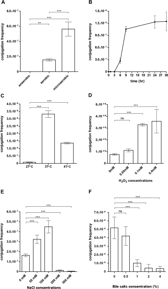
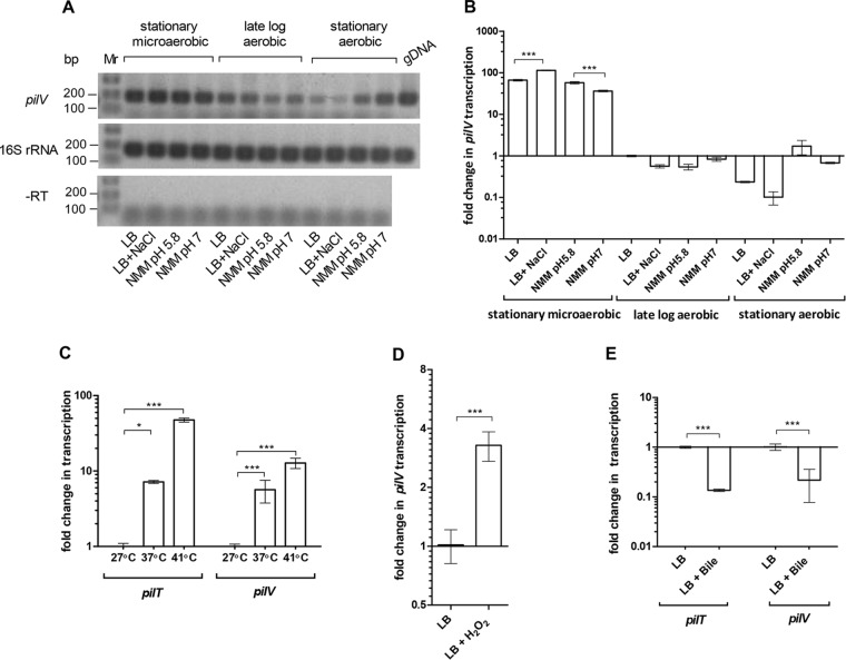
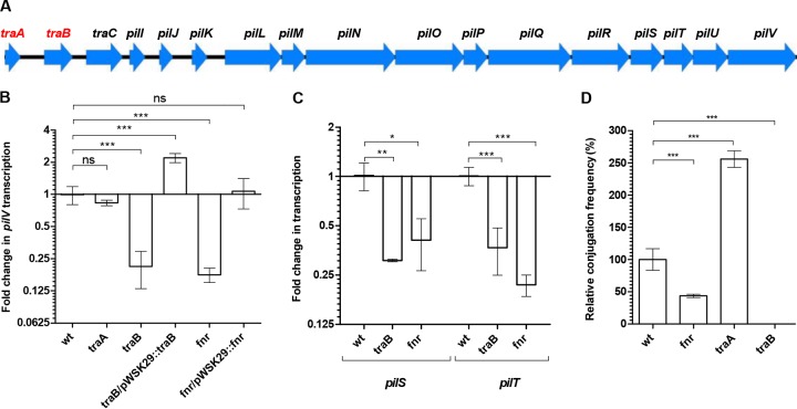
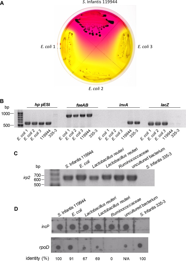
Salmonella enterica serovar Infantis is one of the prevalent salmonellae worldwide. Recently, we showed that the emergence of S Infantis in Israel was facilitated by the acquisition of a unique megaplasmid (pESI) conferring multidrug resistance and increased virulence phenotypes. Here we elucidate the ecology, transmission properties, and regulation of pESI. We show that despite its large size (~280 kb), pESI does not impose a significant metabolic burden in vitro and that it has been recently fixed in the domestic S Infantis population. pESI conjugation and the transcription of its pilus (pil) genes are inhibited at the ambient temperature (27°C) and by ≥1% bile but increased under temperatures of 37 to 41°C, oxidative stress, moderate osmolarity, and the microaerobic conditions characterizing the intestinal environment of warm-blooded animals. The pESI-encoded protein TraB and the oxygen homeostasis regulator Fnr were identified as transcriptional regulators of pESI conjugation. Using the mouse model, we show that following S Infantis infection, pESI can be horizontally transferred to the gut microbiota, including to commensal Escherichia coli strains. Possible transfer, but not persistence, of pESI was also observed into Gram-positive mouse microbiota species, especially Lactobacillus reuteri Moreover, pESI was demonstrated to further disseminate from gut microbiota to S. enterica serovar Typhimurium, in the context of gastrointestinal infection. These findings exhibit the ability of a selfish clinically relevant megaplasmid to distribute to and from the microbiota and suggest an overlooked role of the microbiota as a reservoir of mobile genetic elements and intermediator in the spread of resistance and virulence genes between commensals and pathogenic bacteria.

Importance: Plasmid conjugation plays a key role in microbial evolution, enabling the acquisition of new phenotypes, including resistance and virulence. Salmonella enterica serovar Infantis is one of the ubiquitous salmonellae worldwide and a major cause of foodborne infections. Previously, we showed that the emergence of S Infantis in Israel has involved the acquisition of a unique megaplasmid (pESI) conferring multidrug resistance and increased virulence phenotypes. Recently, the emergence of another S Infantis strain carrying a pESI-like plasmid was identified in Italy, suggesting that the acquisition of pESI may be common to different emergent S Infantis populations globally. Transmission of this plasmid to other strains or bacterial species is an alarming scenario. Understanding the ecology, regulation, and transmission properties of clinically relevant plasmids and the role of the microbiota in their spreading offers a new mechanism explaining the emergence of new pathogenic and resistant biotypes and may assist in the development of appropriate surveillance and prevention measures.


Figures

FIG 1
FIG 1
A modular formation of pESI. (A) Plasmid linearization using S1 nuclease followed by PFGE analysis was performed to determine the plasmid size of p120100 found in a 2008 S. Infantis food isolate (120100) in comparison to pESI. Linearized plasmids are indicated by arrowheads. (B) Pairwise comparison of a DNA fragment of about 120 kb corresponding to the leading and transfer region from pESI and p120100 was performed using the Easyfig tool. The colored bar indicates sequence homology between the overlapped regions. A region of about 21 kb containing the MGEs Tn21-like and Tn1721-like, which are absent from p120100, is indicated. (C) PCR analysis of chromosomal (tcfD), pESI backbone-carried (traC, hp, faeAB, ipfA, and irp2), and MGE-carried (int, sulI, qacEΔ1, merA, and tetA) genes. Template DNA was extracted from S. Infantis isolates 119944 (harboring pESI), 120100 (harboring p120100), and 335-3 (a plasmidless preemergent strain).
FIG 2
FIG 2
pESI is maintained in a single copy number and does not confer a significant metabolic burden in vitro. (A) Ten-fold serial dilutions of genomic DNA extracted from S. Infantis 335-3 harboring pESI were subjected to real-time PCR using specific primers of the pESI-carried gene pilV and the chromosome-carried gene rpoD. Bars represent the mean threshold cycle (CT) values and the standard deviation (SD) from three biological repeats. The CT values of each gene at the different dilutions were used to create a standard curve. The amplicon size (in base pairs), amplification efficiency (percentage), the slope of the standard curve, and the coefficient of determination (R2), showing the linearity of the standard curve, are indicated in the bottom table. (B) Competitive index of S. Infantis 335-3 carrying pESI and its isogenic strain lacking pESI. Both strains were grown together in LB and N-minimal medium for 24 h at 27°C. Competitive index values were determined as (335-3-pESI/335-3)output/(335-3-pESI/335-3)input. Dots represent the results of single (out of 12) independent competition experiments; the mean and SD are shown by the horizontal line and error bars, respectively. A D’Agostino and Pearson omnibus normality test confirmed Gaussian distributions, and a one-sample t test against a theoretical mean of 1 was used to determine statistical significance of the mean. ns, not significant (P > 0.05).
FIG 3
FIG 3
pESI conjugation is increased in response to microaerobiosis, physiological temperature, and moderate osmolarity. The pESI conjugation frequency (obtained transconjugants/donor CFU) between S. Infantis 119944 (donor) and E. coli ORN172 (recipient) was determined under (A) different oxygen conditions, (B) over time under a microaerobic environment, and at different (C) temperatures, (D) hydrogen-peroxide concentrations, (E) sodium chloride concentrations, and (F) bile salt concentrations. Bars show the means and SDs from at least four independent mating experiments. One-way analysis of variance (ANOVA) with Tukey’s (for panels A and C) or Dunnett’s (for panels D to F) multiple comparison tests was implemented to determine statistical significance. ns, not significant; **, P < 0.001; ***, P < 0.0001.
FIG 4
FIG 4
The transcription of pESI pilus is induced in response to microaerobiosis and physiological temperature and repressed by bile. (A) Semiquantitative reverse transcription-PCR analyses of pilV and 16S rRNA transcripts. RNA was extracted from S. Infantis 119944 cultures grown in LB, LB supplemented with 0.3 M NaCl, N-minimal medium (NMM) at pH 5.8, and N-minimal medium at pH 7.0. Templates of genomic DNA (gDNA) or purified RNA without reverse transcriptase treatment (−RT) were included as positive and negative controls, respectively. (B) qRT-PCR shows the fold change in pilV transcription under the same growth conditions as in panel A relative to the transcription of pilV in LB late-logarithmic culture grown under aerobic conditions. (C) Fold change in the transcription of pilV and pilT grown in LB under microaerobic conditions at 37 and 41°C relative to 27°C. (D) RNA was extracted from S. Infantis 119944 grown in LB and in LB supplemented with 0.1 mM H2O2 under microaerobic conditions at 37°C. qRT-PCR analysis was conducted to determine the fold change in the transcription of pilV. (E) Fold change in the transcription of pilV and pilT grown under microaerobic conditions at 37°C in LB supplemented with 4% bile salts (sodium choleate). All RT-PCR results show the mean and SD from three to six biological repeats. Two-tailed t test (B, D, and E) or one-way ANOVA with Tukey’s multiple comparison test (C) was implemented to determine statistical significance. *, P < 0.05; ***, P < 0.0001.
FIG 5
FIG 5
pESI conjugation and pilus transcription are regulated by TraB and FNR. (A) Gene organization of the pil operon carried on pESI. Arrowheads show the location and orientation of the different open reading frames (ORFs). Putative regulatory genes are shown in red. (B and C) RNA was extracted from cultures of S. Infantis 119944 (wild type [wt]) and its derivative mutants (traA, traB, and fnr) and traB/pWSK29::traB and fnr/pWSK29::fnr complemented strains grown in LB under microaerobic conditions at 37°C. qRT-PCR analyses was conducted to determine the fold change in the transcription of pilV (B) and pilS and pilT (C) in the indicated backgrounds relative to the wild-type strain. (D) pESI conjugation between S. Infantis strain 119944 or its fnr-, traA-, and traB-derived mutant strains and E. coli ORN172 were conducted on LB plates under microaerobic conditions. Results show the mean and SD from at least three biological repeats. One-way ANOVA with Dunnett’s multiple comparison test was implemented to determine statistical significance. *, P < 0.005; **, P < 0.001; ***, P < 0.0001; ns, not significant.
FIG 6
FIG 6
Interspecies transfer of pESI during S. Infantis infection. Female C57BL/6 mice were infected with 1.5 × 10⁸ CFU of S. Infantis strain 119944 (harboring pESI) and screened for non-Salmonella microbiota that have acquired pESI following the infection. (A) The donor (S. Infantis 119944) and three mouse-isolate microbiota E. coli transconjugants were plated on XLD plates supplemented with tetracycline. (B) To confirm the presence of pESI in the above E. coli transconjugants, PCR amplification of pESI-specific genes (hp pESI and faeAB) and Salmonella-specific (invA) and E. coli-specific (lacZ) genes was conducted. S. Infantis 119944 and S. Infantis 335-3 lacking pESI were used as positive and negative controls, respectively. (C) Five representative microbiota isolates, including E. coli (isolate 481-49), L. reuteri (isolates 480-44 and 482-46), Ruminococcaceae (isolate 482-50), and an unknown bacterium (isolate 481-27) were subjected to PCR using primers from the pESI-specific gene irp2. S. Infantis 119944 and a pESI-negative S. Infantis isolate (335-3) were used as positive and negative controls, respectively. (D) Total DNA (100 ng) that was extracted from the above microbiota isolates was subjected to a dot blot hybridization using a DIG-labeled incP pESI backbone probe. A DIG-labeled rpoD probe was used as a hybridization control, and the identity (in percentage) between the rpoD sequence of S. Infantis (used to synthesize the rpoD probe) and its homolog in the tested genome is shown at the bottom of the panel.
FIG 7
FIG 7
pESI transfer from microbiota to a naive pathogen during infection. (A) Three E. coli mouse isolates that have acquired pESI following S. Infantis 119944 infection were used as donor strains for conjugation with S. Typhimurium SL1344 carrying a chloramphenicol chromosomally carried marker as a recipient strain. pESI conjugation frequency was determined after 16 h at 37°C under microaerobic conditions, by the number of the obtained transconjugants (cmr tetr S. Typhimurium) per donor CFU. (B) Experimental workflow used to detect pESI transfer from microbiota to S. Typhimurium during infection. Five 7-week-old female C57BL/6 mice were treated with streptomycin 1 day before infection with 1.3 × 10⁷ CFU of mouse isolate E. coli harboring pESI (donor). Two days p.i., these mice were reinfected with 1.6 × 10⁷ CFU of S. Typhimurium SL1344 (STM) carrying the chloramphenicol marker (recipient). At day 6 p.i., mice were sacrificed and GI tissues were homogenized. Feces and tissue homogenates were plated onto XLD plates supplemented with tetracycline (tet), trimethoprim (trim), and chloramphenicol (cm). (C) To confirm the presence of pESI in three S. Typhimurium SL1344 transconjugants (STM TC1-3), PCR amplification of pESI-specific (hp pESI and faeAB), Salmonella-specific (invA), and E. coli-specific (lacZ) genes was conducted. The microbiota E. coli strain (donor) and S. Typhimurium (recipient) were also included as positive and negative controls, respectively. (D) The E. coli microbiota donor, the S. Typhimurium (STM) recipient, and the obtained three STM transconjugants were plated on XLD plates and LB plates supplemented with chloramphenicol, tetracycline, and chloramphenicol+tetracycline+trimethoprim.

References

    1. Galanis E, Lo Fo Wong DM, Patrick ME, Binsztein N, Cieslik A, Chalermchikit T, Aidara-Kane A, Ellis A, Angulo FJ, Wegener HC, World Health Organization Global Salm-Surv . 2006. Web-based surveillance and global Salmonella distribution, 2000–2002. Emerg Infect Dis 12:381–388. doi: 10.3201/eid1205.050854. - DOI - PMC - PubMed
    1. Majowicz SE, Musto J, Scallan E, Angulo FJ, Kirk M, O’Brien SJ, Jones TF, Fazil A, Hoekstra RM, International Collaboration on Enteric Disease Burden of Illness Studies . 2010. The global burden of nontyphoidal Salmonella gastroenteritis. Clin Infect Dis 50:882–889. doi: 10.1086/650733. - DOI - PubMed
    1. Desai PT, Porwollik S, Long F, Cheng P, Wollam A, Bhonagiri-Palsikar V, Hallsworth-Pepin K, Clifton SW, Weinstock GM, McClelland M. 2013. Evolutionary genomics of Salmonella enterica subspecies. mBio 4:e00579-12. doi: 10.1128/mBio.00579-12. - DOI - PMC - PubMed
    1. Edwards RA, Olsen GJ, Maloy SR. 2002. Comparative genomics of closely related salmonellae. Trends Microbiol 10:94–99. doi: 10.1016/S0966-842X(01)02293-4. - DOI - PubMed
    1. Crim SM, Griffin PM, Tauxe R, Marder EP, Gilliss D, Conquist AB, Cartter M, Tobin-D’Angelo M, Blythe D, Smith K, Lathrop S, Zansky S, Cieslak PR, Dunn J, Holt KG, Wolpert B, Henao OL, Centers for Disease Control and Prevention . 2015. Preliminary incidence and trends of infection with pathogens transmitted commonly through food—Foodborne Diseases Active Surveillance Network, 10 U.S. sites, 2006–2014. MMWR Morb Mortal Wkly Rep 64:495–499. - PMC - PubMed

Publication types