Review
. 2017 Jan 3;9(1):a028076.
doi: 10.1101/cshperspect.a028076.

Axoneme Structure from Motile Cilia

Affiliations
Review

Axoneme Structure from Motile Cilia

Takashi Ishikawa. Cold Spring Harb Perspect Biol. .

Abstract

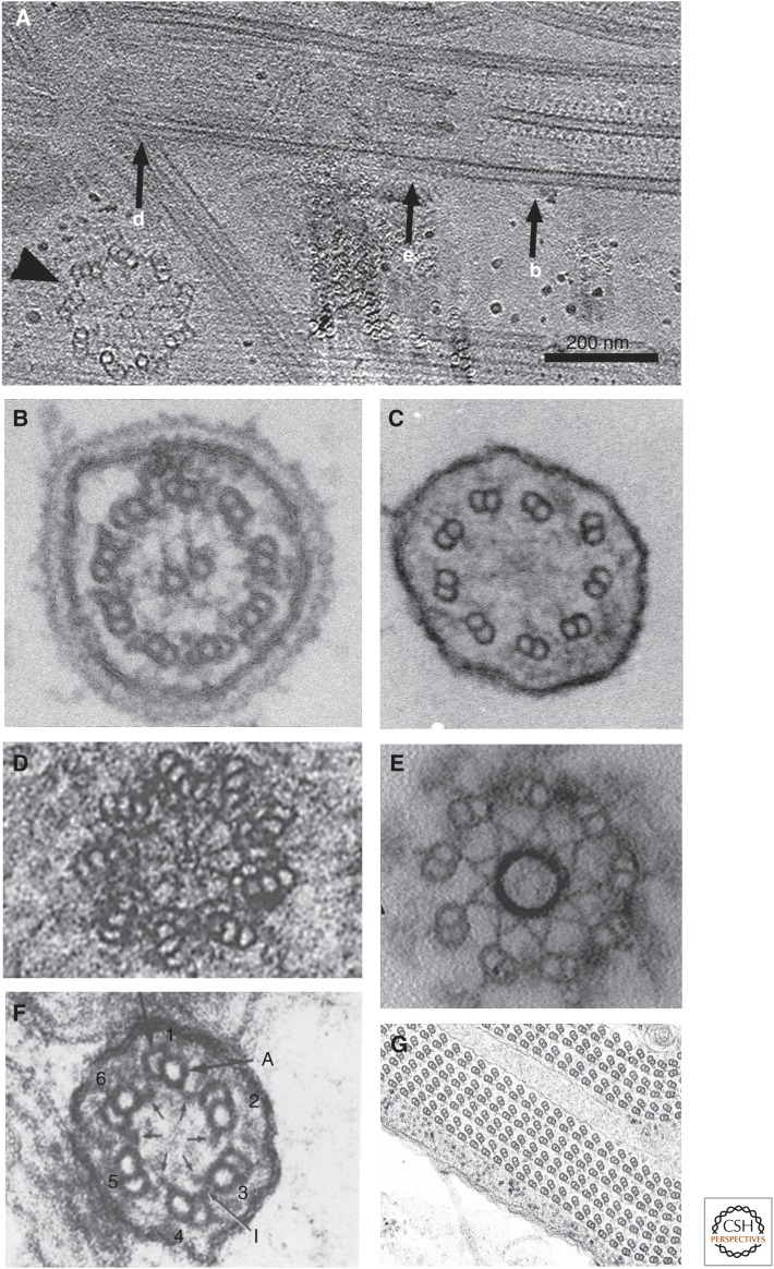
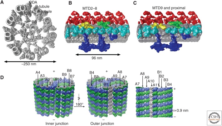
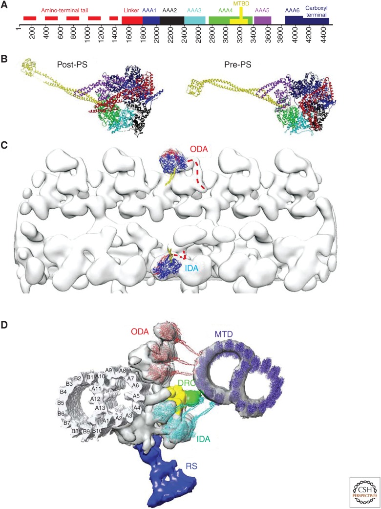
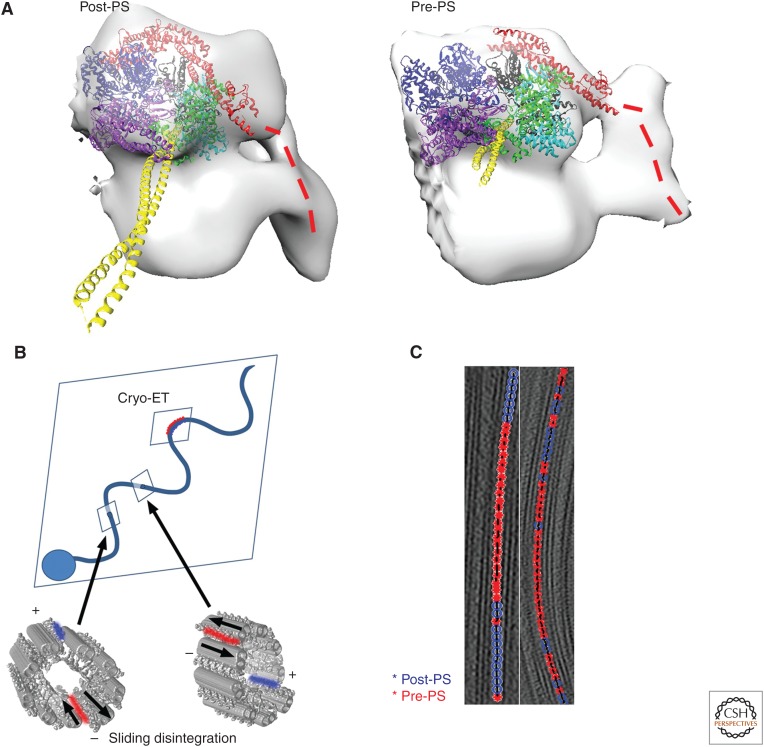
The axoneme is the main extracellular part of cilia and flagella in eukaryotes. It consists of a microtubule cytoskeleton, which normally comprises nine doublets. In motile cilia, dynein ATPase motor proteins generate sliding motions between adjacent microtubules, which are integrated into a well-orchestrated beating or rotational motion. In primary cilia, there are a number of sensory proteins functioning on membranes surrounding the axoneme. In both cases, as the study of proteomics has elucidated, hundreds of proteins exist in this compartmentalized biomolecular system. In this article, we review the recent progress of structural studies of the axoneme and its components using electron microscopy and X-ray crystallography, mainly focusing on motile cilia. Structural biology presents snapshots (but not live imaging) of dynamic structural change and gives insights into the force generation mechanism of dynein, ciliary bending mechanism, ciliogenesis, and evolution of the axoneme.


Figures

Figure 1.
Figure 1.
Axoneme structure from various species and locations of cilia. (A) Horizontal section from tomographic reconstruction of ice-embedded cilia including the basal body (centriole) and the transition zone (TZ) (M Hirono and T Ishikawa, unpubl.). A probasal body (a daughter centriole), which is positioned perpendicular to the basal body, is indicated by an arrowhead. (BG) Electron micrographs of cross sections from plastic embedded cilia. (B) Typical 9+2 structure of the axoneme from Chlamydomonas. (Figure courtesy of Dr. Dennis Diener, Yale University.) (C) 9+0 structure from mouse nodal cilia. (Figure courtesy of Dr. Svetlana Markova, Dr. Dennis Diener, and Prof. Martina Brueckner, Yale University.) (D) Section of a centriole, showing microtubule triplets and the cartwheel (Berns et al. 1977). (E) The TZ from Chlamydomonas flagella. (Image modified from Awata et al. 2014.) (F) Unusual 6+0 structure (Schrevel and Besse 1975). (G) Abnormal axoneme with more than 100 microtubule doublets (MTDs) from the gall-midge fly Asphondylia ruebsaameeni Kertesz (Mencarelli et al. 2000). (Figure courtesy of Prof. Romano Dallai and Prof. Pietro Lupetti, University of Siena.) Corresponding position of cross sections in B, D, and E are indicated in A by b, d, and e, respectively.
Figure 2.
Figure 2.
Overall structure of the 96-nm periodic unit from the axoneme. (A) Structure of the “9+2” axoneme based on cryoelectron tomography (cryo-ET). Outer and inner dynein arms (ODA and IDA, respectively), radial spokes (RS), A- and B-tubules from a microtubule doublet (MTD) as well as a central pair (CP) are indicated. (Figure modified from Bui and Ishikawa 2013.) (B,C) Enlarged views of one 96-nm periodic unit of regular MTDs. MTD2–8 in the distal region and MTD9 are shown (Bui et al. 2009, 2012). Dynein isoforms are indicated in B. (Image based on Bui et al. 2012.) MTD2–8 in the proximal region also lack dynein b. Red, ODA; cyan, IDA; purple, adjacent MTD; green, dynein regulatory complex (DRC); yellow, IC/LC (intermediate chain/light chain); blue, RS. Density maps are available as EMD2113-2130. (D) Molecular arrangement of tubulins in an MTD revealed by single particle cryoelectron microscopy (cryo-EM) (Maheshwari et al. 2015). Green, α-tubulin; blue, β-tubulin. Numbering of the protofilaments in A- and B-tubules is identical to that in Figure 3D.
Figure 3.
Figure 3.
Dynein structure and arrangement in the axoneme. (A) Sequence motif of dynein. (B) Atomic structure of dynein in the post–power stroke (post-PS) conformation (PDB ID: 3VKH) (Kon et al. 2012) and in the pre–power stroke (pre-PS) conformation (4RH7) (Schmidt et al. 2014). In A and B, the color code is as follows: red, amino terminus and linker; blue, AAA1; black, AAA2; cyan, AAA3; green, AAA4; yellow, coiled-coil stalk (which is a part of AAA4); purple, AAA5; dark blue, AAA6 and carboxyl terminus. (C) Atomic models (3VKH) fitted to cryoelectron tomography (cryo-ET) structure (EMDB ID: 2117) (Bui et al. 2012) to show the front and back of dyneins in outer dynein arms (ODAs) and inner dynein arms (IDAs), respectively. The linker (red) is above the AAA ring (blue) in ODAs, whereas it is below the ring in IDAs. Yellow, stalks. (D) Atomic models in which density maps from cryo-ET (Bui et al. 2012) and single-particle analysis (ciliary microtubule doublet [MTD)] (Maheshwari et al. 2015) are fitted. Red, ODA; cyan, IDA; purple, adjacent MTD; green, dynein regulatory complex (DRC); yellow, intermediate and light chains (IC/LC); blue, radial spoke (RS).
Figure 4.
Figure 4.
Dynein structural change induced by nucleotides. (A) Atomic model fitting to cryoelectron tomography (cryo-ET) (Ueno et al. 2014). The overall orientation of the linker at pre–power stroke (pre-PS) and post–power stroke (post-PS) condition (red) is the same as crystallography (Fig. 3B). The color code is the same as in Figure 3A,B. (B) Switching models based on sliding disintegration (Hayashi and Shingyoji 2008) and cryo-ET (Lin et al. 2014) of sea urchin sperm. Sliding disintegration occurs by sliding between two adjacent microtubule doublets (MTDs) (red in the surface-rendered models of two axonemes), whereas dyneins at the opposite side of the axoneme are relaxed (blue). In intact cilia, straight areas between principle and reverse bending (indicated by arrows) should be where sliding takes place. If sliding moves toward the opposite directions at these two sites, bending can happen. The sliding plane is perpendicular to the bending plane—switching occurs between the upper and lower sides of the bending plane. However, switching was found at the area of curvature by cryo-ET (Lin et al. 2014). Image classification of outer arm dynein structure showed localization of two dynein conformations at external and internal sides of curved cilia, suggesting switch within the bending plane. (C) Distribution of structure of outer dyneins along MTDs in the axoneme in the presence of the nonhydrolyzable ATP analog, ADP.Vi, show clustering, in which 10 to 20 dyneins in the post-PS conformation make a row (blue), followed by a row of dyneins in the pre-PS conformation (red). Under the same ADP.Vi concentration, almost all the dyneins turn to the pre-PS conformation without coexistence of multiple forms. This suggests cooperative conformational change of neighboring dyneins in the axoneme (Movassagh et al. 2010).
Figure 5.
Figure 5.
Rulers, microtubule inner proteins (MIPs), and radial spokes (RSs). (A) Location of FAP59 and FAP172, two coiled-coil proteins extending along the microtubule doublet (MTD) as proved by genetically tagged labels, which determine the periodic length, 96 nm. When amino-terminal subdomains of these proteins are duplicated by genetic engineering, the MTD has a 128-nm periodicity, allowing the third RS. Surface-rendered from EMD6108 and EMD6115 (Oda et al. 2014a). (B) Unidentified proteins binding to the inside of MTDs. Most of these MIPs bind two or more protofilaments. (From Maheshwari et al. 2015; reproduced, with permission, from the authors.) (C) Comparison of structure of RSs from Tetrahymena (left) and Chlamydomonas (right). The common structural motif at the base of RS3 is shown by arrows. (Modified from Pigino et al. 2011.)

References

    1. Avolio J, Lebduska S, Sati P. 1984. Dynein arm substructure and the orientation of arm-microtubule attachments. J Mol Biol 173: 389–401. - PubMed
    1. Awata J, Takada S, Standley C, Lechtreck KF, Bellvé KD, Pazour GJ, Fogarty KE, Witman GB. 2014. NPHP4 controls ciliary trafficking of membrane proteins and large soluble proteins at the transition zone. J Cell Sci 127: 4714–4727. - PMC - PubMed
    1. Berns MW, Rattner JB, Brenner S, Meredith S. 1977. The role of the centriolar region in animal cell mitosis. A laser microbeam study. J Cell Biol 72: 351–367. - PMC - PubMed
    1. Bower R, Tritschler D, Vanderwaal K, Perrone CA, Mueller J, Fox L, Sale WS, Porter ME. 2013. The N-DRC forms a conserved biochemical complex that maintains outer doublet alignment and limits microtubule sliding in motile axonemes. Mol Biol Cell 24: 1134–1152. - PMC - PubMed
    1. Bui KH, Ishikawa T. 2013. 3D structural analysis of flagella/cilia by cryo-electron tomography. Methods Enzymol 524: 305–323. - PubMed

LinkOut - more resources