. 2016 Sep 20;113(38):10571-6.
doi: 10.1073/pnas.1522478113. Epub 2016 Sep 6.

In B cells, phosphatidylinositol 5-phosphate 4-kinase-α synthesizes PI(4,5)P2 to impact mTORC2 and Akt signaling

Affiliations

In B cells, phosphatidylinositol 5-phosphate 4-kinase-α synthesizes PI(4,5)P2 to impact mTORC2 and Akt signaling

Simon J Bulley et al. Proc Natl Acad Sci U S A. .

Abstract

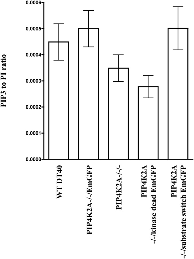
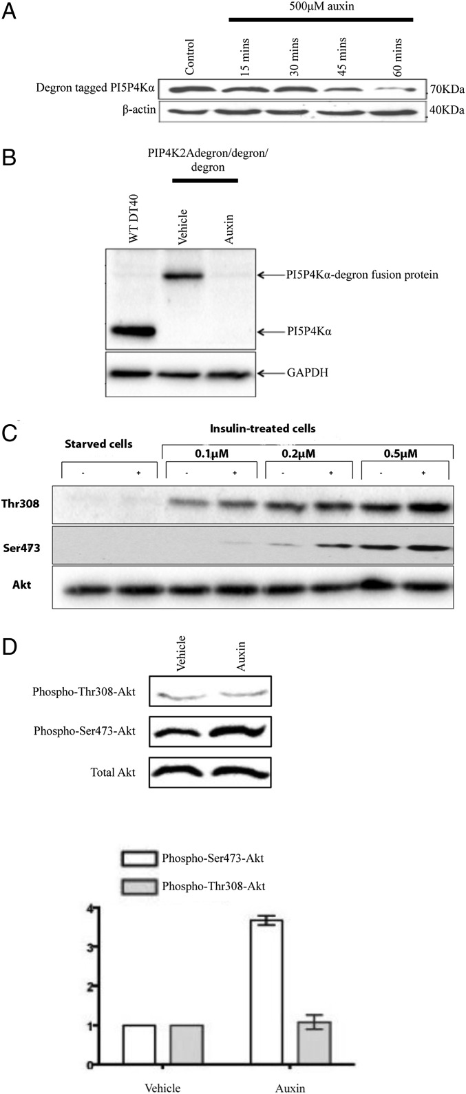
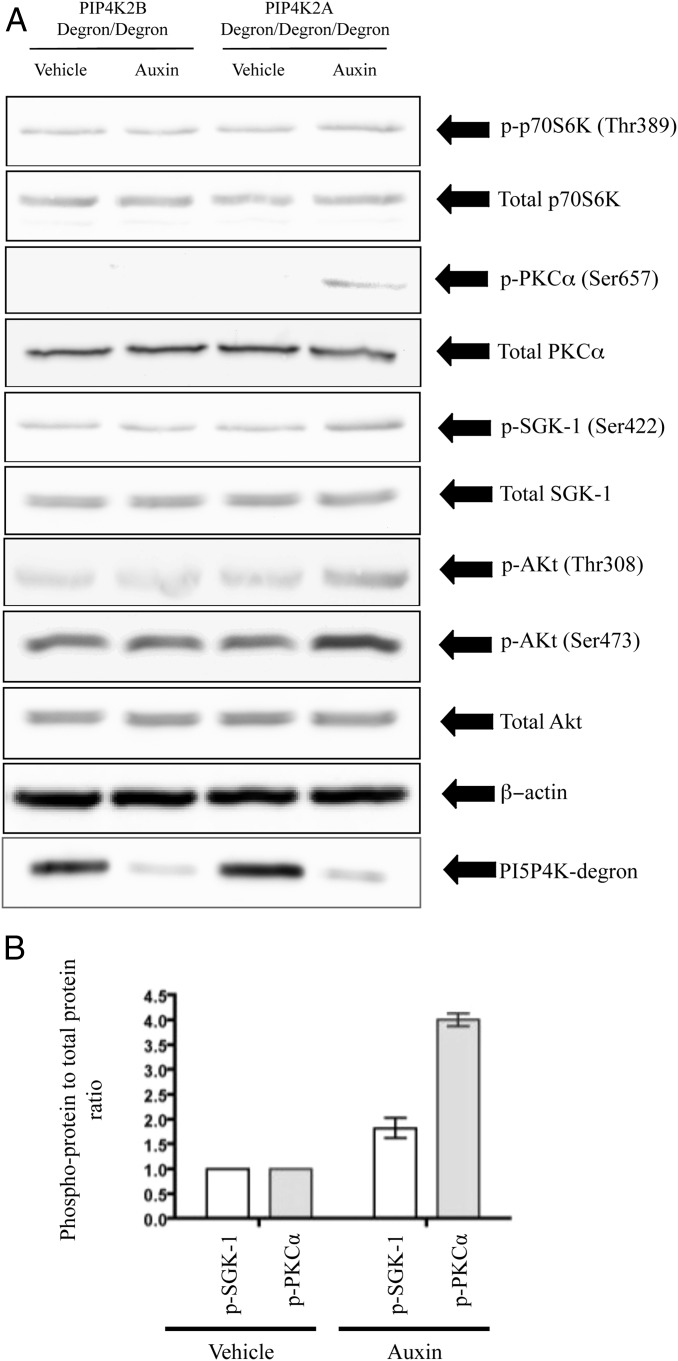
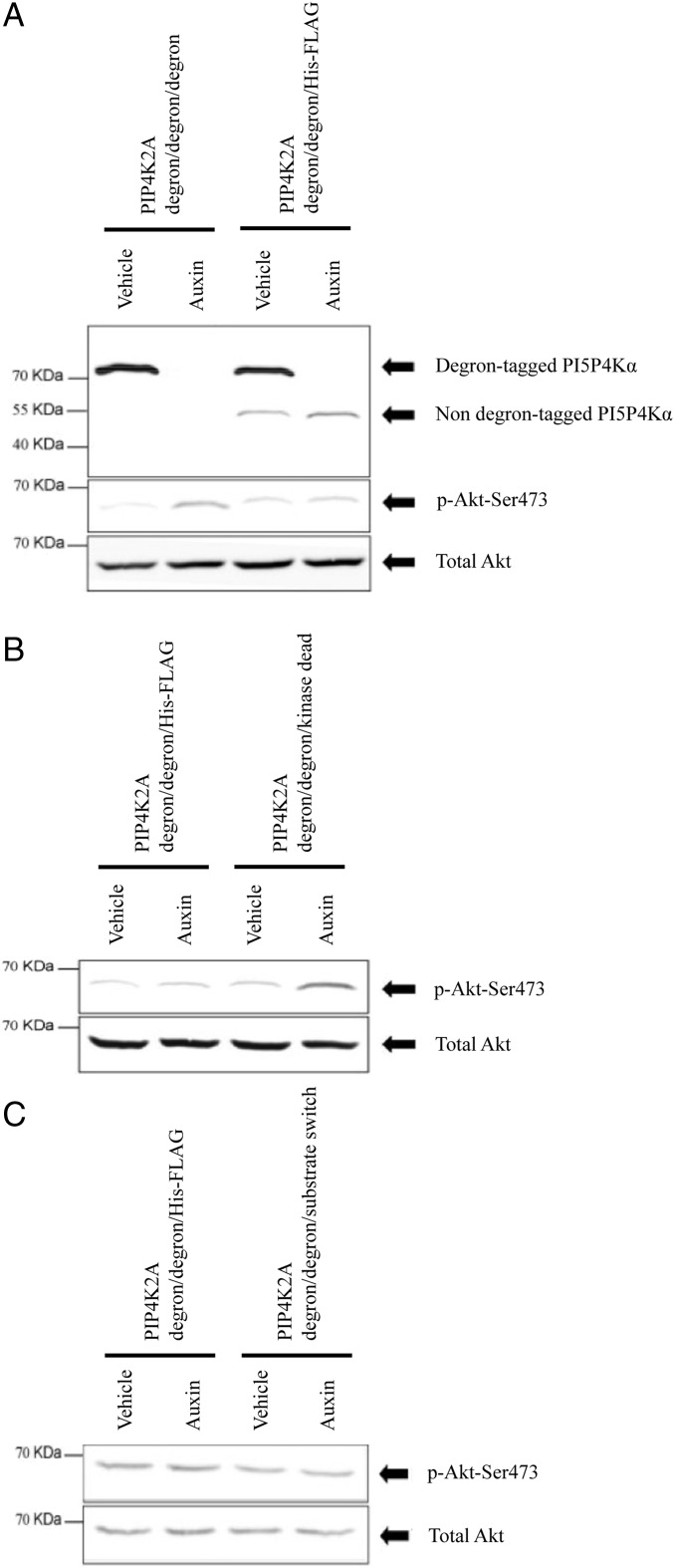
Phosphatidylinositol 5-phosphate 4-kinases (PI5P4Ks) are enigmatic lipid kinases with physiological functions that are incompletely understood, not the least because genetic deletion and cell transfection have led to contradictory data. Here, we used the genetic tractability of DT40 cells to create cell lines in which endogenous PI5P4Kα was removed, either stably by genetic deletion or transiently (within 1 h) by tagging the endogenous protein genomically with the auxin degron. In both cases, removal impacted Akt phosphorylation, and by leaving one PI5P4Kα allele present but mutating it to be kinase-dead or have PI4P 5-kinase activity, we show that all of the effects on Akt phosphorylation were dependent on the ability of PI5P4Kα to synthesize phosphatidylinositol (4,5)-bisphosphate [PI(4,5)P2] rather than to remove PI5P. Although stable removal of PI5P4Kα resulted in a pronounced decrease in Akt phosphorylation at Thr308 and Ser473, in part because of reduced plasma membrane PIP3, its acute removal led to an increase in Akt phosphorylation only at Ser473. This process invokes activation primarily of mammalian target of rapamycin complex 2 (mTORC2), which was confirmed by increased phosphorylation of other mTORC2 substrates. These findings establish PI5P4Kα as a kinase that synthesizes a physiologically relevant pool of PI(4,5)P2 and as a regulator of mTORC2, and show a phenomenon similar to the "butterfly effect" described for phosphatidylinositol 3-kinase Iα [Hart JR, et al. (2015) Proc Natl Acad Sci USA 112(4):1131-1136], whereby through apparently the same underlying mechanism, the removal of a protein's activity from a cell can have widely divergent effects depending on the time course of that removal.

Keywords: Akt; mTOR; phosphatidylinositol (4,5)-bisphosphate; phosphatidylinositol 5-phosphate; phosphatidylinositol 5-phosphate 4-kinase.


Conflict of interest statement

The authors declare no conflict of interest.

Figures

Fig. 1.
Fig. 1.
The effect of endogenous PIP4K2A mutations on Akt phosphorylation at Thr308 and Ser473. (A) Cells were synchronized at the same density in exponential growth before analysis. Complete PIP4K2A deletion results in reduced phospho-Akt at both Thr308 and Ser473. Deletion of two of three PIP4K2A alleles, while leaving the third unaffected aside from tagging with EmGFP, has no effect on Akt phosphorylation. This process acts as a control for the fourth and fifth lanes, where the undeleted allele is either rendered kinase-dead or mutated to encode a PI4P5K (SI Appendix, SI Materials and Methods) along with EmGFP tagging in both cases. A kinase-dead third allele does not support normal Akt phosphorylation in the way that a kinase-live one does, and it makes no difference to Akt phosphorylation whether the PI(4,5)P2 here is generated from PI5P or PI4P. Representative blot from three biological replicates. Note that these blots were captured electronically by a program that generates a negative image. To avoid any unnecessary image manipulation, the original negative image is presented. (B) The cell lines shown in A were serum starved and insulin stimulated (SI Appendix, SI Materials and Methods). The pattern of deficient Akt phosphorylation was found to be the same as when cells were in exponential growth. (C) Substrate specificity of wild-type PI5P4Kα and the A370E (substrate switch) mutant. Recombinant enzymes were assayed as described in SI Appendix, SI Materials and Methods using either PI5P or PI4P substrates. Note the logarithmic y axis. Wild-type PI5P4Kα has two orders of magnitude less activity toward PI4P than toward PI5P, whereas the A370E mutant prefers PI4P by almost three orders of magnitude. The activity of chicken PI5P4Kβ is included for comparison. The activity of chicken PI5P4Kα toward PI5P is about three orders of magnitude greater than that of chicken PI5P4Kβ, very similar to the human enzymes (3). Quantification is of four technical replicates.
Fig. 2.
Fig. 2.
The effect of endogenous PIP4K2A mutations on PIP3 mass levels. Cells were synchronized at the same density in exponential growth before analysis by MS. PIP3 levels are expressed relative to phosphatidylinositol (PI) to correct for cell number. PIP3 and PI internal standards are present in the assay. Data were combined for 38:4, 38:3, and 36:2 PIP3 species. The chart displays means and 95% confidence intervals. Bonferroni’s multiple comparison tests are as follows: WT DT40 vs. PIP4K2A−/−/−: P = 0.065; PIP4K2A−/−/EmGFP vs. PIP4K2A−/−/kinase-dead EmGFP: P < 0.001; and PIP4K2A−/−/EmGFP vs. PIP4K2A−/−/substrate switch EmGFP: P = 0.97. Pooled data are from three replicates. The difference in PIP3 levels between WT and complete PIP4K2A knockout cells does not reach statistical significance, despite the central estimate of PIP3 levels in the latter cell lines being 25% lower than that in the former. This result may be because of an inadequate number of replicates, and therefore, we also pooled data for the cell lines exhibiting normal Akt phosphorylation and compared these with pooled data for the cell lines exhibiting abnormal Akt phosphorylation (SI Appendix, Fig. S6).
Fig. 3.
Fig. 3.
Acute removal of PI5P4Kα with the auxin degron system. (A) Cells (PIP4K2Adegron/degron/degron) were treated with auxin as indicated, and any remaining PI5P4Kα degron fusion protein was immunoprecipitated against its FLAG tag and blotted against its poly-His tag. (B) Whole-cell lysates blotted with an anti-PI5P4Kα antibody. (C) PIP4K2Adegron/degron/degron cells were serum starved with or without 500 μM auxin for 60 min to remove PI5P4K. Insulin was added at the concentrations shown; after 10 min, the cells were lysed, and lysates were blotted sequentially (stripping in-between) for Akt phosphorylation at Thr308 and Ser473. (D) PIP4K2Adegron/degron/degron cells were synchronized in exponential growth and treated with either 500 μM auxin for 60 min to remove PI5P4Kα or vehicle. The effect on Akt phosphorylation at Thr308 and Ser473 sites was examined. Quantitation of four such blots by densitometry is shown in the graph. Bars show means and SEs.
Fig. 4.
Fig. 4.
The effect of acute removal of PI5P4Kα or PI5P4Kβ on mTORC1 and mTORC2 phosphorylation targets. (A) Cells were synchronized at the same density in exponential growth before being treated for 60 min with 500 μM auxin and harvested for analysis. All blots use total cell-free extract, except those against the PI5P4K degron fusion protein, which are antipoly-His blots of anti-FLAG immunoprecipitates as in Fig. 3A. (B) Quantification of the phosphorylation status of the mTORC2 substrates SGK-1 and PKCα in response to acute PI5P4Kα removal by densitometry measurements from three such blots as in A. Results normalized to the phosphorylation status of these substrates in the presence of PI5P4Kα.
Fig. 5.
Fig. 5.
The requirement for PI5P4Kα to synthesize a pool of PI(4,5)P2 to regulate mTORC2 signaling. Cells were synchronized at the same density in exponential growth before being treated for 60 min with 500 μM auxin and harvested for analysis. As shown previously, blots of Akt and phospho-Akt are on total cell lysates, whereas blots of PI5P4Kα are antipoly-His blots of anti-FLAG immunoprecipitates. (A) As previously shown, acute removal of PI5P4Kα from PIP4K2Adegron/degron/degron cells results in increased p-Ser473-Akt. In PIP4K2Adegron/degron/His-FLAG cells, removal of the protein product from the two degron-tagged alleles leaves the nondegron-tagged protein present, and this remaining protein is sufficient to support normal Akt phosphorylation. (B) When PIP4K2Adegron/degron/kinase-dead cells are treated with auxin so as to leave the cells with only kinase-dead PI5P4Kα, an increase in p-Ser473-Akt results. PI5P4Kα, therefore, has to be kinase-live to promote normal Akt phosphorylation. (C) By a similar strategy, treating PIP4K2Adegron/degron/substrate switch cells with auxin so that the cells are left with PI5P4Kα that has been mutated into a PI4P5K has no effect on Akt phosphorylation. Therefore, synthesis of PI(4,5)P2 rather than PI5P removal is the most likely function of PI5P4Kα here.

References

    1. Balla T. Phosphoinositides: Tiny lipids with giant impact on cell regulation. Physiol Rev. 2013;93(3):1019–1137. - PMC - PubMed
    1. Viaud J, Boal F, Tronchère H, Gaits-Iacovoni F, Payrastre B. Phosphatidylinositol 5-phosphate: A nuclear stress lipid and a tuner of membranes and cytoskeleton dynamics. BioEssays. 2014;36(3):260–272. - PubMed
    1. Bulley SJ, Clarke JH, Droubi A, Giudici ML, Irvine RF. Exploring phosphatidylinositol 5-phosphate 4-kinase function. Adv Biol Regul. 2015;57:193–202. - PMC - PubMed
    1. Hart JR, et al. The butterfly effect in cancer: A single base mutation can remodel the cell. Proc Natl Acad Sci USA. 2015;112(4):1131–1136. - PMC - PubMed
    1. Rozenvayn N, Flaumenhaft R. Phosphatidylinositol 4,5-bisphosphate mediates Ca2+-induced platelet alpha-granule secretion: Evidence for type II phosphatidylinositol 5-phosphate 4-kinase function. J Biol Chem. 2001;276(25):22410–22419. - PubMed

Publication types

MeSH terms

Substances

LinkOut - more resources