. 2016 Sep 20;113(38):10583-8.
doi: 10.1073/pnas.1609417113. Epub 2016 Sep 6.

Minibrain and Wings apart control organ growth and tissue patterning through down-regulation of Capicua

Affiliations

Minibrain and Wings apart control organ growth and tissue patterning through down-regulation of Capicua

Liu Yang et al. Proc Natl Acad Sci U S A. .

Abstract

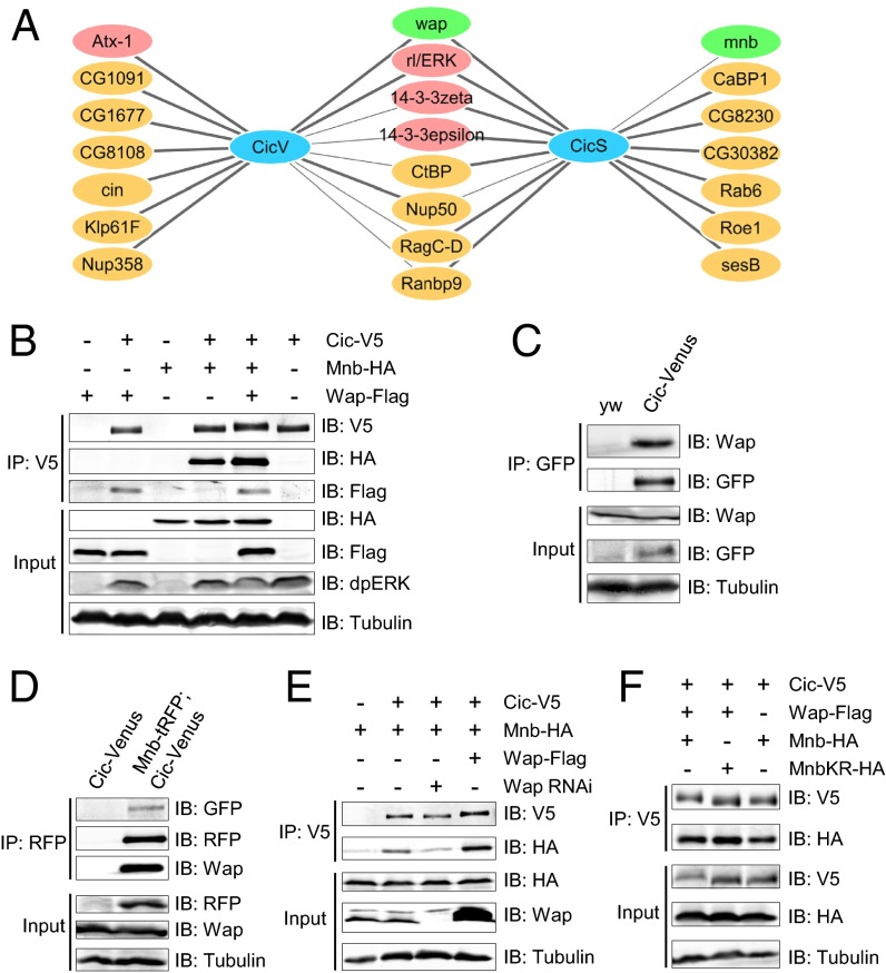
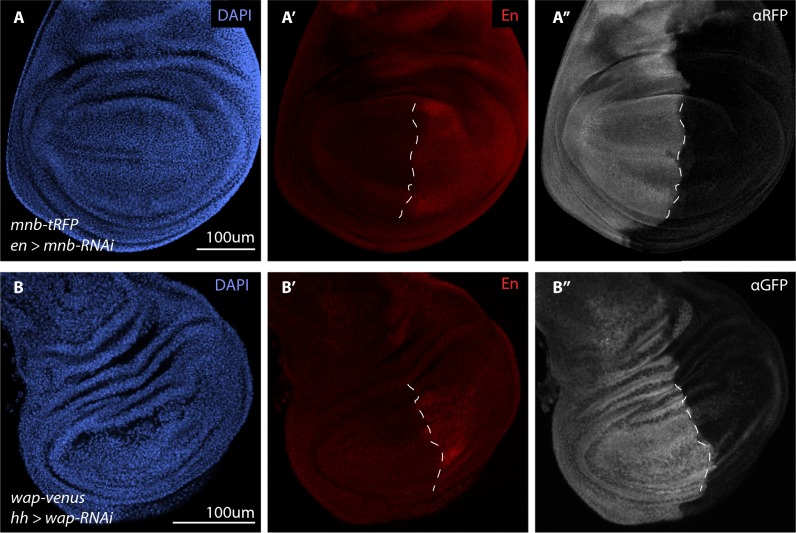
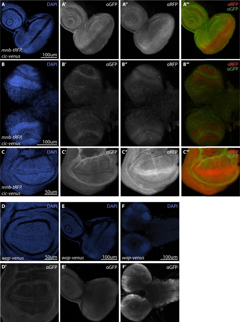
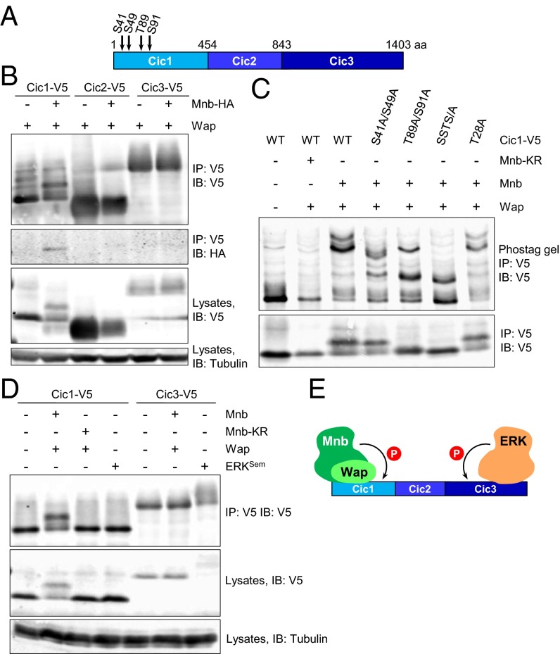
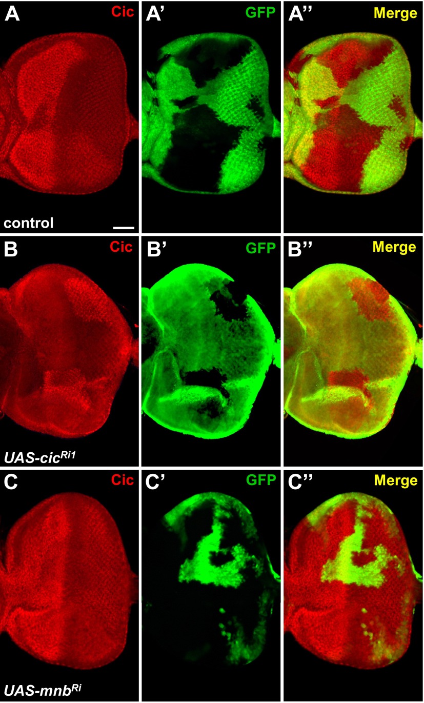
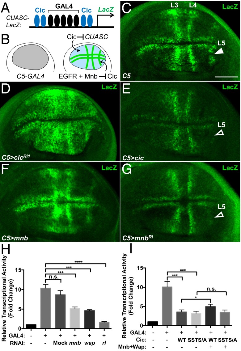
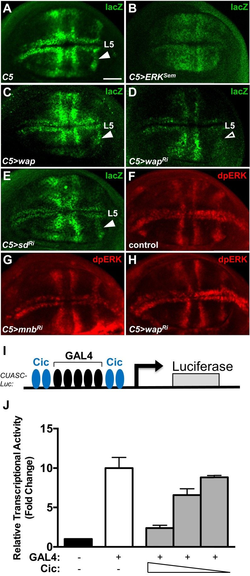
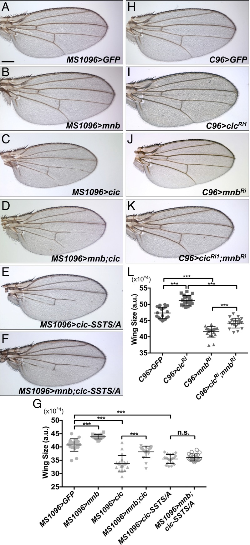
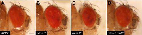
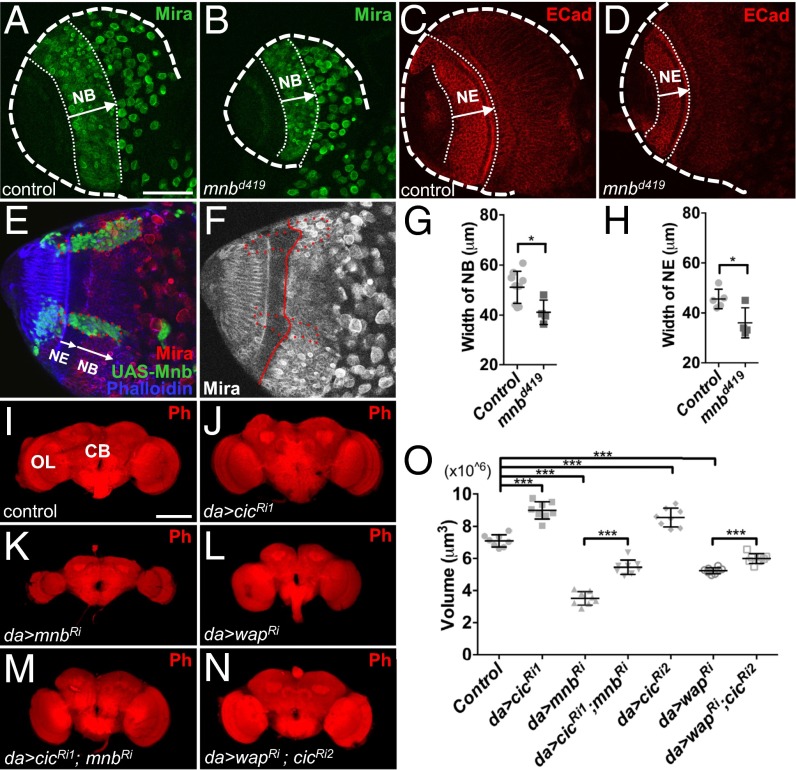
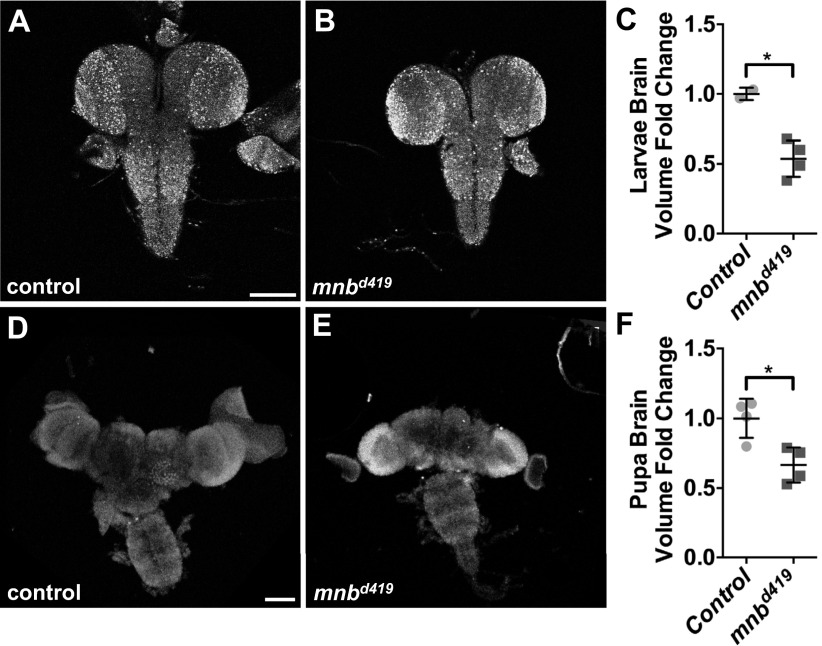
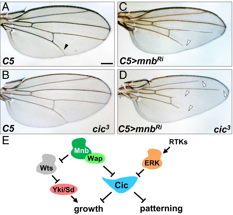
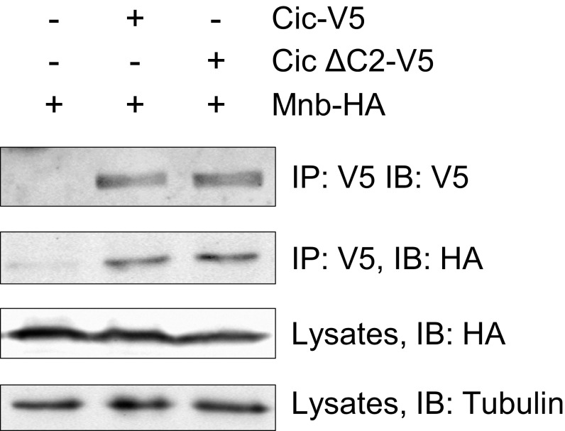
The transcriptional repressor Capicua (Cic) controls tissue patterning and restricts organ growth, and has been recently implicated in several cancers. Cic has emerged as a primary sensor of signaling downstream of the receptor tyrosine kinase (RTK)/extracellular signal-regulated kinase (ERK) pathway, but how Cic activity is regulated in different cellular contexts remains poorly understood. We found that the kinase Minibrain (Mnb, ortholog of mammalian DYRK1A), acting through the adaptor protein Wings apart (Wap), physically interacts with and phosphorylates the Cic protein. Mnb and Wap inhibit Cic function by limiting its transcriptional repressor activity. Down-regulation of Cic by Mnb/Wap is necessary for promoting the growth of multiple organs, including the wings, eyes, and the brain, and for proper tissue patterning in the wing. We have thus uncovered a previously unknown mechanism of down-regulation of Cic activity by Mnb and Wap, which operates independently from the ERK-mediated control of Cic. Therefore, Cic functions as an integrator of upstream signals that are essential for tissue patterning and organ growth. Finally, because DYRK1A and CIC exhibit, respectively, prooncogenic vs. tumor suppressor activities in human oligodendroglioma, our results raise the possibility that DYRK1A may also down-regulate CIC in human cells.

Keywords: DYRK1A; capicua; minibrain; organ growth; tissue patterning.


Conflict of interest statement

The authors declare no conflict of interest.

Figures

Fig. 1.
Fig. 1.
Mnb and Wap physically interact with Cic, and Wap promotes the binding of Mnb to Cic. (A) The Cic protein interactome identified in Drosophila S2 cells (CicS) and embryos (CicV). Thick lines, highly significant interactions. A complete dataset is in Dataset S1. (B) Western blots showing coimmunoprecipitation of Cic, Mnb, and Wap in S2 cells. Endogenous dpERK is stabilized by Cic expression. (C and D) Coimmunoprecipitation of Cic, Mnb, and Wap in vivo using embryo lysates from yw (control), cic-Venus, or cic-Venus crossed with mnb-tRFP. (E) Wap is required and sufficient to bridge Cic and Mnb. (F) Cic mobility was changed when Cic was coexpressed with Wap and wild-type Mnb, but not kinase-dead Mnb (MnbKR).
Fig. S1.
Fig. S1.
Depletion of fluorescently tagged endogenous Mnb and Wap confirms the on-target effect of mnb-RNAi and wap-RNAi. (AB′′) Wing discs from third instar larvae of the indicated genotypes. The en-GAL4 and hh-GAL4 drivers are expressed in the posterior compartment of wing discs (red in A’ and B’) and were used to drive the expression of UAS-mnb RNAi (AA′′) and UAS-wap RNAi (BB′′) respectively. Cell nuclei were detected with DAPI (blue in A and B) whereas Mnb-tRFP (gray in A′′) and Wap-Venus (gray in B′′) were detected by anti-tRFP and anti-GFP antibodies, respectively. (Scale bars: 100 µm.) Dashed lines (A′ and B′) indicate the anterior-posterior compartment boundary of the wing pouch.
Fig. S2.
Fig. S2.
Mnb-RFP, Cic-Venus, and Wap-Venus expression in third instar larval tissues. (AF) Cic-Venus and Wap-Venus expression was detected by GFP antibody, Mnb-tRFP was detected by tRFP antibody and cell nuclei were detected by DAPI (blue). (A′–C′′′) Cic-Venus and Mnb-tRFP expression in eye imaginal discs (AA’’’), brain optic lobes (B’–B’’’), and the pouch region of wing imaginal discs (C’–C’’’). For merged panels (A’’’–C’’’), Cic-Venus is in green and Mnb-tRFP is in red. (DF’) Wap-Venus expression (gray) in wing imaginal discs (D’), eye imaginal discs (E’) and larval brain (F’). (Scale bars: AB′′′, E, and F, 100 µm; C and D, 50 μm.)
Fig. 2.
Fig. 2.
Mnb and ERK target different regions of Cic for phosphorylation. (A) Schematic diagram of the three Cic fragments (Cic1, Cic2, and Cic3) with locations of phosphorylation sites. (B) Mnb interacts with and phosphorylates only the amino-terminal Cic fragment, Cic1. (C) Phos-tag gel analysis of Cic1 phosphorylation. (C Bottom) Regular SDS/PAGE. (D) Mnb phosphorylates region Cic1, whereas activated ERK (ERKSem) phosphorylates region Cic3. (E) Summary of Cic binding and phosphorylation data.
Fig. S3.
Fig. S3.
Locations of Cic phosphorylation sites identified by mass spectrometry. A Cic1 region is shown (amino acids 1–453). The putative DYRK1A consensus is underlined, and the corresponding residue (T28) is highlighted in blue. T28 phosphorylation was not detected by mass spectrometry. S41 and S49 (green) were more highly phosphorylated in wild-type Mnb samples compared with MnbKR. T89 and S91 phosphorylations (red) were found exclusively in the wild-type Mnb samples.
Fig. S4.
Fig. S4.
RNAi depletion of Mnb does not increase Cic levels in imaginal eye discs. (AC′′) Mosaic eye discs of the indicated genotypes generated by using CoinFLP-GAL4 and stained with anti-Cic antibody (red). GAL4-positive cells are marked with UAS-GFP (green). (A–A′′) Control clones do not affect Cic protein levels. (B–B′′) Knockdown of cic reduces Cic protein levels. (CC′′) Knockdown of mnb does not increase Cic protein level or change Cic subcellular localization. (Scale bar: 50 μm.)
Fig. 3.
Fig. 3.
Mnb reduces Cic repressor activity. (A) Diagram of the CUASC-lacZ reporter. (B) Summary diagram of expression patterns. (CG) LacZ expression pattern resulting from C5-GAL4–directed activation of CUASC-lacZ in wing discs from control (C), UAS-cicRNAi1 (D), UAS-cic (E), UAS-mnb (F), and UAS-mnbRNAi (G) larvae. (Scale bar: 50 μm.) (H and I) Luciferase assays using CUASC-Luc reporter in S2 cells. (H) mnb, wap, and rl (ERK) are required to limit the activity of Cic. (I) Mnb and Wap reduce transcriptional repressor activity of wild-type Cic, but not of the phosphorylation site mutant, Cic-SSTS/A. n.s., not significant, *P < 0.05, **P < 0.01, ***P < 0.001, statistical significance was analyzed by using unpaired Student’s t test. Error bars represent SD.
Fig. S5.
Fig. S5.
Modulation of Cic repressor activity by Mnb and Wap does not require Sd and does not affect activity of ERK. (AE) LacZ expression pattern resulting from C5-GAL4–directed activation of CUASC-lacZ in wing imaginal discs from control (A), UAS-ERKSem (B), UAS-wap (C), UAS-wapRNAi (D), and UAS-sdRNAi (E) larvae. Expression of ERKSem and Wap led to a broader activation of the reporter (B and C), whereas knockdown of wap resulted in lower LacZ expession, particularly in presumptive vein L5 (D). Knockdown of sd did not alter the normal pattern of expression in presumptive veins. (FH) dpERK expression pattern in wing discs from control (F), C5-GAL4 > UAS-mnbRNAi (G), and C5-GAL4 > UAS-wapRNAi (H) larvae. (I) Schematic diagram of the CUASC-Luc reporter construct. (J) Dose-dependent repression of CUASC-Luc expression by Cic. S2 cells were cotransfected with the reporter CUASC-Luc, pMT-GAL4, and decreasing amounts of Cic-expressing plasmid (500 ng, 250 ng, 125 ng). The values shown are fold changes over the negative control set at 1. (Scale bar: 50 μm.)
Fig. 4.
Fig. 4.
Mnb opposes Cic function in controlling wing growth. (AF) Wings from adult female flies expressing UAS-GFP as a control (A), UAS-mnb (B), UAS-cic (C), UAS-mnb together with UAS-cic (D), UAS-cic-SSTS/A (E), and UAS-mnb together with UAS-cic-SSTS/A (F) using the MS1096-GAL4 driver. (G) Quantification of the wing areas for the genotypes shown in AF (n = 20 for each genotype). (HK) Wings from adult female flies expressing UAS-GFP as a control (H), UAS-cicRNAi1 (I), UAS-mnbRNAi (J), and UAS-cicRNAi1 together with UAS-mnbRNAi (K) using the C96-GAL4 driver. (L) Quantification of the wing areas for the genotypes shown in HK (n = 20 for each genotype). *P < 0.05, **P < 0.01, ***P < 0.001. Statistical significance was analyzed by using Student’s t test. Error bars represent SD. (Scale bar: 200 μm.)
Fig. S6.
Fig. S6.
Mnb opposes Cic function in controlling eye growth. Adult female flies expressing UAS-GFP (A), UAS-cicRNAi1 (B), UAS-mnb-RNAi (C), and UAS-mnb-RNAi together with UAS-cicRNAi1 (D) under the control of the da-GAL4 driver. Knockdown of mnb results in a smaller eye (C), which is reversed by a concomitant knockdown of cic (D). (Scale bar: 100 μm.)
Fig. 5.
Fig. 5.
Reduction in cic level restores adult brain size in mnb mutants. (A and B) Neuroblast (NB) regions (Mira-positive cells) in larval CNS from control (w1118) (A) and mnbd419 animals (B). (C and D) Neuroepithelium (NE) regions (E-cad positive cells) in larval CNS from control (w1118) (C) and mnbd419 animals (D). (E and F) NE region is expanded cell-autonomously in UAS-mnb overexpression clones (marked in green in E). Dotted red line, clone areas; solid red line, boundary between NB and NE. (G) Quantification of results in A and B (n = 9, 4). (H) Quantification of results in C and D (n = 5, 4). (IN) Brains from adult female flies with the indicated genotypes. da-GAL4 driver was used to drive the expression of UAS-GFP (I), UAS-cicRNAi1 (J), UAS-mnbRNAi (K), UAS-wapRNAi (L), UAS-cicRNAi1 together with UAS-mnbRNAi (M), or UAS-cicRNAi2 together with UAS-wapRNAi (N). Ph, phalloidin stain. (O) Quantification of brain volumes for the genotypes shown in IN (n = 8 for each genotype). (Scale bars: AH, 50 μm; IN, 100 μm.) *P < 0.05, **P < 0.01, ***P < 0.001. Error bars represent SD. Statistical significance was analyzed by using Student’s t test.
Fig. S7.
Fig. S7.
Loss of mnb leads to smaller brain size in larvae and pupae. (A and B) Third instar larval brains from control (w1118) (A) and mnbd419 (B) animals. (C) Quantification of the larval brain volumes in A and B (n = 2, 4). (D and E) Pupal brains from control (w1118) (D) and mnbd419 (E) animals. (F) Quantification of the pupal brain volumes in D and E (n = 4, 4). *P < 0.05. Statistical significance was analyzed by using Student’s t test. (Scale bars: 100 μm.)
Fig. 6.
Fig. 6.
Mnb and ERK function additively to inhibit Cic. Wings from adult female flies of the following genotypes: C5-GAL4 (A), C5-GAL4 cic3/+ (B), C5-GAL4/UAS-mnbRNAi (C), C5-GAL4 cic3/UAS-mnbRNAi (D). (Scale bar: 200 μm.) (E) Cic integrates upstream signals to control organ growth and tissue patterning.
Fig. S8.
Fig. S8.
Deletion of the C2 motif does not affect the binding between Cic and Mnb. Protein lysates from S2 cells transfected with the indicated plasmids were incubated with anti-V5 beads, and immunocomplexes were analyzed on Western blots probed with anti-V5, anti-HA, and anti-tubulin antibodies.

References

    1. Jiménez G, Guichet A, Ephrussi A, Casanova J. Relief of gene repression by torso RTK signaling: Role of capicua in Drosophila terminal and dorsoventral patterning. Genes Dev. 2000;14(2):224–231. - PMC - PubMed
    1. Jiménez G, Shvartsman SY, Paroush Z. The Capicua repressor--a general sensor of RTK signaling in development and disease. J Cell Sci. 2012;125(Pt 6):1383–1391. - PMC - PubMed
    1. Goff DJ, Nilson LA, Morisato D. Establishment of dorsal-ventral polarity of the Drosophila egg requires capicua action in ovarian follicle cells. Development. 2001;128(22):4553–4562. - PubMed
    1. Roch F, Jiménez G, Casanova J. EGFR signalling inhibits Capicua-dependent repression during specification of Drosophila wing veins. Development. 2002;129(4):993–1002. - PubMed
    1. Astigarraga S, et al. A MAPK docking site is critical for downregulation of Capicua by Torso and EGFR RTK signaling. EMBO J. 2007;26(3):668–677. - PMC - PubMed

Publication types

MeSH terms