. 2016 Aug 23:7:273.
doi: 10.3389/fphar.2016.00273. eCollection 2016.

Adenosine Receptor Stimulation by Polydeoxyribonucleotide Improves Tissue Repair and Symptomology in Experimental Colitis

Affiliations

Adenosine Receptor Stimulation by Polydeoxyribonucleotide Improves Tissue Repair and Symptomology in Experimental Colitis

Giovanni Pallio et al. Front Pharmacol. .

Erratum in

Abstract

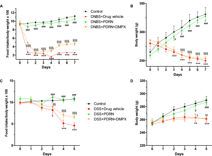
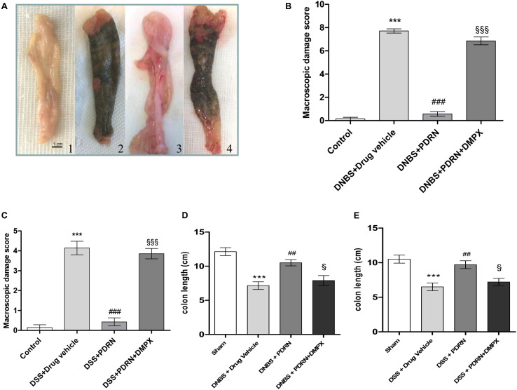
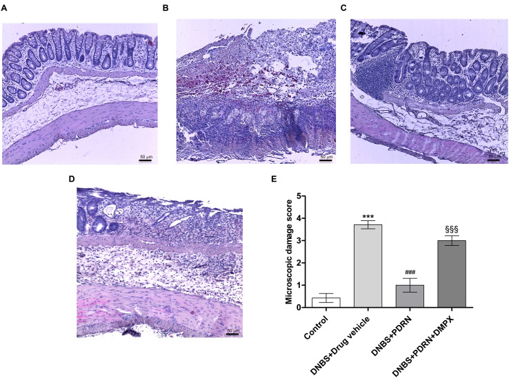
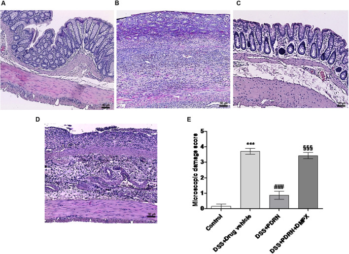
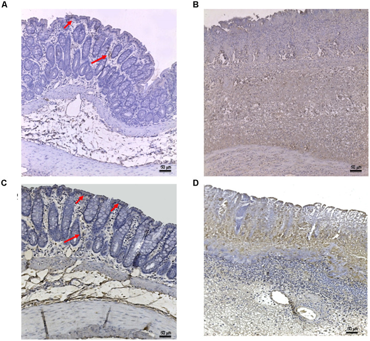
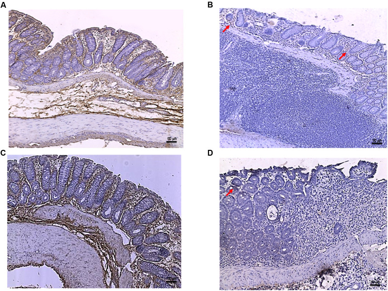
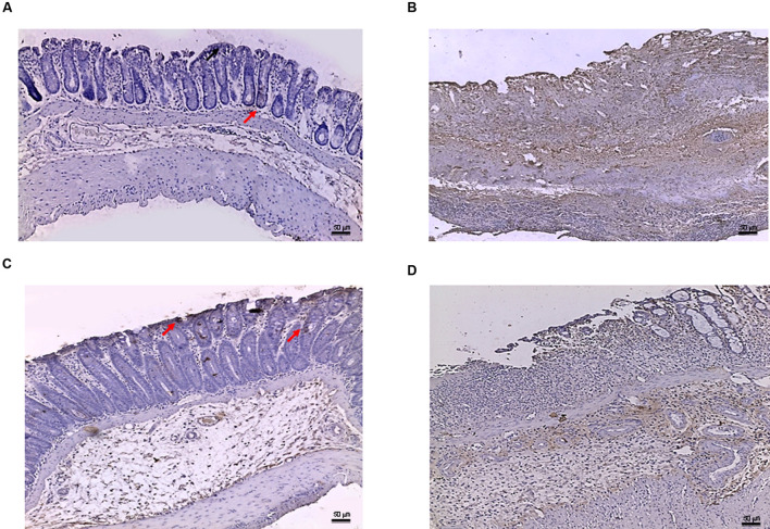
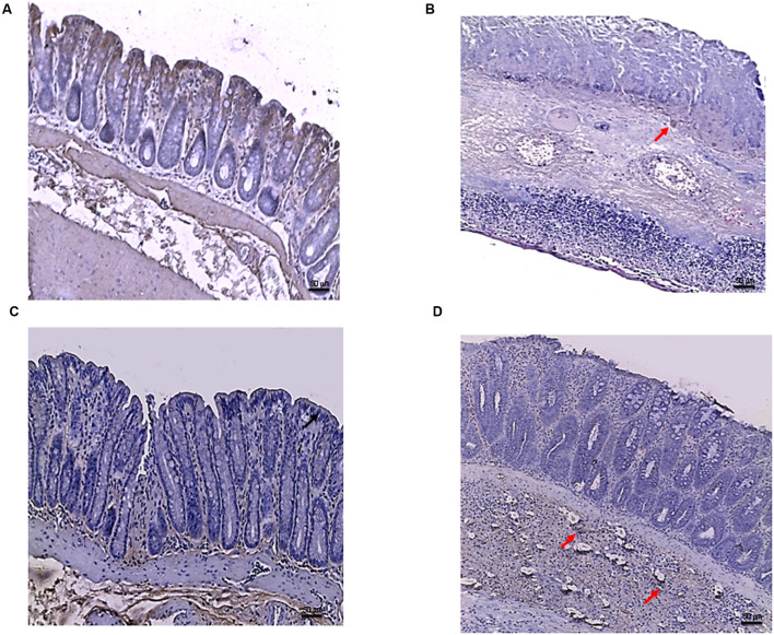
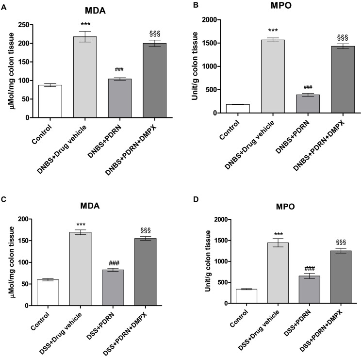
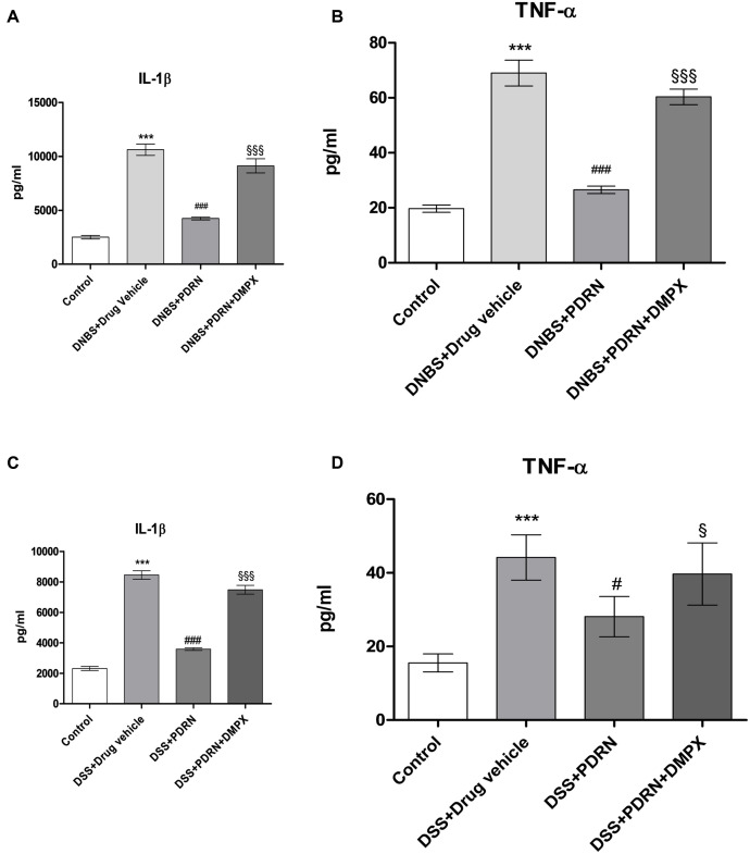
Activation of the adenosine receptor pathway has been demonstrated to be effective in improving tissue remodeling and blunting the inflammatory response. Active colitis is characterized by an intense inflammatory reaction resulting in extensive tissue damage. Symptomatic improvement requires both control of the inflammatory process and repair and remodeling of damaged tissues. We investigated the ability of an A2A receptor agonist, polydeoxyribonucleotide (PDRN), to restore tissue structural integrity in two experimental colitis models using male Sprague-Dawley rats. In the first model, colitis was induced with a single intra-colonic instillation of dinitrobenzenesulfonic acid (DNBS), 25 mg diluted in 0.8 ml 50% ethanol. After 6 h, animals were randomized to receive either PDRN (8 mg/kg/i.p.), or PDRN + the A2A antagonist [3,7-dimethyl-1-propargylxanthine (DMPX); 10 mg/kg/i.p.], or vehicle (0.8 ml saline solution) daily. In the second model, dextran sulfate sodium (DSS) was dissolved in drinking water at a concentration of 8%. Control animals received standard drinking water. After 24 h animals were randomized to receive PDRN or PDRN+DMPX as described above. Rats were sacrificed 7 days after receiving DNBS or 5 days after DSS. In both experimental models of colitis, PDRN ameliorated the clinical symptoms and weight loss associated with disease as well as promoted the histological repair of damaged tissues. Moreover, PDRN reduced expression of inflammatory cytokines, myeloperoxidase activity, and malondialdehyde. All these effects were abolished by the concomitant administration of the A2A antagonist DMPX. Our study suggests that PDRN may represent a promising treatment for improving tissue repair during inflammatory bowel diseases.

Keywords: A2A agonist; PDRN; apoptosis; colitis; inflammation.


Figures

FIGURE 1
FIGURE 1
Clinical evaluations. Food intake in DNBS-treated animals (A), weight loss in DNBS-treated animals (B), food intake in DSS-treated animals (C), and weight loss in DSS-treated animals (D) was recorded. Values were obtained from seven animals per group and are expressed as means and SEM. p < 0.01, ∗∗p < 0.001, ∗∗∗p < 0.0001 vs control group; #p < 0.01, ###p < 0.0001 vs DNBS/DSS+drug vehicle group; § p < 0.01, §§ p < 0.001, §§§ p < 0.0001 vs DNBS/DSS+PDRN group.
FIGURE 2
FIGURE 2
Macroscopic evaluations. Colons (A) from control (1), DNBS+drug vehicle (2), DNBS+PDRN (3), DNBS+PDRN+DMPX (4) are depicted. Macroscopic damage scores from the DNBS model and the DSS model are summarized in (B) and (C), respectively, while colon length is shown in (D) and (E) for these models. Values were obtained from seven animals per group, and are expressed as the means and SEM. ∗∗∗p < 0.0001 vs control group; ##p < 0.001, ###p < 0.0001 vs DNBS/DSS+drug vehicle group; § p < 0.01, §§§ p < 0.0001 vs DNBS/DSS+PDRN group.
FIGURE 3
FIGURE 3
Histological evaluations from DNBS-treated animals. Representative photomicrographs of H&E stained tissues (original magnification 10×) derived from control (A), DNBS+drug vehicle (B), DNBS+PDRN (C), or DNBS+PDRN+DMPX (D) animals are shown. The graph represents the microscopic damage score (E), where values are expressed as the means and SEM of seven animals. ∗∗∗p < 0.0001 vs control group; ###p < 0.0001 vs DNBS+drug vehicle group; §§§p < 0.0001 vs DNBS+PDRN group. Scale bar 50 μm.
FIGURE 4
FIGURE 4
Histological evaluations from DSS-treated animals. Representative H&E images (original magnification 10×) of tissues derived from control (A), DSS+drug vehicle (B), DSS+PDRN (C), or DSS+PDRN+DMPX (D) treated animals are shown here. The graph in (E) represents the microscopic damage score values expressed as means and SEM derived from seven animals for each condition. ∗∗∗p < 0.0001 vs control group; ###p < 0.0001 vs DSS+drug vehicle group; §§§ p < 0.0001 vs DSS+PDRN group. Scale bar 50 μm.
FIGURE 5
FIGURE 5
Bax immunostaining from DNBS-treated animals. Representative Bax immunostaining (original magnification 10×) of colons derived from control (A), DNBS+drug vehicle (B), DNBS+PDRN (C), or DNBS+PDRN+DMPX (D) treated animals are depicted. The arrows in (A) and (C) point to areas of slight positivity. The pictures in (B,D) demonstrate a diffuse staining for the apoptotic factor. Scale bar 50 μm.
FIGURE 6
FIGURE 6
Bcl-2 immunostaining from DNBS-treated animals. Representative Bcl-2 immunostaining (original magnification 10×) of tissues derived from control (A), DNBS+drug vehicle (B), DNBS+PDRN (C), or DNBS+PDRN+DMPX (D) treated animals are depicted. The images in (A,C) demonstrate diffuse Bcl-2 staining, while the arrows in (B,D) point to the residual Bcl-2 staining in the mucosal layer. Scale bar 50 μm.
FIGURE 7
FIGURE 7
Bax immunostaining from DSS-treated animals. Representative Bax immunostaining (original magnification 10×) of colons derived from control (A), DSS+drug vehicle (B), DSS+PDRN (C), or DSS+PDRN+DMPX (D) treated animals are shown. The arrows in (A,C) point to regions of slight positivity. The pictures in (B,D) demonstrate a diffuse staining for the apoptotic factor. Scale bar 50 μm.
FIGURE 8
FIGURE 8
Bcl-2 immunostaining from DSS-treated animals. Representative images of Bcl-2 immunostaining (original magnification 10×) of colons derived from control (A), DSS+drug vehicle (B), DSS+PDRN (C), or DSS+PDRN+DMPX (D) treated animals are shown. The image in (A,C) demonstrate diffuse staining for Bcl-2, while the arrows in (B,D) point to residual Bcl-2 staining in the mucosal layer. Scale bar 50 μm.
FIGURE 9
FIGURE 9
Evaluation of inflammatory markers. The effects of PDRN on malondialdehyde levels and myeloperoxidase activity in DNBS-treated animals are shown in (A) and (B), respectively, while the consequences of PDRN treatment on malondialdehyde levels and myeloperoxidase activity in the DSS model are summarized in (C) and (D), respectively. All values are expressed as means and SEM based on observations made on seven animals per group. ∗∗∗p < 0.0001 vs control group; ###p < 0.0001 vs DNBS/DSS+drug vehicle group; §§§ p < 0.0001 vs DNBS/DSS+PDRN group.
FIGURE 10
FIGURE 10
Evaluation of inflammatory cytokines. Serum of DNBS- or DSS-challenged animals was extracted, and Interleukin 1β (IL-1β) (A,C) and tumor necrosis factor-alpha (TNF-α) (B,D) levels were determined by enzyme linked immunosorbent assay (ELISA). Values shown here are expressed as the means and SEM of seven animals per group. ∗∗∗p < 0.0001 vs control group; #p < 0.01, ###p < 0.0001 vs DNBS/DSS+drug vehicle group; § p < 0.01, §§§ p < 0.0001 vs DNBS/DSS+PDRN group.

References

    1. Aherne C. M., Saeedi B., Collins C. B., Masterson J. C., McNamee E. N., Perrenoud L., et al. (2015). Epithelial-specific A2B adenosine receptor signaling protects the colonic epithelial barrier during acute colitis. Mucosal Immunol. 8 1324–1338. - PMC - PubMed
    1. Alam M. S., Costales M. G., Cavanaugh C., Williams K. (2015). Extracellular adenosine generation in the regulation of pro-inflammatory responses and pathogencolonization. Biomolecules 5 775–792. 10.3390/biom5020775 - DOI - PMC - PubMed
    1. Altavilla D., Squadrito F., Polito F., Irrera N., Calò M., Lo Cascio P., et al. (2011). Activation of adenosine A2A receptors restores the altered cell-cycle machinery during impaired wound healing in genetically diabetic mice. Surgery 149 253–261. 10.1016/j.surg.2010.04.024 - DOI - PubMed
    1. Antonioli L., Csóka B., Fornai M., Colucci R., Kókai E., Blandizzi C., et al. (2014). Adenosine and inflammation: what’s new on the horizon? Drug Discov. Today 19 1051–1068. 10.1016/j.drudis.2014.02.010 - DOI - PubMed
    1. Antonioli L., Fornai M., Colucci R., Awwad O., Ghisu N., Tuccori M., et al. (2010). The blockade of adenosine deaminase ameliorates chronic experimental colitis through the recruitment of adenosine A2A and A3 receptors. J. Pharmacol. Exp. Ther. 335 434–442. 10.1124/jpet.110.171223 - DOI - PubMed