. 2016 Sep;12(3):2087-2091.
doi: 10.3892/ol.2016.4907. Epub 2016 Jul 22.

Branchiogenic carcinoma with high-risk-type human papillomavirus infection: A case report

Affiliations

Branchiogenic carcinoma with high-risk-type human papillomavirus infection: A case report

Hiroyuki Maeda et al. Oncol Lett. 2016 Sep.

Abstract

Branchiogenic carcinoma (BC) usually appears as a mass lesion with a predominant cystic component. Since lymph node metastasis from oropharyngeal carcinoma (OPC) has a cystic appearance, it is occasionally difficult to distinguish between BC and nodal metastases from clinically silent OPC. Factors associated with the malignant transformation process in BC remain obscure. The present study reports the case of a 56-year-old man with a right cystic cervical mass that was diagnosed as squamous cell carcinoma based on examination by fine-needle aspiration biopsy. The primary tumor could not be detected despite several imaging examinations, a pan-endoscopy of the head and neck, esophagus and stomach, biopsies of the head and neck regions, and bilateral tonsillectomies. The pathological findings of the surgical specimens from a radical neck dissection were consistent with the histological characteristics of BC, with evidence of transition from dysplasia through intraepithelial carcinoma to invasive carcinoma. Normal squamous epithelium and dysplastic and cancerous portions in the BC showed strong p16INK4a immunoreactivity. The expression of p16INK4a was also observed in all 9 nodal metastases in the neck dissection specimens. The cystic formation observed in the BC was not observed in the nodal metastases. As the presence of human papillomavirus-16 in the tumor was confirmed by polymerase chain reaction, quantitative polymerase chain reaction was employed for the measurement of human papillomavirus-16 viral load and integration. The results showed that the viral load of human papillomavirus-16 was 3.01×10⁷/50 ng genomic DNA, and the E2/E6 ratio was 0.13, so the integration state was judged to be the mixed type. To the best of our knowledge, this is the first report of BC associated with high-risk-type human papillomavirus infection. The study indicates that a human papillomavirus-positive neck mass may not necessarily be OPC, but that it could be BC with a poor prognosis. This report lends support to the existence of BC and proposes that the etiology is human papillomavirus infection.

Keywords: branchiogenic carcinoma; high-risk-type; human papillomavirus; integration; p16INK4a expression.


Figures

Figure 1.
Figure 1.
Computed tomography (CT) and magnetic resonance imaging of the neck. (A) CT with contrast enhancement. The tumor was anterior to the sternocleidomastoid muscle. The tumor contained a prominent cystic portion and a solid portion. (B) T1-weighted image with gadolinium enhancement (axial view). The cyst wall and solid portion (medial region of the tumor) showed high signal intensity on magnetic resonance imaging. (C) T1-weighted image with gadolinium enhancement (coronal view). The solid portion was located at the superiomedial aspect of the tumor.
Figure 2.
Figure 2.
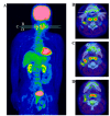
Images of 18F-fluorodeoxyglucose (FDG) positron emission tomography combined with computed tomography. (A) Whole-body scan. FDG uptake was observed in the right cervical mass (maximum standardized uptake value, 9.2). Physiological FDG uptake was also observed in the brain, tonsils, heart, kidneys and urinary bladder. (B) Axial slice at level B in (A). (C) Axial slice at level C in (A). (D) Axial slice at level D in (A). FDG uptake was observed at the cyst wall and in the solid region of the tumor. The palatine tonsil and tongue base also exhibited physiological FDG uptake.
Figure 3.
Figure 3.
Hematoxylin-eosin staining of the tumor. (A) Low magnification view of the tumor. The tumor formed a cyst consisting of normal squamous epithelium and invasive cancer. Scattered lymphoid cells were observed in the subepithelial layer. Bar, 100 µm. (B) High magnification view of the boxed region in (A), showing squamous epithelium with a few areas of dysplasia. The epithelial cells showed marked koilocytosis. Bar, 25 µm.
Figure 4.
Figure 4.
Transition from severe dysplasia to squamous cell carcinoma (hematoxylin-eosin staining). (A) Low magnification view of the transition area. Highly dysplastic epithelium (arrow B) with a change to squamous cell carcinoma (arrow C). Bar, 100 µm. (B) High magnification view of the region indicated by arrow B in (A). The epithelium has retained a distinct basal cell layer and cellular differentiation. Epithelial cell atypia was minimal. Bar, 25 µm. (C) High magnification view of the region indicated by arrow C in (A). The epithelium has lost cellular differentiation and polarity. Bar, 25 µm.
Figure 5.
Figure 5.
Expression of p16INK4a. (A) The solid portion of the tumor. The tumor cells showed strong p16INK4a expression. Bar, 100 µm. (B) The squamous epithelium also showed strong p16INK4a expression. Bar, 100 µm. (C) Nodal metastasis. Tumor cells in the lymph node showed strong p16INK4a expression. There was no cystic formation in the nodal metastasis. Bar, 100 µm. Lum, lumen of the tumor.
Figure 6.
Figure 6.
Polymerase chain reaction (PCR) for detection of human papilloma virus (HPV). (A) PCR results using GP5+/GP6+ consensus primer sets (6). (B) PCR results using MY09/MY11 consensus primer sets (6). (C) PCR results of the internal control, β-globin (6). Lane 1, tumor sample; lane 2, oropharyngeal cancer sample without HPV infection as the negative control; lane 3, CaSki cells as the positive control; and lane 4, no sample DNA in the reaction solution. The expression of HPV DNA was observed in the tumor sample and direct sequencing of the PCR product indicated HPV-16.

References

    1. Martin H, Morfit HM, Ehrlich H. The case for branchiogenic cancer (malignant branchioma) Ann Surg. 1950;132:867–887. doi: 10.1097/00000658-195011000-00002. - DOI - PMC - PubMed
    1. Khafif RA, Prichep R, Minkowitz S. Primary branchiogenic carcinoma. Head Neck. 1989;11:153–163. doi: 10.1002/hed.2880110209. - DOI - PubMed
    1. Hasegawa M, Maeda H, Deng Z, Kiyuna A, Ganaha A, Yamashita Y, Matayoshi S, Agena S, Toita T, Uehara T, Suzuki M. Prediction of concurrent chemoradiotherapy outcome in advanced oropharyngeal cancer. Int J Oncol. 2014;45:1017–1026. - PMC - PubMed
    1. Deng Z, Hasegawa M, Aoki K, Matayoshi S, Kiyuna A, Yamashita Y, Uehara T, Agena S, Maeda H, Xie M, Suzuki M. A comprehensive evaluation of human papillomavirus positive status and p16INK4a overexpression as a prognostic biomarker in head and neck squamous cell carcinoma. Int J Oncol. 2014;45:67–76. - PMC - PubMed
    1. Deng Z, Hasegawa M, Yamashita Y, Matayoshi S, Kiyuna A, Agena S, Uehara T, Maeda H, Suzuki M. Prognostic value of human papillomavirus and squamous cell carcinoma antigen in head and neck squamous cell carcinoma. Cancer Sci. 2012;103:2127–2134. doi: 10.1111/cas.12009. - DOI - PMC - PubMed

LinkOut - more resources